|
Turricephaly
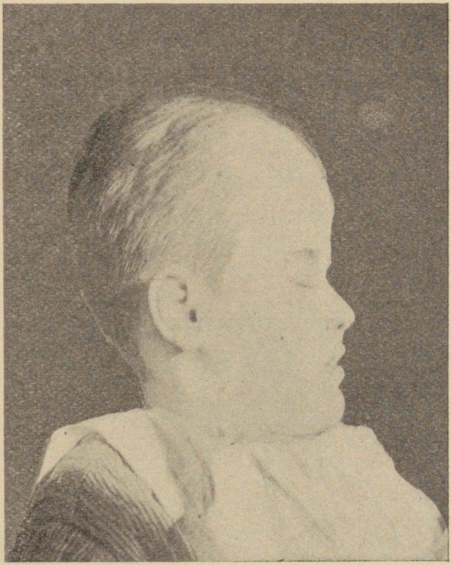
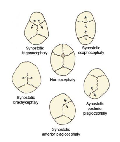
Turricephaly is a type of cephalic disorder where the head appears tall with a small length and width. It is due to premature closure of the coronal suture plus any other suture, like the lambdoid, or it may be used to describe the premature fusion of all sutures. It should be differentiated from Crouzon syndrome. Oxycephaly (or acrocephaly) is a form of turricephaly where the head is cone-shaped, and is the most severe of the craniosynostoses. Presentation Common associations It may be associated with: * 8th cranial nerve lesion * Optic nerve compression * Intellectual disability * Syndactyly Diagnosis Treatment See also * Acrocephalosyndactylia Acrocephalosyndactyly is a group of autosomal dominant congenital disorders characterized by craniofacial (craniosynostosis) and hand and foot (syndactyly) abnormalities. When polydactyly is present, the classification is acrocephalopolysyndactyl ... References Further reading NINDS Overview* External links Congenit ... [...More Info...] [...Related Items...] OR: [Wikipedia] [Google] [Baidu] |
Syphilis And Marriage 5 (Acrocephalus)
Syphilis () is a sexually transmitted infection caused by the bacterium ''Treponema pallidum'' subspecies ''pallidum''. The signs and symptoms of syphilis vary depending in which of the four stages it presents (primary, secondary, latent, and tertiary). The primary stage classically presents with a single chancre (a firm, painless, non-itchy skin ulceration usually between 1 cm and 2 cm in diameter) though there may be multiple sores. In secondary syphilis, a diffuse rash occurs, which frequently involves the palms of the hands and soles of the feet. There may also be sores in the mouth or vagina. In latent syphilis, which can last for years, there are few or no symptoms. In tertiary syphilis, there are gummas (soft, non-cancerous growths), neurological problems, or heart symptoms. Syphilis has been known as "the great imitator" as it may cause symptoms similar to many other diseases. Syphilis is most commonly spread through sexual activity. It may also be transmitte ... [...More Info...] [...Related Items...] OR: [Wikipedia] [Google] [Baidu] |
Cephalic Disorder
Cephalic disorders () are congenital conditions that stem from damage to, or abnormal development of, the budding nervous system. Cephalic disorders are not necessarily caused by a single factor, but may be influenced by hereditary or genetic conditions, nutritional deficiencies, or by environmental exposures during pregnancy, such as medication taken by the mother, maternal infection, or exposure to radiation. Some cephalic disorders occur when the cranial sutures (the fibrous joints that connect the bones of the skull) join prematurely. Most cephalic disorders are caused by a disturbance that occurs very early in the development of the fetal nervous system. The human nervous system develops from a small, specialized plate of cells on the surface of the embryo. Early in development, this plate of cells forms the neural tube, a narrow sheath that closes between the third and fourth weeks of pregnancy to form the brain and spinal cord of the embryo. Four main processes are resp ... [...More Info...] [...Related Items...] OR: [Wikipedia] [Google] [Baidu] |
Coronal Suture
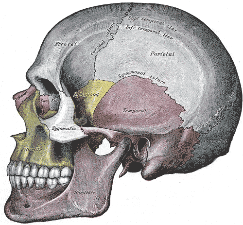
The coronal suture is a dense, fibrous connective tissue joint that separates the two parietal bones from the frontal bone of the skull. Structure The coronal suture lies between the paired parietal bones and the frontal bone of the skull. It runs from the pterion on each side. Nerve supply The coronal suture is likely supplied by a branch of the trigeminal nerve. Development The coronal suture is derived from the paraxial mesoderm. Clinical significance If certain bones of the skull grow too fast then premature fusion of the sutures may occur. This can result in skull deformities. There are two possible deformities that can be caused by the premature closure of the coronal suture: * a high, tower-like skull called "oxycephaly" or "turret skull". * a twisted and asymmetrical skull called "plagiocephaly". References * "Sagittal suture." ''Stedman's Medical Dictionary, 27th ed.'' (2000). * Moore, Keith L., and T.V.N. Persaud. ''The Developing Human: Clinically Orien ... [...More Info...] [...Related Items...] OR: [Wikipedia] [Google] [Baidu] |
Suture (anatomy)
In anatomy, a suture is a fairly rigid joint between two or more hard elements of an organism, with or without significant overlap of the elements. Sutures are found in the skeletons or exoskeletons of a wide range of animals, in both invertebrates and vertebrates. Sutures are found in animals with hard parts from the Cambrian period to the present day. Sutures were and are formed by several different methods, and they exist between hard parts that are made from several different materials. Vertebrate skeletons The skeletons of vertebrate animals (fish, amphibians, reptiles, birds, and mammals) are made of bone, in which the main rigid ingredient is calcium phosphate. Cranial sutures The skulls of most vertebrates consist of sets of bony plates held together by cranial sutures. These sutures are held together mainly by Sharpey's fibers which grow from each bone into the adjoining one. Sutures in the ankles of land vertebrates In the type of crurotarsal ankle which is found ... [...More Info...] [...Related Items...] OR: [Wikipedia] [Google] [Baidu] |
Lambdoid Suture
The lambdoid suture (or lambdoidal suture) is a dense, fibrous connective tissue joint on the posterior aspect of the skull that connects the parietal bones with the occipital bone. It is continuous with the occipitomastoid suture. Structure The lambdoid suture is between the paired parietal bones and the occipital bone of the skull. It runs from the asterion on each side. Nerve supply The lambdoid suture may be supplied by a branch of the supraorbital nerve, a branch of the frontal branch of the trigeminal nerve. Clinical significance At birth, the bones of the skull do not meet. If certain bones of the skull grow too fast, then craniosynostosis (premature closure of the sutures) may occur. This can result in skull deformities. If the lambdoid suture closes too soon on one side, the skull will appear twisted and asymmetrical, a condition called "plagiocephaly". Plagiocephaly refers to the shape and not the condition. The condition is craniosynostosis. The lambdoid sut ... [...More Info...] [...Related Items...] OR: [Wikipedia] [Google] [Baidu] |
Crouzon Syndrome
Crouzon syndrome is an autosomal dominant genetic disorder known as a branchial arch syndrome. Specifically, this syndrome affects the first branchial (or pharyngeal) arch, which is the precursor of the maxilla and mandible. Since the branchial arches are important developmental features in a growing embryo, disturbances in their development create lasting and widespread effects. This syndrome is named after Octave Crouzon, a French physician who first described this disorder. First called "craniofacial dysostosis" ("craniofacial" refers to the skull and face, and " dysostosis" refers to malformation of bone), the disorder was characterized by a number of clinical features which can be described by the rudimentary meanings of its former name. This syndrome is caused by a mutation in the fibroblast growth factor receptor 2 (''FGFR2''), located on chromosome 10. The developing fetus's skull and facial bones fuse early or are unable to expand. Thus, normal bone growth cannot occur. ... [...More Info...] [...Related Items...] OR: [Wikipedia] [Google] [Baidu] |
Craniosynostosis
Craniosynostosis is a condition in which one or more of the fibrous sutures in a young infant's skull prematurely fuses by turning into bone (ossification), thereby changing the growth pattern of the skull. Because the skull cannot expand perpendicular to the fused suture, it compensates by growing more in the direction parallel to the closed sutures. Sometimes the resulting growth pattern provides the necessary space for the growing brain, but results in an abnormal head shape and abnormal facial features. In cases in which the compensation does not effectively provide enough space for the growing brain, craniosynostosis results in increased intracranial pressure leading possibly to visual impairment, sleeping impairment, eating difficulties, or an impairment of mental development combined with a significant reduction in IQ. Craniosynostosis occurs in one in 2000 births. Craniosynostosis is part of a syndrome in 15% to 40% of affected patients, but it usually occurs as an isol ... [...More Info...] [...Related Items...] OR: [Wikipedia] [Google] [Baidu] |
Vestibulocochlear Nerve
The vestibulocochlear nerve or auditory vestibular nerve, also known as the eighth cranial nerve, cranial nerve VIII, or simply CN VIII, is a cranial nerve that transmits sound and equilibrium (balance) information from the inner ear to the brain. Through olivocochlear fibers, it also transmits motor and modulatory information from the superior olivary complex in the brainstem to the cochlea. Structure The vestibulocochlear nerve consists mostly of bipolar neurons and splits into two large divisions: the cochlear nerve and the vestibular nerve. Cranial nerve 8, the vestibulocochlear nerve, goes to the middle portion of the brainstem called the pons (which then is largely composed of fibers going to the cerebellum). The 8th cranial nerve runs between the base of the pons and medulla oblongata (the lower portion of the brainstem). This junction between the pons, medulla, and cerebellum that contains the 8th nerve is called the cerebellopontine angle. The vestibulocochlear nerv ... [...More Info...] [...Related Items...] OR: [Wikipedia] [Google] [Baidu] |
Optic Nerve
In neuroanatomy, the optic nerve, also known as the second cranial nerve, cranial nerve II, or simply CN II, is a paired cranial nerve that transmits visual system, visual information from the retina to the brain. In humans, the optic nerve is derived from optic stalks during the seventh week of development and is composed of retinal ganglion cell axons and glial cells; it extends from the optic disc to the optic chiasma and continues as the optic tract to the lateral geniculate nucleus, Pretectal area, pretectal nuclei, and superior colliculus. Structure The optic nerve has been classified as the second of twelve paired cranial nerves, but it is technically part of the central nervous system, rather than the peripheral nervous system because it is derived from an out-pouching of the diencephalon (optic stalks) during embryonic development. As a consequence, the fibers of the optic nerve are covered with myelin produced by oligodendrocytes, rather than Schwann cells of the per ... [...More Info...] [...Related Items...] OR: [Wikipedia] [Google] [Baidu] |
Intellectual Disability
Intellectual disability (ID), also known as general learning disability in the United Kingdom and formerly mental retardation,Rosa's Law, Pub. L. 111-256124 Stat. 2643(2010). is a generalized neurodevelopmental disorder characterized by significantly impaired intellectual and adaptive functioning. It is defined by an IQ under 70, in addition to deficits in two or more adaptive behaviors that affect everyday, general living. Intellectual functions are defined under DSM-V as reasoning, problem‑solving, planning, abstract thinking, judgment, academic learning, and learning from instruction and experience, and practical understanding confirmed by both clinical assessment and standardized tests. Adaptive behavior is defined in terms of conceptual, social, and practical skills involving tasks performed by people in their everyday lives. Intellectual disability is subdivided into syndromic intellectual disability, in which intellectual deficits associated with other medical and be ... [...More Info...] [...Related Items...] OR: [Wikipedia] [Google] [Baidu] |
Syndactyly
Syndactyly is a condition wherein two or more digits are fused together. It occurs normally in some mammals, such as the siamang and diprotodontia, but is an unusual condition in humans. The term is from Greek σύν, ''syn'' 'together' and δάκτυλος, ''daktulos'' 'finger'. Classification Syndactyly can be simple or complex. * In simple syndactyly, adjacent fingers or toes are joined by soft tissue. * In complex syndactyly, the bones of adjacent digits are fused. The kangaroo exhibits complex syndactyly. Syndactyly can be complete or incomplete. * In complete syndactyly, the skin is joined all the way to the tip of the involved digits. * In incomplete syndactyly, the skin is only joined part of the distance to the tip of the involved digits. Complex syndactyly occurs as part of a syndrome (such as Apert syndrome) and typically involves more digits than simple syndactyly. Fenestrated syndactyly, also known as acrosyndactyly or terminal syndactyly, means the skin is joine ... [...More Info...] [...Related Items...] OR: [Wikipedia] [Google] [Baidu] |


.jpg)

