|
Turbinates
In anatomy, a nasal concha (), plural conchae (), also called a nasal turbinate or turbinal, is a long, narrow, curled shelf of bone tissue, bone that protrudes into the breathing passage of the nose in humans and various animals. The conchae are shaped like an elongated seashell, which gave them their name (Latin ''concha'' from Greek ''κόγχη''). A concha is any of the scrolled spongy bones of the nasal cavity, nasal passages in vertebrates.''Anatomy of the Human Body'' Gray, Henry (1918) The Nasal Cavity. In humans, the conchae divide the nasal airway into four groove-like air passages, and are responsible for forcing inhaled air to flow in a steady, regular pattern around the largest possible surface area of nasal mucosa. As a cilium, ciliated mucous membrane with shallow blood supply, the nasal mucosa cleans and warms t ... [...More Info...] [...Related Items...] OR: [Wikipedia] [Google] [Baidu] |
Nasal Cavity
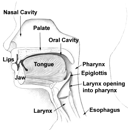
The nasal cavity is a large, air-filled space above and behind the nose in the middle of the face. The nasal septum divides the cavity into two cavities, also known as fossae. Each cavity is the continuation of one of the two nostrils. The nasal cavity is the uppermost part of the respiratory system and provides the nasal passage for inhaled air from the nostrils to the nasopharynx and rest of the respiratory tract. The paranasal sinuses surround and drain into the nasal cavity. Structure The term "nasal cavity" can refer to each of the two cavities of the nose, or to the two sides combined. The lateral wall of each nasal cavity mainly consists of the maxilla. However, there is a deficiency that is compensated for by the perpendicular plate of the palatine bone, the medial pterygoid plate, the labyrinth of ethmoid and the inferior concha. The paranasal sinuses are connected to the nasal cavity through small orifices called ostia. Most of these ostia communicate with the n ... [...More Info...] [...Related Items...] OR: [Wikipedia] [Google] [Baidu] |
Nose
A nose is a protuberance in vertebrates that houses the nostrils, or nares, which receive and expel air for respiration alongside the mouth. Behind the nose are the olfactory mucosa and the sinuses. Behind the nasal cavity, air next passes through the pharynx, shared with the digestive system, and then into the rest of the respiratory system. In humans, the nose is located centrally on the face and serves as an alternative respiratory passage especially during suckling for infants. The protruding nose that completely separate from the mouth part is a characteristic found only in therian mammals. It has been theorized that this unique mammalian nose evolved from the anterior part of the upper jaw of the reptilian-like ancestors ( synapsids). Air treatment Acting as the first interface between the external environment and an animal's delicate internal lungs, a nose conditions incoming air, both as a function of thermal regulation and filtration during respiration, as well as ... [...More Info...] [...Related Items...] OR: [Wikipedia] [Google] [Baidu] |
Inferior Nasal Concha
The inferior nasal concha (inferior turbinated bone or inferior turbinal/turbinate) is one of the three paired nasal conchae in the nose. It extends horizontally along the lateral wall of the nasal cavity and consists of a lamina of spongy bone, curled upon itself like a scroll, (''turbinate'' meaning inverted cone). The inferior nasal conchae are considered a pair of facial bones. As the air passes through the turbinates, the air is churned against these mucosa-lined bones in order to receive warmth, moisture and cleansing. Superior to inferior nasal concha are the middle nasal concha and superior nasal concha which both arise from the ethmoid bone, of the cranial portion of the skull. Hence, these two are considered as a part of the cranial bones. It has two surfaces, two borders, and two extremities. Structure Surfaces The medial surface is convex, perforated by numerous apertures, and traversed by longitudinal grooves for the lodgement of vessels. The lateral surface is con ... [...More Info...] [...Related Items...] OR: [Wikipedia] [Google] [Baidu] |
Ethmoid Bone
The ethmoid bone (; from grc, ἡθμός, hēthmós, sieve) is an unpaired bone in the skull that separates the nasal cavity from the brain. It is located at the roof of the nose, between the two orbits. The cubical bone is lightweight due to a spongy construction. The ethmoid bone is one of the bones that make up the orbit of the eye. Structure The ethmoid bone is an anterior cranial bone located between the eyes. It contributes to the medial wall of the orbit, the nasal cavity, and the nasal septum. The ethmoid has three parts: cribriform plate, ethmoidal labyrinth, and perpendicular plate. The cribriform plate forms the roof of the nasal cavity and also contributes to formation of the anterior cranial fossa, the ethmoidal labyrinth consists of a large mass on either side of the perpendicular plate, and the perpendicular plate forms the superior two-thirds of the nasal septum. Between the orbital plate and the nasal conchae are the ethmoidal sinuses or ethmoidal air cells, w ... [...More Info...] [...Related Items...] OR: [Wikipedia] [Google] [Baidu] |
Ciliated
The cilium, plural cilia (), is a membrane-bound organelle found on most types of eukaryotic cell, and certain microorganisms known as ciliates. Cilia are absent in bacteria and archaea. The cilium has the shape of a slender threadlike projection that extends from the surface of the much larger cell body. Eukaryotic flagella found on sperm cells and many protozoans have a similar structure to motile cilia that enables swimming through liquids; they are longer than cilia and have a different undulating motion. There are two major classes of cilia: ''motile'' and ''non-motile'' cilia, each with a subtype, giving four types in all. A cell will typically have one primary cilium or many motile cilia. The structure of the cilium core called the axoneme determines the cilium class. Most motile cilia have a central pair of single microtubules surrounded by nine pairs of double microtubules called a 9+2 axoneme. Most non-motile cilia have a 9+0 axoneme that lacks the central pair of mi ... [...More Info...] [...Related Items...] OR: [Wikipedia] [Google] [Baidu] |
Ethmoid Sinus
The ethmoid sinuses or ethmoid air cells of the ethmoid bone are one of the four paired paranasal sinuses. The cells are variable in both size and number in the lateral mass of each of the ethmoid bones and cannot be palpated during an extraoral examination. They are divided into anterior and posterior groups. The ethmoid air cells are numerous thin-walled cavities situated in the ethmoidal labyrinth and completed by the frontal, maxilla, lacrimal, sphenoidal, and palatine bones. They lie between the upper parts of the nasal cavities and the orbits, and are separated from these cavities by thin bony lamellae. Groups of sinuses The groups of the ethmoidal air cells drain into the nasal meatuses.Otorhinolaryngology, Head and Neck Surgery, Anniko, Springer, 2010, page 188 * The posterior group the ''posterior ethmoidal sinus'' drains into the superior meatus above the middle nasal concha; sometimes one or more opens into the sphenoidal sinus. * The anterior group the ''anterior ethmo ... [...More Info...] [...Related Items...] OR: [Wikipedia] [Google] [Baidu] |
Maxillary Sinus
The pyramid-shaped maxillary sinus (or antrum of Highmore) is the largest of the paranasal sinuses, and drains into the middle meatus of the nose through the osteomeatal complex.Human Anatomy, Jacobs, Elsevier, 2008, page 209-210 Structure It is the largest air sinus in the body. Found in the body of the maxilla, this sinus has three recesses: an alveolar recess pointed inferiorly, bounded by the alveolar process of the maxilla; a zygomatic recess pointed laterally, bounded by the zygomatic bone; and an infraorbital recess pointed superiorly, bounded by the inferior orbital surface of the maxilla. The medial wall is composed primarily of cartilage. The ostia for drainage are located high on the medial wall and open into the semilunar hiatus of the lateral nasal cavity; because of the position of the ostia, gravity cannot drain the maxillary sinus contents when the head is erect (see pathology). The ostium of the maxillary sinus is high up on the medial wall and on average is 2. ... [...More Info...] [...Related Items...] OR: [Wikipedia] [Google] [Baidu] |
Little Finger
The little finger, or pinkie, also known as the baby finger, fifth digit, or pinky finger, is the most ulnar and smallest digit of the human hand, and next to the ring finger. Etymology The word "pinkie" is derived from the Dutch word ''pink'', meaning "little finger". The earliest recorded use of the term "pinkie" is from Scotland in 1808. The term (sometimes spelled "pinky") is common in Scottish English and American English, and is also used extensively in other Commonwealth countries such as New Zealand, Canada, and Australia. Nerves and muscles The little finger is nearly impossible for most people to bend independently (without also bending the ring finger), due to the nerves for each digit being intertwined. There are also nine muscles that control the fifth digit: Three in the hypothenar eminence, two extrinsic flexors, two extrinsic extensors, and two more intrinsic muscles: * Hypothenar eminence: ** Opponens digiti minimi muscle ** Abductor minimi digiti muscle (a ... [...More Info...] [...Related Items...] OR: [Wikipedia] [Google] [Baidu] |
Middle Nasal Concha
The medial surface of the labyrinth of ethmoid consists of a thin lamella, which descends from the under surface of the cribriform plate, and ends below in a free, convoluted margin, the middle nasal concha (middle nasal turbinate). It is rough, and marked above by numerous grooves, directed nearly vertically downward from the cribriform plate; they lodge branches of the olfactory nerves, which are distributed to the mucous membrane covering the superior nasal concha. Additional images File:Illu nose nasal cavities.jpg, Nose and nasal cavities File:Gray152.png, Ethmoid bone from the right side. File:Gray196.png, Roof, floor, and lateral wall of left nasal cavity. File:Gray780.png, The sphenopalatine ganglion and its branches. File:Gray859.png, Coronal section of nasal cavities. File:Gray994.png, Sagittal section of nose, mouth, pharynx, and larynx. File:Nasenmuscheln1.JPG, Nasal conchae See also * Nasal concha In anatomy, a nasal concha (), plural conchae (), also cal ... [...More Info...] [...Related Items...] OR: [Wikipedia] [Google] [Baidu] |
Olfactory Bulb
The olfactory bulb (Latin: ''bulbus olfactorius'') is a grey matter, neural structure of the vertebrate forebrain involved in olfaction, the sense of odor, smell. It sends olfactory information to be further processed in the amygdala, the orbitofrontal cortex (OFC) and the hippocampus where it plays a role in emotion, memory and learning. The bulb is divided into two distinct structures: the main olfactory bulb and the accessory olfactory bulb. The main olfactory bulb connects to the amygdala via the piriform cortex of the primary olfactory cortex and directly projects from the main olfactory bulb to specific amygdala areas. The accessory olfactory bulb resides on the dorsal-posterior region of the main olfactory bulb and forms a parallel pathway. Destruction of the olfactory bulb results in ipsilateral anosmia, while irritative lesions of the uncus can result in olfactory and gustatory hallucinations. Structure In most vertebrates, the olfactory bulb is the most Anatomical term ... [...More Info...] [...Related Items...] OR: [Wikipedia] [Google] [Baidu] |
Superior Nasal Concha
The superior nasal concha is a small, curved plate of bone representing a medial bony process of the labyrinth of the ethmoid bone. The superior nasal concha forms the roof of the superior nasal meatus. Anatomy Anatomical relations The superior nasal concha is situated posterosuperiorly to the middle nasal concha. It forms the superior boundary of the superior nasal meatus. Superior to the superior nasal concha is the sphenoethmoidal recess where the sphenoid sinus communicates with the nasal cavity; the sphenoethmoidal recess is interposed between the superior nasal concha, and (the anterior aspect of) the body of sphenoid bone. See also * Nasal concha In anatomy, a nasal concha (), plural conchae (), also called a nasal turbinate or turbinal, is a long, narrow, curled shelf of bone that protrudes into the breathing passage of the nose in humans and various animals. The conchae are shaped lik ... Additional images File:Gray152.png, Ethmoid bone from the right side. ... [...More Info...] [...Related Items...] OR: [Wikipedia] [Google] [Baidu] |
Nasal Septum
The nasal septum () separates the left and right airways of the Human nose, nasal cavity, dividing the two nostrils. It is Depression (kinesiology), depressed by the depressor septi nasi muscle. Structure The fleshy external end of the nasal septum is called the Human nose#Cartilages, columella or columella nasi, and is made up of cartilage and soft tissue. The nasal septum contains bone and hyaline cartilage. It is normally about 2 mm thick. The nasal septum is composed of four structures: * Perpendicular plate of ethmoid bone * Vomer bone * Septal nasal cartilage * Maxillary bone (the crest) The lowest part of the septum is a narrow strip of bone that projects from the maxilla and the Palatine bone, palatine bones, and is the length of the septum. This strip of bone is called the maxillary crest; it articulates in front with the septal nasal cartilage, and at the back with the vomer. The maxillary crest is described in the anatomy of the nasal septum as having a maxill ... [...More Info...] [...Related Items...] OR: [Wikipedia] [Google] [Baidu] |


