|
Tubuloglomerular Feedback
In the physiology of the kidney, tubuloglomerular feedback (TGF) is a feedback system inside the kidneys. Within each nephron, information from the renal tubules (a downstream area of the tubular fluid) is signaled to the glomerulus (an upstream area). Tubuloglomerular feedback is one of several mechanisms the kidney uses to regulate glomerular filtration rate (GFR). It involves the concept of purinergic signaling, in which an increased distal tubular sodium chloride concentration causes a basolateral release of adenosine from the macula densa cells. This initiates a cascade of events that ultimately brings GFR to an appropriate level. Background The kidney maintains the electrolyte concentrations, osmolality, and acid-base balance of blood plasma within the narrow limits that are compatible with effective cellular function; and the kidney participates in blood pressure regulation and in the maintenance of steady whole-organism water volume Fluid flow through the nephron must be ... [...More Info...] [...Related Items...] OR: [Wikipedia] [Google] [Baidu] |
Renal Physiology
Renal physiology (Latin ''rēnēs'', "kidneys") is the study of the physiology of the kidney. This encompasses all functions of the kidney, including maintenance of acid-base balance; regulation of fluid balance; regulation of sodium, potassium, and other electrolytes; clearance of toxins; absorption of glucose, amino acids, and other small molecules; regulation of blood pressure; production of various hormones, such as erythropoietin; and activation of vitamin D. Much of renal physiology is studied at the level of the nephron, the smallest functional unit of the kidney. Each nephron begins with a filtration component that filters the blood entering the kidney. This filtrate then flows along the length of the nephron, which is a tubular structure lined by a single layer of specialized cells and surrounded by capillaries. The major functions of these lining cells are the reabsorption of water and small molecules from the filtrate into the blood, and the secretion of wastes f ... [...More Info...] [...Related Items...] OR: [Wikipedia] [Google] [Baidu] |
Loop Diuretics
Loop diuretics are diuretics that act on the Na-K-Cl cotransporter along the thick ascending limb of the loop of Henle in the kidney. They are primarily used in medicine to treat hypertension and edema often due to congestive heart failure or chronic kidney disease. While thiazide diuretics are more effective in patients with normal kidney function, loop diuretics are more effective in patients with impaired kidney function. Mechanism of action Loop diuretics are 90% bonded to proteins and are secreted into the proximal convoluted tubule through organic anion transporter 1 (OAT-1), OAT-2, and ABCC4. Loop diuretics act on the Na+-K+-2Cl− symporter (NKCC2) in the thick ascending limb of the loop of Henle to inhibit sodium, chloride and potassium reabsorption. This is achieved by competing for the Cl− binding site. Loop diuretics also inhibits NKCC2 at macula densa, reducing sodium transported into macula densa cells. This stimulates the release of renin, which through renin� ... [...More Info...] [...Related Items...] OR: [Wikipedia] [Google] [Baidu] |
Inositol Trisphosphate
Inositol trisphosphate or inositol 1,4,5-trisphosphate abbreviated InsP3 or Ins3P or IP3 is an inositol phosphate signaling molecule. It is made by hydrolysis of phosphatidylinositol 4,5-bisphosphate (PIP2), a phospholipid that is located in the plasma membrane, by phospholipase C (PLC). Together with diacylglycerol (DAG), IP3 is a second messenger molecule used in signal transduction in biological cells. While DAG stays inside the membrane, IP3 is soluble and diffuses through the cell, where it binds to its receptor, which is a calcium channel located in the endoplasmic reticulum. When IP3 binds its receptor, calcium is released into the cytosol, thereby activating various calcium regulated intracellular signals. Properties Chemical formula and molecular weight IP3 is an organic molecule with a molecular mass of 420.10 g/mol. Its empirical formula is C6H15O15P3. It is composed of an inositol ring with three phosphate groups bound at the 1, 4, and 5 carbon positions, and three h ... [...More Info...] [...Related Items...] OR: [Wikipedia] [Google] [Baidu] |
Phospholipase C
Phospholipase C (PLC) is a class of membrane-associated enzymes that cleave phospholipids just before the phosphate group (see figure). It is most commonly taken to be synonymous with the human forms of this enzyme, which play an important role in eukaryotic cell physiology, in particular signal transduction pathways. Phospholipase C's role in signal transduction is its cleavage of phosphatidylinositol 4,5-bisphosphate (PIP2) into diacyl glycerol (DAG) and inositol 1,4,5-trisphosphate (IP3), which serve as second messengers. Activators of each PLC vary, but typically include heterotrimeric G protein subunits, protein tyrosine kinases, small G proteins, Ca2+, and phospholipids. There are thirteen kinds of mammalian phospholipase C that are classified into six isotypes (β, γ, δ, ε, ζ, η) according to structure. Each PLC has unique and overlapping controls over expression and subcellular distribution. Variants Mammalian variants The extensive number of functions exerte ... [...More Info...] [...Related Items...] OR: [Wikipedia] [Google] [Baidu] |
Adenylyl Cyclase
Adenylate cyclase (EC 4.6.1.1, also commonly known as adenyl cyclase and adenylyl cyclase, abbreviated AC) is an enzyme with systematic name ATP diphosphate-lyase (cyclizing; 3′,5′-cyclic-AMP-forming). It catalyzes the following reaction: :ATP = 3′,5′-cyclic AMP + diphosphate It has key regulatory roles in essentially all cells. It is the most polyphyletic known enzyme: six distinct classes have been described, all catalyzing the same reaction but representing unrelated gene families with no known sequence or structural homology. The best known class of adenylyl cyclases is class III or AC-III (Roman numerals are used for classes). AC-III occurs widely in eukaryotes and has important roles in many human tissues. All classes of adenylyl cyclase catalyse the conversion of adenosine triphosphate (ATP) to 3',5'-cyclic AMP (cAMP) and pyrophosphate.Magnesium ions are generally required and appear to be closely involved in the enzymatic mechanism. The cAMP produced by AC ... [...More Info...] [...Related Items...] OR: [Wikipedia] [Google] [Baidu] |
Adenosine A2B Receptor
The adenosine A2B receptor, also known as ADORA2B, is a G-protein coupled adenosine receptor, and also denotes the human adenosine A2b receptor gene In biology, the word gene (from , ; "... Wilhelm Johannsen coined the word gene to describe the Mendelian units of heredity..." meaning ''generation'' or ''birth'' or ''gender'') can have several different meanings. The Mendelian gene is a b ... which encodes it. Mechanism This integral membrane protein stimulates adenylate cyclase activity in the presence of adenosine. This protein also interacts with netrin-1, which is involved in axon elongation. Gene The gene is located near the Smith-Magenis syndrome region on chromosome 17. Ligands Research into selective A2B ligands has lagged somewhat behind the development of ligands for the other three adenosine receptor subtypes, but a number of A2B-selective compounds have now been developed, and research into their potential therapeutic applications is ongoing. Agonists * BAY ... [...More Info...] [...Related Items...] OR: [Wikipedia] [Google] [Baidu] |
Adenosine A2A Receptor
The adenosine A2A receptor, also known as ADORA2A, is an adenosine receptor, and also denotes the human gene encoding it. Structure This protein is a member of the G protein-coupled receptor (GPCR) family which possess seven transmembrane alpha helices, as well as an extracellular N-terminus and an intracellular C-terminus. Furthermore, located in the intracellular side close to the membrane is a small alpha helix, often referred to as helix 8 (H8). The crystallographic structure of the adenosine A2A receptor reveals a ligand binding pocket distinct from that of other structurally determined GPCRs (i.e., the beta-2 adrenergic receptor and rhodopsin).; Below this primary ( orthosteric) binding pocket lies a secondary ( allosteric) binding pocket. The crystal-structure of A2A bound to the antagonist ZM241385 (PDB code: 4EIY) showed that a sodium-ion can be found in this location of the protein, thus giving it the name 'sodium-ion binding pocket'. Heteromers The actions of the ... [...More Info...] [...Related Items...] OR: [Wikipedia] [Google] [Baidu] |
Gi Alpha Subunit
Gi protein alpha subunit is a family of heterotrimeric G protein alpha subunits. This family is also commonly called the Gi/o (Gi /Go ) family or Gi/o/z/t family to include closely related family members. G alpha subunits may be referred to as Gi alpha, Gαi, or Giα. Family members There are four distinct subtypes of alpha subunits in the Gi/o/z/t alpha subunit family that define four families of heterotrimeric G proteins: * Gi proteins: Gi1α, Gi2α, and Gi3α * Go protein: Goα (in mouse there is alternative splicing to generate Go1α and Go2α) * Gz protein: Gzα * Transducins (Gt proteins): Gt1α, Gt2α, Gt3α Giα proteins Gi1α Gi1α is encoded by the gene GNAI1. Gi2α Gi2α is encoded by the gene GNAI2. Gi3α Gi3α is encoded by the gene GNAI3. Goα protein Go1α is encoded by the gene GNAO1. Gzα protein Gzα is encoded by the gene GNAZ. Transducin proteins Gt1α Transducin/Gt1α is encoded by the gene GNAT1. Gt2α Transducin 2/Gt ... [...More Info...] [...Related Items...] OR: [Wikipedia] [Google] [Baidu] |
Adenosine A1 Receptor
The adenosine A1 receptor is one member of the adenosine receptor group of G protein-coupled receptors with adenosine as endogenous ligand. Biochemistry A1 receptors are implicated in sleep promotion by inhibiting wake-promoting cholinergic neurons in the basal forebrain. A1 receptors are also present in smooth muscle throughout the vascular system. The adenosine A1 receptor has been found to be ubiquitous throughout the entire body. Signalling Activation of the adenosine A1 receptor by an agonist causes binding of Gi1/2/3 or Go protein. Binding of Gi1/2/3 causes an inhibition of adenylate cyclase and, therefore, a decrease in the cAMP concentration. An increase of the inositol triphosphate/diacylglycerol concentration is caused by an activation of phospholipase C, whereas the elevated levels of arachidonic acid are mediated by DAG lipase, which cleaves DAG to form arachidonic acid. Several types of potassium channels are activated but N-, P-, and Q-type calcium channels are in ... [...More Info...] [...Related Items...] OR: [Wikipedia] [Google] [Baidu] |
NT5E
5′-nucleotidase (5′-NT), also known as ecto-5′-nucleotidase or CD73 (cluster of differentiation 73), is an enzyme that in humans is encoded by the ''NT5E'' gene. CD73 commonly serves to convert AMP to adenosine. Transcription factor binding sites NT5E contains binding sites for transcription factors AP-2, SMAD proteins, SP-1 and elements responsive to c-AMP , which can be found in c-AMP promoter parts. SMADs 2, 3, 4 and 5 and SP-1 are binding to the NT5E promoter in rats, as was proven in chromatin immunoprecipitation assays. Due to the fact, that the human and rat NT5E transcripts are 89% identical, human NT5E could be also regulated by SMAD proteins. Function Ecto-5-prime-nucleotidase (5-prime-ribonucleotide phosphohydrolase; EC 3.1.3.5) catalyzes the conversion at neutral pH of purine 5-prime mononucleotides to nucleosides, the preferred substrate being AMP. The enzyme consists of a dimer of 2 identical 70-kD subunits bound by a glycosyl phosphatidyl inositol link ... [...More Info...] [...Related Items...] OR: [Wikipedia] [Google] [Baidu] |
Adenosine Triphosphate
Adenosine triphosphate (ATP) is an organic compound that provides energy to drive many processes in living cells, such as muscle contraction, nerve impulse propagation, condensate dissolution, and chemical synthesis. Found in all known forms of life, ATP is often referred to as the "molecular unit of currency" of intracellular energy transfer. When consumed in metabolic processes, it converts either to adenosine diphosphate (ADP) or to adenosine monophosphate (AMP). Other processes regenerate ATP. The human body recycles its own body weight equivalent in ATP each day. It is also a precursor to DNA and RNA, and is used as a coenzyme. From the perspective of biochemistry, ATP is classified as a nucleoside triphosphate, which indicates that it consists of three components: a nitrogenous base (adenine), the sugar ribose, and the Polyphosphate, triphosphate. Structure ATP consists of an adenine attached by the 9-nitrogen atom to the 1′ carbon atom of a sugar (ribose), which i ... [...More Info...] [...Related Items...] OR: [Wikipedia] [Google] [Baidu] |
Osmolarity
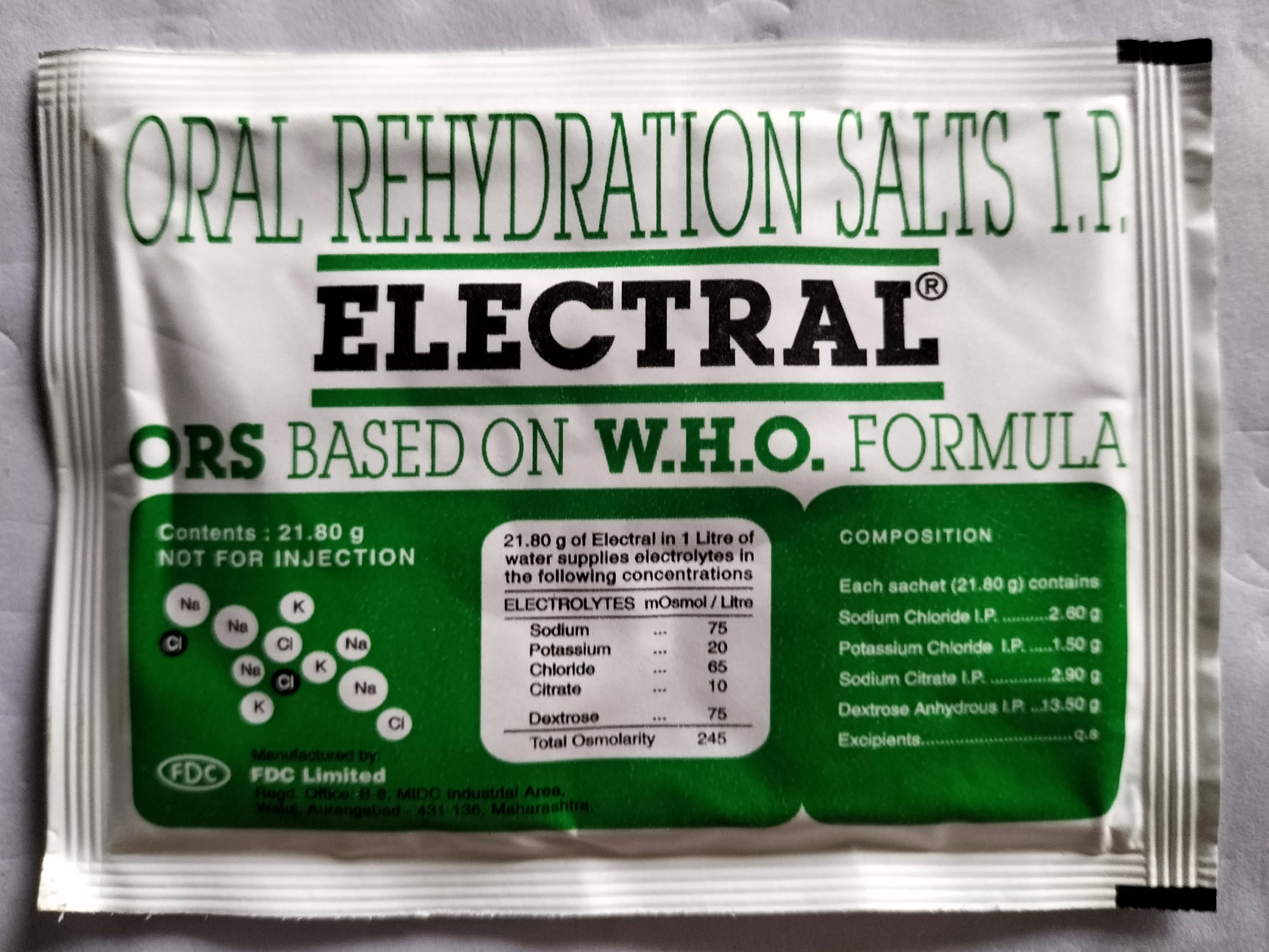
Osmotic concentration, formerly known as osmolarity, is the measure of solute concentration, defined as the number of osmoles (Osm) of solute per litre (L) of solution (osmol/L or Osm/L). The osmolarity of a solution is usually expressed as Osm/L (pronounced "osmolar"), in the same way that the molarity of a solution is expressed as "M" (pronounced "molar"). Whereas molarity measures the number of moles of solute per unit volume of solution, osmolarity measures the number of ''osmoles of solute particles'' per unit volume of solution. This value allows the measurement of the osmotic pressure of a solution and the determination of how the solvent will diffuse across a semipermeable membrane (osmosis) separating two solutions of different osmotic concentration. Unit The unit of osmotic concentration is the osmole. This is a non- SI unit of measurement that defines the number of moles of solute that contribute to the osmotic pressure of a solution. A milliosmole (mOsm) is 1/1,000 ... [...More Info...] [...Related Items...] OR: [Wikipedia] [Google] [Baidu] |


