|
Trigonocephaly
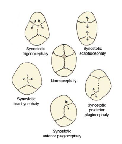
Trigonocephaly is a congenital condition of premature fusion of the metopic suture (from the Greek , "forehead"), leading to a triangular forehead. The merging of the two frontal bones leads to transverse growth restriction and parallel growth expansion. It may occur syndromic, involving other abnormalities, or isolated. The term is from the Greek , "triangle", and , "head". Cause Trigonocephaly can either occur syndromatic or isolated. Trigonocephaly is associated with the following syndromes: Opitz syndrome, Muenke syndrome, Jacobsen syndrome, Baller–Gerold syndrome and Say–Meyer syndrome. The etiology of trigonocephaly is mostly unknown although there are three main theories. Trigonocephaly is probably a multifactorial congenital condition, but due to limited proof of these theories this cannot safely be concluded. Intrinsic bone malformation The first theory assumes that the origin of pathological synostosis lies within disturbed bone formation early on in the pregnan ... [...More Info...] [...Related Items...] OR: [Wikipedia] [Google] [Baidu] |
Opitz Syndrome
Trigonocephaly is a congenital condition of premature fusion of the metopic suture (from the Greek , "forehead"), leading to a triangular forehead. The merging of the two frontal bones leads to transverse growth restriction and parallel growth expansion. It may occur syndromic, involving other abnormalities, or isolated. The term is from the Greek , "triangle", and , "head". Cause Trigonocephaly can either occur syndromatic or isolated. Trigonocephaly is associated with the following syndromes: Opitz syndrome, Muenke syndrome, Jacobsen syndrome, Baller–Gerold syndrome and Say–Meyer syndrome. The etiology of trigonocephaly is mostly unknown although there are three main theories. Trigonocephaly is probably a multifactorial congenital condition, but due to limited proof of these theories this cannot safely be concluded. Intrinsic bone malformation The first theory assumes that the origin of pathological synostosis lies within disturbed bone formation early on in the pregnan ... [...More Info...] [...Related Items...] OR: [Wikipedia] [Google] [Baidu] |
Say–Meyer Syndrome
Say–Meyer syndrome is a rare X-linked genetic disorder that is mostly characterized as developmental delay. It is one of the rare causes of short stature. It is closely related with trigonocephaly (a misshapen forehead due to premature fusion of bones in the skull). People with Say–Meyer syndrome have impaired growth, deficits in motor skills development and mental state. It is suggested that it is from a X-linked transmission. Signs and symptoms Common signs of Say–Meyer syndrome are trigonocephaly as well as head and neck symptoms. The head and neck symptoms come in the form of craniosynostosis affecting the metopic suture (the dense connective tissue structure that divides the two halves of the skull in children which usually fuse together by the age of six). Symptoms of Say–Meyer syndrome other than developmental delay and short stature include * Intellectual disability. * Low-set ears/posteriorly rotated ears * Intellectual deficit as well as learning disabilit ... [...More Info...] [...Related Items...] OR: [Wikipedia] [Google] [Baidu] |
FGFR1
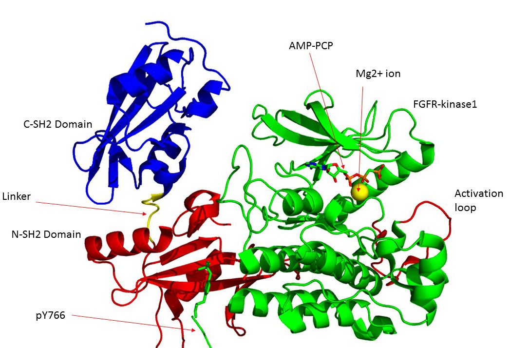
Fibroblast growth factor receptor 1 (FGFR1), also known as basic fibroblast growth factor receptor 1, fms-related tyrosine kinase-2 / Pfeiffer syndrome, and CD331, is a receptor tyrosine kinase whose ligands are specific members of the fibroblast growth factor family. FGFR1 has been shown to be associated with Pfeiffer syndrome, and clonal eosinophilias. Gene The ''FGFR1'' gene is located on human chromosome 8 at position p11.23 (i.e. 8p11.23), has 24 exons, and codes for a Precursor mRNA that is alternatively spliced at exons 8A or 8B thereby generating two mRNAs coding for two FGFR1 isoforms, FGFR1-IIIb (also termed FGFR1b) and FGFR1-IIIc (also termed FGFR1c), respectively. Although these two isoforms have different tissue distributions and FGF-binding affinities, FGFR1-IIIc appears responsible for most of functions of the FGFR1 gene while FGFR1-IIIb appears to have only a minor, somewhat redundant functional role. There are four other members of the ''FGFR1'' gene family: FGF ... [...More Info...] [...Related Items...] OR: [Wikipedia] [Google] [Baidu] |
Medical Genetics
Medical genetics is the branch tics in that human genetics is a field of scientific research that may or may not apply to medicine, while medical genetics refers to the application of genetics to medical care. For example, research on the causes and inheritance of genetic disorders would be considered within both human genetics and medical genetics, while the diagnosis, management, and counselling people with genetic disorders would be considered part of medical genetics. In contrast, the study of typically non-medical phenotypes such as the genetics of eye color would be considered part of human genetics, but not necessarily relevant to medical genetics (except in situations such as albinism). ''Genetic medicine'' is a newer term for medical genetics and incorporates areas such as gene therapy, personalized medicine, and the rapidly emerging new medical specialty, predictive medicine. Scope Medical genetics encompasses many different areas, including clinical practice of ... [...More Info...] [...Related Items...] OR: [Wikipedia] [Google] [Baidu] |
Röntgenography
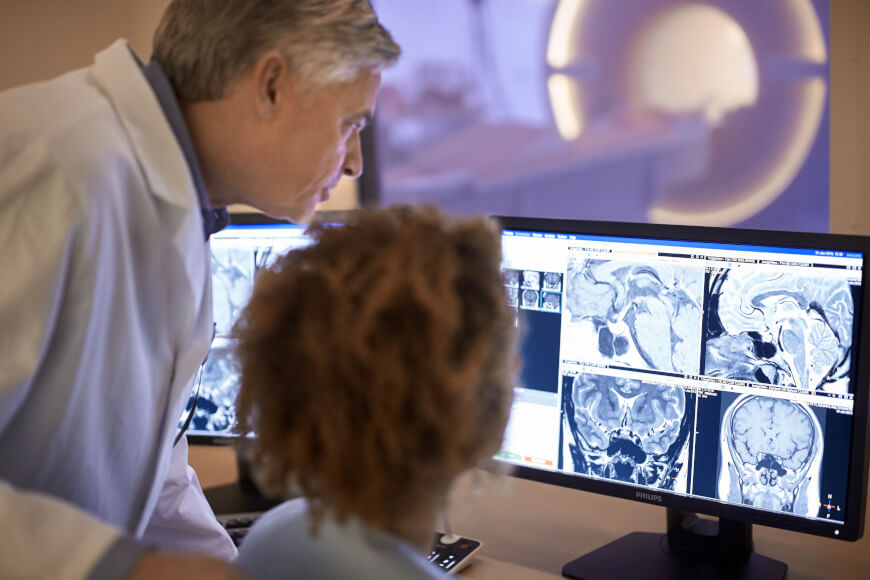
Radiology ( ) is the medical discipline that uses medical imaging to diagnose diseases and guide their treatment, within the bodies of humans and other animals. It began with radiography (which is why its name has a root referring to radiation), but today it includes all imaging modalities, including those that use no electromagnetic radiation (such as ultrasonography and magnetic resonance imaging), as well as others that do, such as computed tomography (CT), fluoroscopy, and nuclear medicine including positron emission tomography (PET). Interventional radiology is the performance of usually minimally invasive medical procedures with the guidance of imaging technologies such as those mentioned above. The modern practice of radiology involves several different healthcare professions working as a team. The radiologist is a medical doctor who has completed the appropriate post-graduate training and interprets medical images, communicates these findings to other physicians by mea ... [...More Info...] [...Related Items...] OR: [Wikipedia] [Google] [Baidu] |
Ethmoidal
The ethmoid sinuses or ethmoid air cells of the ethmoid bone are one of the four paired paranasal sinuses. The cells are variable in both size and number in the lateral mass of each of the ethmoid bones and cannot be palpated during an extraoral examination. They are divided into anterior and posterior groups. The ethmoid air cells are numerous thin-walled cavities situated in the ethmoidal labyrinth and completed by the frontal, maxilla, lacrimal, sphenoidal, and palatine bones. They lie between the upper parts of the nasal cavities and the orbits, and are separated from these cavities by thin bony lamellae. Groups of sinuses The groups of the ethmoidal air cells drain into the nasal meatuses.Otorhinolaryngology, Head and Neck Surgery, Anniko, Springer, 2010, page 188 * The posterior group the ''posterior ethmoidal sinus'' drains into the superior meatus above the middle nasal concha; sometimes one or more opens into the sphenoidal sinus. * The anterior group the ''anterior ethmo ... [...More Info...] [...Related Items...] OR: [Wikipedia] [Google] [Baidu] |
Hypoplasia
Hypoplasia (from Ancient Greek ὑπo- ''hypo-'' 'under' + πλάσις ''plasis'' 'formation'; adjective form ''hypoplastic'') is underdevelopment or incomplete development of a tissue or organ. Dictionary of Cell and Molecular Biology (11 March 2008) Although the term is not always used precisely, it properly refers to an inadequate or below-normal number of cells.Hypoplasia Stedman's Medical Dictionary. lww.com Hypoplasia is similar to |
Lateral (anatomy)
Standard anatomical terms of location are used to unambiguously describe the anatomy of animals, including humans. The terms, typically derived from Latin or Greek roots, describe something in its standard anatomical position. This position provides a definition of what is at the front ("anterior"), behind ("posterior") and so on. As part of defining and describing terms, the body is described through the use of anatomical planes and anatomical axes. The meaning of terms that are used can change depending on whether an organism is bipedal or quadrupedal. Additionally, for some animals such as invertebrates, some terms may not have any meaning at all; for example, an animal that is radially symmetrical will have no anterior surface, but can still have a description that a part is close to the middle ("proximal") or further from the middle ("distal"). International organisations have determined vocabularies that are often used as standard vocabularies for subdisciplines of anatom ... [...More Info...] [...Related Items...] OR: [Wikipedia] [Google] [Baidu] |
Epicanthal Folds
An epicanthic fold or epicanthus is a skin fold of the upper eyelid that covers the inner corner (medial canthus) of the eye. However, variation occurs in the nature of this feature and the possession of "partial epicanthic folds" or "slight epicanthic folds" is noted in the relevant literature.Lang, Berel (ed.) (2000) ''Race and Racism in Theory and Practice'', Rowman & Littlefield, p. 10 Various factors influence whether epicanthic folds form, including ancestry, age, and certain medical conditions. Etymology ''Epicanthus'' means 'above the canthus', with epi-canthus being the Latinized form of the Ancient Greek : 'corner of the eye'. Classification Variation in the shape of the epicanthic fold has led to four types being recognised: * ''Epicanthus supraciliaris'' runs from the brow, curving downwards towards the lachrymal sac. * ''Epicanthus palpebralis'' begins above the upper tarsus and extends to the inferior orbital rim. * ''Epicanthus tarsalis'' originates at th ... [...More Info...] [...Related Items...] OR: [Wikipedia] [Google] [Baidu] |
Anterior Cranial Fossa
The anterior cranial fossa is a depression in the floor of the cranial base which houses the projecting frontal lobes of the brain. It is formed by the orbital part of frontal bone, orbital plates of the Frontal bone, frontal, the cribriform plate of the ethmoid, and the small wings and front part of the body of the Sphenoid bone, sphenoid; it is limited behind by the posterior borders of the small wings of the sphenoid and by the anterior margin of the chiasmatic groove. The lesser wings of the sphenoid separate the anterior and middle cranial fossa, middle fossae. Structure It is traversed by the frontoethmoidal, sphenoethmoidal, and sphenofrontal sutures. Its lateral portions roof in the orbital cavities and support the frontal lobes of the cerebrum; they are convex and marked by depressions for the brain convolutions, and grooves for branches of the meningeal vessels. The central portion corresponds with the roof of the nasal cavity, and is markedly depressed on either side o ... [...More Info...] [...Related Items...] OR: [Wikipedia] [Google] [Baidu] |


