|
Tricuspid Atresia
Tricuspid atresia is a form of congenital heart disease whereby there is a complete absence of the tricuspid valve. Therefore, there is an absence of right atrioventricular connection. This leads to a hypoplastic (undersized) or absent right ventricle. This defect is contracted during prenatal development, when the heart does not finish developing. It causes the systemic circulation to be filled with relatively deoxygenated blood. The causes of tricuspid atresia are unknown. In most cases of tricuspid atresia, additional defects exist to allow exchange of blood between the loops of systematic circulation and pulmonary circulation, filling in the role of the missing atrioventricular connection. An atrial septal defect (ASD) must be present to fill the left atrium and the left ventricle with blood. Since there is a lack of a right ventricle, there must also be a way to pump blood into the pulmonary artery. This can be accomplished by a ventricular septal defect (VSD) connecting the l ... [...More Info...] [...Related Items...] OR: [Wikipedia] [Google] [Baidu] |
Congenital Heart Disease
A congenital heart defect (CHD), also known as a congenital heart anomaly and congenital heart disease, is a defect in the structure of the heart or great vessels that is present at birth. A congenital heart defect is classed as a cardiovascular disease. Signs and symptoms depend on the specific type of defect. Symptoms can vary from none to life-threatening. When present, symptoms may include rapid breathing, bluish skin (cyanosis), poor weight gain, and feeling tired. CHD does not cause chest pain. Most congenital heart defects are not associated with other diseases. A complication of CHD is heart failure. The cause of a congenital heart defect is often unknown. Risk factors include certain infections during pregnancy such as rubella, use of certain medications or drugs such as alcohol or tobacco, parents being closely related, or poor nutritional status or obesity in the mother. Having a parent with a congenital heart defect is also a risk factor. A number of genetic condition ... [...More Info...] [...Related Items...] OR: [Wikipedia] [Google] [Baidu] |
Holosystolic
Systolic heart murmurs are heart murmurs heard during systole, i.e. they begin and end between S1 and S2. Many involve stenosis of the semilunar valves or regurgitation of the atrioventricular valves. Types * Mid-systolic ejection murmurs are due to blood flow through the semilunar valves. They occur at the start of blood ejection — which starts after S1 — and ends with the cessation of the blood flow — which is before S2. Therefore, the onset of a midsystolic ejection murmur is separated from S1 by the isovolumic contraction phase; the cessation of the murmur and the S2 interval is the aortic or pulmonary hangout time. The resultant configuration of this murmur is a crescendo-decrescendo murmur. Causes of midsystolic ejection murmurs include outflow obstruction, increased flow through normal semilunar valves, dilation of aortic root or pulmonary trunk, or structural changes in the semilunar valves without obstruction. * Late systolic murmurs start after S1 and, if left s ... [...More Info...] [...Related Items...] OR: [Wikipedia] [Google] [Baidu] |
Subclavian Artery
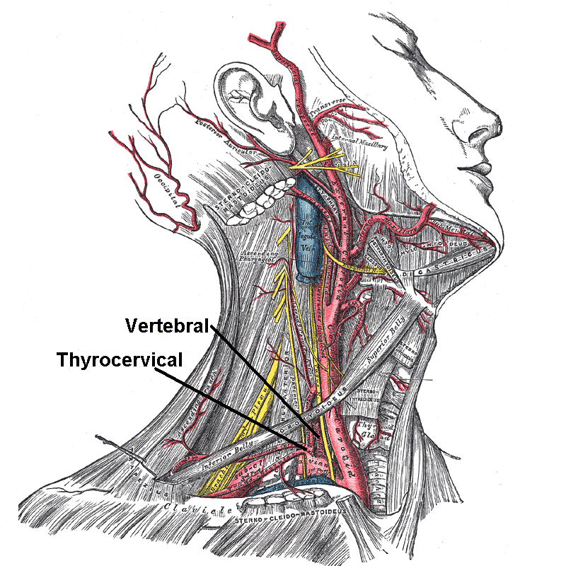
In human anatomy, the subclavian arteries are paired major arteries of the upper thorax, below the clavicle. They receive blood from the aortic arch. The left subclavian artery supplies blood to the left arm and the right subclavian artery supplies blood to the right arm, with some branches supplying the head and thorax. On the left side of the body, the subclavian comes directly off the aortic arch, while on the right side it arises from the relatively short brachiocephalic artery when it bifurcates into the subclavian and the right common carotid artery. The usual branches of the subclavian on both sides of the body are the vertebral artery, the internal thoracic artery, the thyrocervical trunk, the costocervical trunk and the dorsal scapular artery, which may branch off the transverse cervical artery, which is a branch of the thyrocervical trunk. The subclavian becomes the axillary artery at the lateral border of the first rib. Structure From its origin, the subclavian artery t ... [...More Info...] [...Related Items...] OR: [Wikipedia] [Google] [Baidu] |
Gore-Tex
Gore-Tex is a waterproof, breathable fabric membrane and registered trademark of W. L. Gore & Associates. Invented in 1969, Gore-Tex can repel liquid water while allowing water vapor to pass through and is designed to be a lightweight, waterproof fabric for all-weather use. It is composed of stretched polytetrafluoroethylene (PTFE), which is more commonly known by the generic trademark Teflon. The material is formally known as the generic term expanded PTFE (ePTFE). History Gore-Tex was co-invented by Wilbert L. Gore and Gore's son, Robert W. Gore. In 1969, Bob Gore stretched heated rods of polytetrafluoroethylene (PTFE) and created expanded polytetrafluoroethylene (ePTFE). His discovery of the right conditions for stretching PTFE was a happy accident, born partly of frustration. Instead of slowly stretching the heated material, he applied a sudden, accelerating yank. The solid PTFE unexpectedly stretched about 800%, forming a microporous structure that was about 70% air. ... [...More Info...] [...Related Items...] OR: [Wikipedia] [Google] [Baidu] |
PGE1
Prostaglandin E1 (PGE1), also known as alprostadil, is a naturally occurring prostaglandin which is used as a medication. In infants with congenital heart defects, it is delivered by slow injection into a vein to open the ductus arteriosus until surgery can be carried out. By injection into the penis or placement in the urethra, it is used to treat erectile dysfunction. Common side effects when given to babies include decreased breathing, fever, and low blood pressure. When used for erectile dysfunction side effects may include penile pain, bleeding at the site of injection, and prolonged erection (priapism). Prostaglandin E1 is in the vasodilator family of medications. It works by opening blood vessels and relaxing smooth muscle. Prostaglandin E1 was isolated in 1957 and approved for medical use in the United States in 1981. It is on the World Health Organization's List of Essential Medicines. Medical uses Patent ductus arteriosus Alprostadil is also used in maintainin ... [...More Info...] [...Related Items...] OR: [Wikipedia] [Google] [Baidu] |
Chest Radiograph
A chest radiograph, called a chest X-ray (CXR), or chest film, is a projection radiograph of the chest used to diagnose conditions affecting the chest, its contents, and nearby structures. Chest radiographs are the most common film taken in medicine. Like all methods of radiography, chest radiography employs ionizing radiation in the form of X-rays to generate images of the chest. The mean radiation dose to an adult from a chest radiograph is around 0.02 mSv (2 mrem) for a front view (PA, or posteroanterior) and 0.08 mSv (8 mrem) for a side view (LL, or latero-lateral). Together, this corresponds to a background radiation equivalent time of about 10 days. Medical uses Conditions commonly identified by chest radiography * Pneumonia * Pneumothorax * Interstitial lung disease * Heart failure * Bone fracture * Hiatal hernia Chest radiographs are used to diagnose many conditions involving the chest wall, including its bones, and also structures contained within the thoracic ... [...More Info...] [...Related Items...] OR: [Wikipedia] [Google] [Baidu] |
Echocardiogram
An echocardiography, echocardiogram, cardiac echo or simply an echo, is an ultrasound of the heart. It is a type of medical imaging of the heart, using standard ultrasound or Doppler ultrasound. Echocardiography has become routinely used in the diagnosis, management, and follow-up of patients with any suspected or known heart diseases. It is one of the most widely used diagnostic imaging modalities in cardiology. It can provide a wealth of helpful information, including the size and shape of the heart (internal chamber size quantification), pumping capacity, location and extent of any tissue damage, and assessment of valves. An echocardiogram can also give physicians other estimates of heart function, such as a calculation of the cardiac output, ejection fraction, and diastolic function (how well the heart relaxes). Echocardiography is an important tool in assessing wall motion abnormality in patients with suspected cardiac disease. It is a tool which helps in reaching an early ... [...More Info...] [...Related Items...] OR: [Wikipedia] [Google] [Baidu] |
Anomaly Scan
The anomaly scan, also sometimes called the anatomy scan, 20-week ultrasound, or level 2 ultrasound, evaluates anatomic structures of the fetus, placenta, and maternal pelvic organs. This scan is an important and common component of routine prenatal care. The function of the ultrasound is to measure the fetus so that growth abnormalities can be recognized quickly later in pregnancy, to assess for congenital malformations and multiple pregnancies, and to plan method of delivery. Procedure This scan is conducted between 18 and 22 weeks' gestation, but most often performed at 19 weeks, as a component of routine prenatal care. Prior to 18 weeks' gestation, the fetal organs may be of insufficient size and development to allow for ultrasound evaluation. Scans performed beyond 22 weeks' gestation may limit the ability to seek pregnancy termination, depending on local legislation. Two-dimensional (2D) is used to evaluate fetal structures, placenta, and amniotic fluid volume. Maternal pe ... [...More Info...] [...Related Items...] OR: [Wikipedia] [Google] [Baidu] |
Foramen Ovale (heart)
In the fetal heart, the foramen ovale (), also foramen Botalli, or the ostium secundum of Born, allows blood to enter the left atrium from the right atrium. It is one of two fetal cardiac shunts, the other being the ductus arteriosus (which allows blood that still escapes to the right ventricle to bypass the pulmonary circulation). Another similar adaptation in the fetus is the ductus venosus. In most individuals, the foramen ovale closes at birth. It later forms the fossa ovalis. Development The foramen ovale () forms in the late fourth week of gestation, as a small passageway between the septum secundum and the ostium secundum. Initially the atria are separated from one another by the septum primum except for a small opening below the septum, the ostium primum. As the septum primum grows, the ostium primum narrows and eventually closes. Before it does so, bloodflow from the inferior vena cava wears down a portion of the septum primum, forming the ostium secundum. Some embryo ... [...More Info...] [...Related Items...] OR: [Wikipedia] [Google] [Baidu] |
Left Ventricular Hypertrophy
Left ventricular hypertrophy (LVH) is thickening of the heart muscle of the left ventricle of the heart, that is, left-sided ventricular hypertrophy and resulting increased left ventricular mass. Causes While ventricular hypertrophy occurs naturally as a reaction to aerobic exercise and strength training, it is most frequently referred to as a pathological reaction to cardiovascular disease, or high blood pressure. It is one aspect of ventricular remodeling. While LVH itself is not a disease, it is usually a marker for disease involving the heart. Disease processes that can cause LVH include any disease that increases the afterload that the heart has to contract against, and some primary diseases of the muscle of the heart. Causes of increased afterload that can cause LVH include aortic stenosis, aortic insufficiency and hypertension. Primary disease of the muscle of the heart that cause LVH are known as hypertrophic cardiomyopathies, which can lead into heart failure. Lon ... [...More Info...] [...Related Items...] OR: [Wikipedia] [Google] [Baidu] |


