|
Transvaginal Ultrasonography
Vaginal ultrasonography is a medical ultrasonography that applies an ultrasound transducer (or "probe") in the vagina to visualize organs within the pelvic cavity. It is also called transvaginal ultrasonography because the ultrasound waves go ''across'' the vaginal wall to study tissues beyond it. Uses Vaginal ultrasonography is used both as a means of gynecologic ultrasonography and obstetric ultrasonography. It is preferred over abdominal ultrasonography in the diagnosis of ectopic pregnancy. See also * Gynecologic ultrasonography Gynecologic ultrasonography or gynecologic sonography refers to the application of medical ultrasonography to the female pelvic organs (specifically the uterus, the ovaries, and the fallopian tubes) as well as the bladder, the adnexa, and the rec ... References External links * Medical ultrasonography {{medical-equipment-stub ... [...More Info...] [...Related Items...] OR: [Wikipedia] [Google] [Baidu] |
Transvaginal Ultrasonography Device
{{Dab ...
Transvaginal refers to a transluminal procedure through the reproductive tract of the vagina, including: *Transvaginal oocyte retrieval *Transvaginal ultrasonography *Transvaginal mesh Transvaginal mesh, also known as vaginal mesh implant, is a net-like surgical tool that is used to treat Pelvic organ prolapse, pelvic organ prolapse (POP) and Stress incontinence, stress urinary incontinence (SUI) among female patients. The surgic ... [...More Info...] [...Related Items...] OR: [Wikipedia] [Google] [Baidu] |
Medical Ultrasonography
Medical ultrasound includes diagnostic techniques (mainly medical imaging, imaging techniques) using ultrasound, as well as therapeutic ultrasound, therapeutic applications of ultrasound. In diagnosis, it is used to create an image of internal body structures such as tendons, muscles, joints, blood vessels, and internal organs, to measure some characteristics (e.g. distances and velocities) or to generate an informative audible sound. Its aim is usually to find a source of disease or to exclude pathology. The usage of ultrasound to produce visual images for medicine is called medical ultrasonography or simply sonography. The practice of examining pregnant women using ultrasound is called obstetric ultrasonography, and was an early development of clinical ultrasonography. Ultrasound is composed of sound waves with frequency, frequencies which are significantly higher than the range of human hearing (>20,000 Hz). Ultrasonic images, also known as sonograms, are created by se ... [...More Info...] [...Related Items...] OR: [Wikipedia] [Google] [Baidu] |
Vagina
In mammals, the vagina is the elastic, muscular part of the female genital tract. In humans, it extends from the vestibule to the cervix. The outer vaginal opening is normally partly covered by a thin layer of mucosal tissue called the hymen. At the deep end, the cervix (neck of the uterus) bulges into the vagina. The vagina allows for sexual intercourse and birth. It also channels menstrual flow, which occurs in humans and closely related primates as part of the menstrual cycle. Although research on the vagina is especially lacking for different animals, its location, structure and size are documented as varying among species. Female mammals usually have two external openings in the vulva; these are the urethral opening for the urinary tract and the vaginal opening for the genital tract. This is different from male mammals, who usually have a single urethral opening for both urination and reproduction. The vaginal opening is much larger than the nearby urethral opening, an ... [...More Info...] [...Related Items...] OR: [Wikipedia] [Google] [Baidu] |
Pelvic Cavity
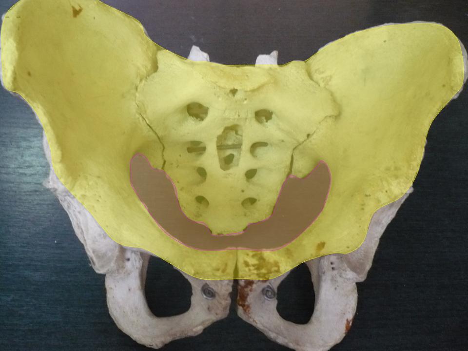
The pelvic cavity is a body cavity that is bounded by the bones of the pelvis. Its oblique roof is the pelvic inlet (the superior opening of the pelvis). Its lower boundary is the pelvic floor. The pelvic cavity primarily contains the reproductive organs, urinary bladder, distal ureters, proximal urethra, terminal sigmoid colon, rectum, and anal canal. In females, the uterus, Fallopian tubes, ovaries and upper vagina occupy the area between the other viscera. The rectum is located at the back of the pelvis, in the curve of the sacrum and coccyx; the bladder is in front, behind the pubic symphysis. The pelvic cavity also contains major arteries, veins, muscles, and nerves. These structures coexist in a crowded space, and disorders of one pelvic component may impact upon another; for example, constipation may overload the rectum and compress the urinary bladder, or childbirth might damage the pudendal nerves and later lead to anal weakness. Structure The pelvis has an antero ... [...More Info...] [...Related Items...] OR: [Wikipedia] [Google] [Baidu] |
Vaginal Ultrasound
Vaginal ultrasonography is a medical ultrasonography that applies an ultrasound transducer (or "probe") in the vagina to visualize organs within the pelvic cavity. It is also called transvaginal ultrasonography because the ultrasound waves go ''across'' the vaginal wall to study tissues beyond it. Uses Vaginal ultrasonography is used both as a means of gynecologic ultrasonography and obstetric ultrasonography. It is preferred over abdominal ultrasonography in the diagnosis of ectopic pregnancy. See also * Gynecologic ultrasonography Gynecologic ultrasonography or gynecologic sonography refers to the application of medical ultrasonography to the female pelvic organs (specifically the uterus, the ovaries, and the fallopian tubes) as well as the bladder, the adnexa, and the rec ... References External links * Medical ultrasonography {{medical-equipment-stub ... [...More Info...] [...Related Items...] OR: [Wikipedia] [Google] [Baidu] |
Gynecologic Ultrasonography
Gynecologic ultrasonography or gynecologic sonography refers to the application of medical ultrasonography to the female pelvic organs (specifically the uterus, the ovaries, and the fallopian tubes) as well as the bladder, the adnexa, and the recto-uterine pouch. The procedure may lead to other medically relevant findings in the pelvis.This technique is useful to detect myomas or mullerian malformations. Routes The examination can be performed by transabdominal ultrasonography, generally with a full bladder which acts as an acoustic window to achieve better visualization of pelvis organs, or by transvaginal ultrasonography with a specifically designed vaginal transducer. Transvaginal imaging utilizes a higher frequency imaging, which gives better resolution of the ovaries, uterus and endometrium (the fallopian tubes are generally not seen unless distended), but is limited to depth of image penetration, whereas larger lesions reaching into the abdomen are better seen trans ... [...More Info...] [...Related Items...] OR: [Wikipedia] [Google] [Baidu] |
Obstetric Ultrasonography
Obstetric ultrasonography, or prenatal ultrasound, is the use of medical ultrasonography in pregnancy, in which sound waves are used to create real-time visual images of the developing embryo or fetus in the uterus (womb). The procedure is a standard part of prenatal care in many countries, as it can provide a variety of information about the health of the mother, the timing and progress of the pregnancy, and the health and development of the embryo or fetus. The International Society of Ultrasound in Obstetrics and Gynecology (ISUOG) recommends that pregnant women have routine obstetric ultrasounds between 18 weeks' and 22 weeks' gestational age (the anatomy scan) in order to confirm pregnancy dating, to measure the fetus so that growth abnormalities can be recognized quickly later in pregnancy, and to assess for congenital malformations and multiple pregnancies (twins, etc). Additionally, the ISUOG recommends that pregnant patients who desire genetic testing have obstetric ultra ... [...More Info...] [...Related Items...] OR: [Wikipedia] [Google] [Baidu] |
Abdominal Ultrasonography
Abdominal ultrasonography (also called abdominal ultrasound imaging or abdominal sonography) is a form of medical ultrasonography (medical application of ultrasound technology) to visualise abdominal anatomical structures. It uses transmission and reflection of ultrasound waves to visualise internal organs through the abdominal wall (with the help of gel, which helps transmission of the sound waves). For this reason, the procedure is also called a transabdominal ultrasound, in contrast to endoscopic ultrasound, the latter combining ultrasound with endoscopy through visualize internal structures from within hollow organs. Abdominal ultrasound examinations are performed by gastroenterologists or other specialists in internal medicine, radiologists, or sonographers trained for this procedure. Medical uses Abdominal ultrasound can be used to diagnose abnormalities in various internal organs, such as the kidneys, liver, gallbladder, pancreas, spleen and abdominal aorta. If Doppler u ... [...More Info...] [...Related Items...] OR: [Wikipedia] [Google] [Baidu] |
Ectopic Pregnancy
Ectopic pregnancy is a complication of pregnancy in which the embryo attaches outside the uterus. Signs and symptoms classically include abdominal pain and vaginal bleeding, but fewer than 50 percent of affected women have both of these symptoms. The pain may be described as sharp, dull, or crampy. Pain may also spread to the shoulder if bleeding into the abdomen has occurred. Severe bleeding may result in a fast heart rate, fainting, or shock. With very rare exceptions the fetus is unable to survive. Overall, ectopic pregnancies are very rare, annually affecting less than 2% of pregnancies worldwide. Risk factors for ectopic pregnancy include pelvic inflammatory disease, often due to chlamydia infection; tobacco smoking; prior tubal surgery; a history of infertility; and the use of assisted reproductive technology. Those who have previously had an ectopic pregnancy are at much higher risk of having another one. Most ectopic pregnancies (90%) occur in the fallopian tube, whic ... [...More Info...] [...Related Items...] OR: [Wikipedia] [Google] [Baidu] |
Gynecologic Ultrasonography
Gynecologic ultrasonography or gynecologic sonography refers to the application of medical ultrasonography to the female pelvic organs (specifically the uterus, the ovaries, and the fallopian tubes) as well as the bladder, the adnexa, and the recto-uterine pouch. The procedure may lead to other medically relevant findings in the pelvis.This technique is useful to detect myomas or mullerian malformations. Routes The examination can be performed by transabdominal ultrasonography, generally with a full bladder which acts as an acoustic window to achieve better visualization of pelvis organs, or by transvaginal ultrasonography with a specifically designed vaginal transducer. Transvaginal imaging utilizes a higher frequency imaging, which gives better resolution of the ovaries, uterus and endometrium (the fallopian tubes are generally not seen unless distended), but is limited to depth of image penetration, whereas larger lesions reaching into the abdomen are better seen trans ... [...More Info...] [...Related Items...] OR: [Wikipedia] [Google] [Baidu] |


