|
Transmediastinal Gunshot Wound
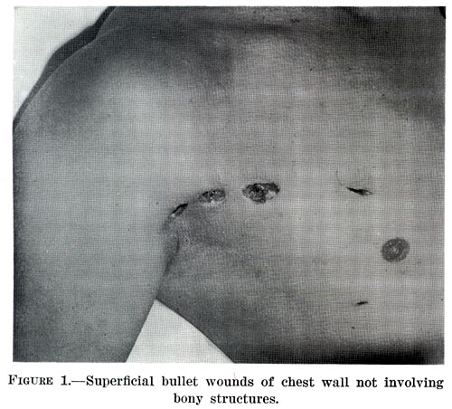
A transmediastinal gunshot wound (TMGSW) is a penetrating injury to a person's thorax in which a bullet enters the mediastinum, possibly damaging some of the major structures in this area. Hemodynamic instability has been reported in approximately fifty percent of cases with a mortality rate ranging from twenty to forty percent. Some studies have shown marked improvement in the mortality rate of patients who survived transfer to the operating room rather than being treated surgically in the ER. Presentation Complications Complications caused by a TMGSW can range from mild to life-threatening depending on which structures are damaged. It can be rapidly lethal if a major structure is involved. Some of the possible complications caused by a TMGSW are: * damage to great vessels such as the vena cava, aorta, pulmonary arteries * damage to cardiac muscle * massive hemorrhage * cardiac tamponade * hemomediastinum * pneumomediastinum * neurologic injury * In many cases there is pneumothor ... [...More Info...] [...Related Items...] OR: [Wikipedia] [Google] [Baidu] |
Penetrating Trauma
Penetrating trauma is an open wound injury that occurs when an object pierces the skin and enters a tissue of the body, creating a deep but relatively narrow entry wound. In contrast, a blunt or ''non-penetrating'' trauma may have some deep damage, but the overlying skin is not necessarily broken and the wound is still closed to the outside environment. The penetrating object may remain in the tissues, come back out the path it entered, or pass through the full thickness of the tissues and exit from another area. A penetrating injury in which an object enters the body or a structure and passes all the way through an exit wound is called a perforating trauma, while the term ''penetrating trauma'' implies that the object does not perforate wholly through. In gunshot wounds, perforating trauma is associated with an entrance wound and an often larger exit wound. Penetrating trauma can be caused by a foreign object or by fragments of a broken bone. Usually occurring in violent cr ... [...More Info...] [...Related Items...] OR: [Wikipedia] [Google] [Baidu] |
Bronchoscopy
Bronchoscopy is an endoscopic technique of visualizing the inside of the airways for diagnostic and therapeutic purposes. An instrument (bronchoscope) is inserted into the airways, usually through the nose or mouth, or occasionally through a tracheostomy. This allows the practitioner to examine the patient's airways for abnormalities such as foreign bodies, bleeding, tumors, or inflammation. Specimens may be taken from inside the lungs. The construction of bronchoscopes ranges from rigid metal tubes with attached lighting devices to flexible optical fiber instruments with realtime video equipment. History The German laryngologist Gustav Killian is attributed with performing the first bronchoscopy in 1897. Killian used a rigid bronchoscope to remove a pork bone. The procedure was done in an awake patient using topical cocaine as a local anesthetic. From this time until the 1970s, rigid bronchoscopes were used exclusively. Chevalier Jackson, refined the rigid bronchoscope in the 19 ... [...More Info...] [...Related Items...] OR: [Wikipedia] [Google] [Baidu] |
Thoracotomy
A thoracotomy is a surgical procedure to gain access into the pleural space of the chest. It is performed by surgeons (emergency physicians or paramedics under certain circumstances) to gain access to the thoracic organs, most commonly the heart, the lungs, or the esophagus, or for access to the thoracic aorta or the anterior spine (the latter may be necessary to access tumors in the spine). A thoracotomy is the first step in thoracic surgeries including lobectomy or pneumonectomy for lung cancer or to gain thoracic access in major trauma. Approaches There are many different surgical approaches to performing a thoracotomy. Some common forms of thoracotomies include: * Median sternotomy provides wide access to the mediastinum and is the incision of choice for most open-heart surgery and access to the anterior mediastinum * Posterolateral thoracotomy is an incision through an intercostal space on the back, and is often widened with rib spreaders. It is a very common approach ... [...More Info...] [...Related Items...] OR: [Wikipedia] [Google] [Baidu] |
Esophagus
The esophagus (American English) or oesophagus (British English; both ), non-technically known also as the food pipe or gullet, is an organ in vertebrates through which food passes, aided by peristaltic contractions, from the pharynx to the stomach. The esophagus is a fibromuscular tube, about long in adults, that travels behind the trachea and heart, passes through the diaphragm, and empties into the uppermost region of the stomach. During swallowing, the epiglottis tilts backwards to prevent food from going down the larynx and lungs. The word ''oesophagus'' is from Ancient Greek οἰσοφάγος (oisophágos), from οἴσω (oísō), future form of φέρω (phérō, “I carry”) + ἔφαγον (éphagon, “I ate”). The wall of the esophagus from the lumen outwards consists of mucosa, submucosa (connective tissue), layers of muscle fibers between layers of fibrous tissue, and an outer layer of connective tissue. The mucosa is a stratified squamous epithel ... [...More Info...] [...Related Items...] OR: [Wikipedia] [Google] [Baidu] |
Trachea
The trachea, also known as the windpipe, is a Cartilage, cartilaginous tube that connects the larynx to the bronchi of the lungs, allowing the passage of air, and so is present in almost all air-breathing animals with lungs. The trachea extends from the larynx and branches into the two primary bronchi. At the top of the trachea the cricoid cartilage attaches it to the larynx. The trachea is formed by a number of horseshoe-shaped rings, joined together vertically by overlying annular ligaments of trachea, ligaments, and by the trachealis muscle at their ends. The epiglottis closes the opening to the larynx during swallowing. The trachea begins to form in the second month of embryo development, becoming longer and more fixed in its position over time. It is epithelium lined with columnar epithelium, column-shaped cells that have hair-like extensions called cilia, with scattered goblet cells that produce protective mucins. The trachea can be affected by inflammation or infection, us ... [...More Info...] [...Related Items...] OR: [Wikipedia] [Google] [Baidu] |
Pleural Cavity
The pleural cavity, pleural space, or interpleural space is the potential space between the pleurae of the pleural sac that surrounds each lung. A small amount of serous pleural fluid is maintained in the pleural cavity to enable lubrication between the membranes, and also to create a pressure gradient. The serous membrane that covers the surface of the lung is the visceral pleura and is separated from the outer membrane the parietal pleura by just the film of pleural fluid in the pleural cavity. The visceral pleura follows the fissures of the lung and the root of the lung structures. The parietal pleura is attached to the mediastinum, the upper surface of the diaphragm, and to the inside of the ribcage. Structure In humans, the left and right lungs are completely separated by the mediastinum, and there is no communication between their pleural cavities. Therefore, in cases of a unilateral pneumothorax, the contralateral lung will remain functioning normally unless there is ... [...More Info...] [...Related Items...] OR: [Wikipedia] [Google] [Baidu] |
Blood Pressure
Blood pressure (BP) is the pressure of circulating blood against the walls of blood vessels. Most of this pressure results from the heart pumping blood through the circulatory system. When used without qualification, the term "blood pressure" refers to the pressure in the large arteries. Blood pressure is usually expressed in terms of the systolic pressure (maximum pressure during one heartbeat) over diastolic pressure (minimum pressure between two heartbeats) in the cardiac cycle. It is measured in millimeters of mercury ( mmHg) above the surrounding atmospheric pressure. Blood pressure is one of the vital signs—together with respiratory rate, heart rate, oxygen saturation, and body temperature—that healthcare professionals use in evaluating a patient's health. Normal resting blood pressure, in an adult is approximately systolic over diastolic, denoted as "120/80 mmHg". Globally, the average blood pressure, age standardized, has remained about the same since 1 ... [...More Info...] [...Related Items...] OR: [Wikipedia] [Google] [Baidu] |
Chest Tube
A chest tube (also chest drain, thoracic catheter, tube thoracostomy or intercostal drain) is a surgical drain that is inserted through the chest wall and into the pleural space or the mediastinum in order to remove clinically undesired substances such as air (pneumothorax), excess fluid (pleural effusion or hydrothorax), blood (hemothorax), chyle ( chylothorax) or pus (empyema) from the intrathoracic space. An intrapleural chest tube is also known as a Bülau drain or an intercostal catheter (ICC), and can either be a thin, flexible silicone tube (known as a "pigtail" drain), or a larger, semi-rigid, fenestrated plastic tube, which often involves a flutter valve or underwater seal. The concept of chest drainage was first advocated by Hippocrates when he described the treatment of empyema by means of incision, cautery and insertion of metal tubes. However, the technique was not widely used until the influenza epidemic of 1918 to evacuate post-pneumonic empyema, which was first ... [...More Info...] [...Related Items...] OR: [Wikipedia] [Google] [Baidu] |
Fluid Replacement
Fluid replacement or fluid resuscitation is the medical practice of replenishing bodily fluid lost through sweating, bleeding, fluid shifts or other pathologic processes. Fluids can be replaced with oral rehydration therapy (drinking), intravenous therapy, rectally such as with a Murphy drip, or by ''hypodermoclysis'', the direct injection of fluid into the subcutaneous tissue. Fluids administered by the oral and hypodermic routes are absorbed more slowly than those given intravenously. By mouth Oral rehydration therapy (ORT) is a simple treatment for dehydration associated with diarrhea, particularly gastroenteritis/gastroenteropathy, such as that caused by cholera or rotavirus. ORT consists of a solution of salts and sugars which is taken by mouth. For most mild to moderate dehydration in children, the preferable treatment in an emergency department is ORT over intravenous replacement of fluid., which cites: * It is used around the world, but is most important in the developing ... [...More Info...] [...Related Items...] OR: [Wikipedia] [Google] [Baidu] |
Shock (circulatory)
Shock is the state of insufficient blood flow to the tissues of the body as a result of problems with the circulatory system. Initial symptoms of shock may include weakness, fast heart rate, fast breathing, sweating, anxiety, and increased thirst. This may be followed by confusion, unconsciousness, or cardiac arrest, as complications worsen. Shock is divided into four main types based on the underlying cause: low volume, cardiogenic, obstructive, and distributive shock. Low volume shock, also known as hypovolemic shock, may be from bleeding, diarrhea, or vomiting. Cardiogenic shock may be due to a heart attack or cardiac contusion. Obstructive shock may be due to cardiac tamponade or a tension pneumothorax. Distributive shock may be due to sepsis, anaphylaxis, injury to the upper spinal cord, or certain overdoses. The diagnosis is generally based on a combination of symptoms, physical examination, and laboratory tests. A decreased pulse pressure (systolic blood pressure m ... [...More Info...] [...Related Items...] OR: [Wikipedia] [Google] [Baidu] |
Cardiac Arrest
Cardiac arrest is when the heart suddenly and unexpectedly stops beating. It is a medical emergency that, without immediate medical intervention, will result in sudden cardiac death within minutes. Cardiopulmonary resuscitation (CPR) and possibly defibrillation are needed until further treatment can be provided. Cardiac arrest results in a rapid loss of consciousness, and breathing may be abnormal or absent. While cardiac arrest may be caused by heart attack or heart failure, these are not the same, and in 15 to 25% of cases, there is a non-cardiac cause. Some individuals may experience chest pain, shortness of breath, nausea, an elevated heart rate, and a light-headed feeling immediately before entering cardiac arrest. The most common cause of cardiac arrest is an underlying heart problem like coronary artery disease that decreases the amount of oxygenated blood supplying the heart muscle. This, in turn, damages the structure of the muscle, which can alter its function. ... [...More Info...] [...Related Items...] OR: [Wikipedia] [Google] [Baidu] |


