|
Transcranial Doppler
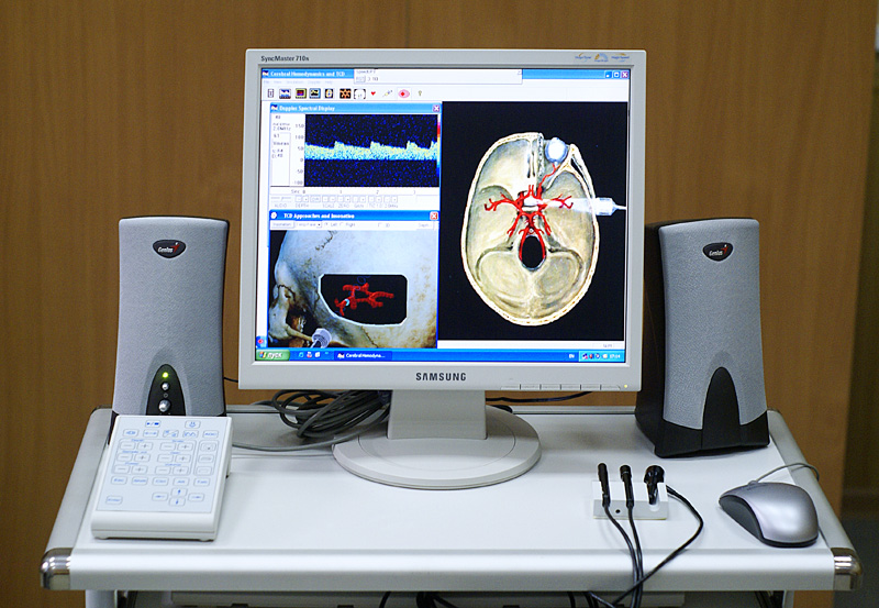
Transcranial Doppler (TCD) and transcranial color Doppler (TCCD) are types of Doppler ultrasonography that measure the velocity of blood flow through the brain's blood vessels by measuring the echoes of ultrasound waves moving transcranially (through the cranium). These modes of medical imaging conduct a spectral analysis of the acoustic signals they receive and can therefore be classified as methods of active acoustocerebrography. They are used as tests to help diagnose emboli, stenosis, vasospasm from a subarachnoid hemorrhage (bleeding from a ruptured aneurysm), and other problems. These relatively quick and inexpensive tests are growing in popularity. The tests are effective for detecting sickle cell disease, ischemic cerebrovascular disease, subarachnoid hemorrhage, arteriovenous malformations, and cerebral circulatory arrest. The tests are possibly useful for perioperative monitoring and meningeal infection. The equipment used for these tests is becoming increasingly portab ... [...More Info...] [...Related Items...] OR: [Wikipedia] [Google] [Baidu] |
Doppler Ultrasound Analyzer Of Blood Velocity
The Doppler effect or Doppler shift (or simply Doppler, when in context) is the change in frequency of a wave in relation to an observer (physics), observer who is moving relative to the wave source. It is named after the Austrian physicist Christian Doppler, who described the phenomenon in 1842. A common example of Doppler shift is the change of pitch (music), pitch heard when a vehicle sounding a horn approaches and recedes from an observer. Compared to the emitted frequency, the received frequency is higher during the approach, identical at the instant of passing by, and lower during the recession. The reason for the Doppler effect is that when the source of the waves is moving towards the observer, each successive wave Crest (physics), crest is emitted from a position closer to the observer than the crest of the previous wave. Therefore, each wave takes slightly less time to reach the observer than the previous wave. Hence, the time between the arrivals of successive wave c ... [...More Info...] [...Related Items...] OR: [Wikipedia] [Google] [Baidu] |
Subarachnoid Hemorrhage
Subarachnoid hemorrhage (SAH) is bleeding into the subarachnoid space—the area between the arachnoid membrane and the pia mater surrounding the brain. Symptoms may include a severe headache of rapid onset, vomiting, decreased level of consciousness, fever, and sometimes seizures. Neck stiffness or neck pain are also relatively common. In about a quarter of people a small bleed with resolving symptoms occurs within a month of a larger bleed. SAH may occur as a result of a head injury or spontaneously, usually from a ruptured cerebral aneurysm. Risk factors for spontaneous cases include high blood pressure, smoking, family history, alcoholism, and cocaine use. Generally, the diagnosis can be determined by a CT scan of the head if done within six hours of symptom onset. Occasionally, a lumbar puncture is also required. After confirmation further tests are usually performed to determine the underlying cause. Treatment is by prompt neurosurgery or endovascular coiling. Medicat ... [...More Info...] [...Related Items...] OR: [Wikipedia] [Google] [Baidu] |
Duplex Ultrasonography
Doppler ultrasonography is medical ultrasonography that employs the Doppler effect to perform imaging of the movement of tissues and body fluids (usually blood), and their relative velocity to the probe. By calculating the frequency shift of a particular sample volume, for example, flow in an artery or a jet of blood flow over a heart valve, its speed and direction can be determined and visualized. Duplex ultrasonography sometimes refers to Doppler ultrasonography or spectral Doppler ultrasonography. Doppler ultrasonography consists of two components: brightness mode (B-mode) showing anatomy of the organs, and Doppler mode (showing blood flow) superimposed on the B-mode. Meanwhile, spectral Doppler ultrasonography consists of three components: B-mode, Doppler mode, and spectral waveform displayed at the lower half of the image. Therefore, "duplex ultrasonography" is a misnomer for spectral Doppler ultrasonography, and more exact name should be "triplex ultrasonography". This is ... [...More Info...] [...Related Items...] OR: [Wikipedia] [Google] [Baidu] |
Doppler Effect
The Doppler effect or Doppler shift (or simply Doppler, when in context) is the change in frequency of a wave in relation to an observer who is moving relative to the wave source. It is named after the Austrian physicist Christian Doppler, who described the phenomenon in 1842. A common example of Doppler shift is the change of pitch heard when a vehicle sounding a horn approaches and recedes from an observer. Compared to the emitted frequency, the received frequency is higher during the approach, identical at the instant of passing by, and lower during the recession. The reason for the Doppler effect is that when the source of the waves is moving towards the observer, each successive wave crest is emitted from a position closer to the observer than the crest of the previous wave. Therefore, each wave takes slightly less time to reach the observer than the previous wave. Hence, the time between the arrivals of successive wave crests at the observer is reduced, causing an increa ... [...More Info...] [...Related Items...] OR: [Wikipedia] [Google] [Baidu] |
Ultrasound Research Interface
An ultrasound research interface (URI) is a software tool loaded onto a diagnostic clinical ultrasound device which provides functionality beyond typical clinical modes of operation. A normal clinical ultrasound user only has access to the ultrasound data in its final processed form, typically a B-Mode image, in DICOM format. For reasons of device usability they also have limited access to the processing parameters that can be modified.Dickie K, Leung C, Zahiri R, Pelissier L. A flexible research interface for collecting clinical ultrasound images. SPIE Multispectral Image Acquisition. 2009 Oct;7494(02)Rohling R, Fung W, Lajevardi P. PUPIL: Programmable Ultrasound Platform and Interface Library. MICCAI. 2003 Nov;(2879);424-431Shamdasani V, Bae U, Sikdar S, Yoo YM, Karadayi K, Managuli R, Kim Y. Research interface on a programmable ultrasound scanner. Ultrasonics. 2008 Jul;48(03);159-168. A URI allows a researcher to achieve different results by either acquiring the image at vari ... [...More Info...] [...Related Items...] OR: [Wikipedia] [Google] [Baidu] |


