|
Touton Giant Cell
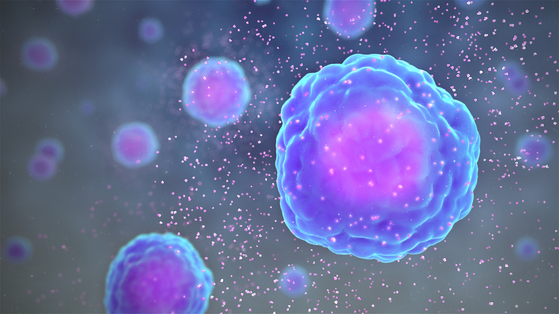
Touton giant cells are a type of multinucleated giant cell seen in lesions with high lipid content such as fat necrosis, xanthoma, and xanthelasma and xanthogranulomas. They are also found in dermatofibroma. History Touton giant cells are named for Karl Touton, a German botanist and dermatologist. Karl Touton first observed these cells in 1885 and named them "xanthelasmatic giant cells", a name which has since fallen out of favor. Appearance Touton giant cells, being multinucleated giant cells, can be distinguished by the presence of several nuclei in a distinct pattern. They contain a ring of nuclei surrounding a central homogeneous cytoplasm, while foamy cytoplasm surrounds the nuclei. The cytoplasm surrounded by the nuclei has been described as both amphophilic and eosinophilic, while the cytoplasm near the periphery of the cell is pale and foamy in appearance. Causes Touton giant cells are formed by the fusion of macrophage-derived foam cells. It has been suggested that ... [...More Info...] [...Related Items...] OR: [Wikipedia] [Google] [Baidu] |
Juvenile Xanthogranuloma - Very High Mag
{{Disambiguation ...
Juvenile may refer to: *Juvenile status, or minor (law), prior to adulthood *Juvenile (organism) *Juvenile (rapper) (born 1975), American rapper * ''Juvenile'' (2000 film), Japanese film * ''Juvenile'' (2017 film) *Juvenile (greyhounds), a greyhound competition *Juvenile particles, a type of volcanic ejecta *A two-year-old horse in horse racing terminology See also *"The Juvenile", a song by Ace of Base *Juvenile novel **Any of "Heinlein juveniles" *Juvenile delinquency *Juvenilia, works by an author while a youth *Juvenal (other) Juvenal was a poet. Juvenal or Juvenals may also refer to: * Juvenal (name), and persons with the name * Juvenals, a student society * An immature bird {{disambiguation ... [...More Info...] [...Related Items...] OR: [Wikipedia] [Google] [Baidu] |
Cell Nucleus
The cell nucleus (pl. nuclei; from Latin or , meaning ''kernel'' or ''seed'') is a membrane-bound organelle found in eukaryotic cells. Eukaryotic cells usually have a single nucleus, but a few cell types, such as mammalian red blood cells, have no nuclei, and a few others including osteoclasts have many. The main structures making up the nucleus are the nuclear envelope, a double membrane that encloses the entire organelle and isolates its contents from the cellular cytoplasm; and the nuclear matrix, a network within the nucleus that adds mechanical support. The cell nucleus contains nearly all of the cell's genome. Nuclear DNA is often organized into multiple chromosomes – long stands of DNA dotted with various proteins, such as histones, that protect and organize the DNA. The genes within these chromosomes are structured in such a way to promote cell function. The nucleus maintains the integrity of genes and controls the activities of the cell by regulating gene expres ... [...More Info...] [...Related Items...] OR: [Wikipedia] [Google] [Baidu] |
M-CSF
The colony stimulating factor 1 (CSF1), also known as macrophage colony-stimulating factor (M-CSF), is a secreted cytokine which causes hematopoietic stem cells to differentiate into macrophages or other related cell types. Eukaryotic cells also produce M-CSF in order to combat intercellular viral infection. It is one of the three experimentally described colony-stimulating factors. M-CSF binds to the colony stimulating factor 1 receptor. It may also be involved in development of the placenta. Structure M-CSF is a cytokine, being a smaller protein involved in cell signaling. The active form of the protein is found extracellularly as a disulfide-linked homodimer, and is thought to be produced by proteolytic cleavage of membrane-bound precursors. Four transcript variants encoding three different isoforms (a proteoglycan, glycoprotein and cell surface protein) have been found for this gene. Function M-CSF (or CSF-1) is a hematopoietic growth factor that is involved in the prol ... [...More Info...] [...Related Items...] OR: [Wikipedia] [Google] [Baidu] |
Interleukin-3
Interleukin 3 (IL-3) is a protein that in humans is encoded by the ''IL3'' gene localized on chromosome 5q31.1. Sometimes also called colony-stimulating factor, multi-CSF, mast cell growth factor, MULTI-CSF, MCGF; MGC79398, MGC79399: the protein contains 152 amino acids and its molecular weight is 17 kDa. IL-3 is produced as a monomer by activated T cells, monocytes/macrophages and stroma cells. The major function of IL-3 cytokine is to regulate the concentrations of various blood-cell types. It induces proliferation and differentiation in both early pluripotent stem cells and committed Progenitor cell, progenitors. It also has many more specific effects like the regeneration of platelets and potentially aids in early antibody Immunoglobulin class switching, isotype switching. Function Interleukin 3 is an interleukin, a type of biological signal (cytokine) that can improve the body's natural response to disease as part of the immune system. In conjunction with other β common ch ... [...More Info...] [...Related Items...] OR: [Wikipedia] [Google] [Baidu] |
Interferon Gamma
Interferon gamma (IFN-γ) is a dimerized soluble cytokine that is the only member of the type II class of interferons. The existence of this interferon, which early in its history was known as immune interferon, was described by E. F. Wheelock as a product of human leukocytes stimulated with phytohemagglutinin, and by others as a product of antigen-stimulated lymphocytes. It was also shown to be produced in human lymphocytes. or tuberculin-sensitized mouse peritoneal lymphocytes challenged with Mantoux test (PPD); the resulting supernatants were shown to inhibit growth of vesicular stomatitis virus. Those reports also contained the basic observation underlying the now widely employed IFN-γ release assay used to test for tuberculosis. In humans, the IFN-γ protein is encoded by the ''IFNG'' gene. Through cell signaling, IFN-γ plays a role in regulating the immune response of its target cell. A key signaling pathway that is activated by type II IFN is the JAK-STAT signal ... [...More Info...] [...Related Items...] OR: [Wikipedia] [Google] [Baidu] |
Cytokines
Cytokines are a broad and loose category of small proteins (~5–25 kDa) important in cell signaling. Cytokines are peptides and cannot cross the lipid bilayer of cells to enter the cytoplasm. Cytokines have been shown to be involved in autocrine, paracrine and endocrine signaling as immunomodulating agents. Cytokines include chemokines, interferons, interleukins, lymphokines, and tumour necrosis factors, but generally not hormones or growth factors (despite some overlap in the terminology). Cytokines are produced by a broad range of cells, including immune cells like macrophages, B lymphocytes, T lymphocytes and mast cells, as well as endothelial cells, fibroblasts, and various stromal cells; a given cytokine may be produced by more than one type of cell. They act through cell surface receptors and are especially important in the immune system; cytokines modulate the balance between humoral and cell-based immune responses, and they regulate the maturation, growth, and resp ... [...More Info...] [...Related Items...] OR: [Wikipedia] [Google] [Baidu] |
Eosinophilic
Eosinophilic (Greek suffix -phil-, meaning ''loves eosin'') is the staining of tissues, cells, or organelles after they have been washed with eosin, a dye. Eosin is an acidic dye for staining cell cytoplasm, collagen, and muscle fibers. ''Eosinophilic'' describes the appearance of cells and structures seen in histological sections that take up the staining dye eosin. Such eosinophilic structures are, in general, composed of protein. Eosin is usually combined with a stain called hematoxylin to produce a hematoxylin- and eosin-stained section (also called an H&E stain, HE or H+E section). It is the most widely used histological stain for a medical diagnosis. When a pathologist examines a biopsy of a suspected cancer, they will stain the biopsy with H&E. Some structures seen inside cells are described as being eosinophilic; for example, Lewy and Mallory bodies. [...More Info...] [...Related Items...] OR: [Wikipedia] [Google] [Baidu] |
Amphophilic
Staining is a technique used to enhance contrast in samples, generally at the microscopic level. Stains and dyes are frequently used in histology (microscopic study of biological tissues), in cytology (microscopic study of cells), and in the medical fields of histopathology, hematology, and cytopathology that focus on the study and diagnoses of diseases at the microscopic level. Stains may be used to define biological tissues (highlighting, for example, muscle fibers or connective tissue), cell populations (classifying different blood cells), or organelles within individual cells. In biochemistry, it involves adding a class-specific ( DNA, proteins, lipids, carbohydrates) dye to a substrate to qualify or quantify the presence of a specific compound. Staining and fluorescent tagging can serve similar purposes. Biological staining is also used to mark cells in flow cytometry, and to flag proteins or nucleic acids in gel electrophoresis. Light microscopes are used for viewi ... [...More Info...] [...Related Items...] OR: [Wikipedia] [Google] [Baidu] |
Cytoplasm
In cell biology, the cytoplasm is all of the material within a eukaryotic cell, enclosed by the cell membrane, except for the cell nucleus. The material inside the nucleus and contained within the nuclear membrane is termed the nucleoplasm. The main components of the cytoplasm are cytosol (a gel-like substance), the organelles (the cell's internal sub-structures), and various cytoplasmic inclusions. The cytoplasm is about 80% water and is usually colorless. The submicroscopic ground cell substance or cytoplasmic matrix which remains after exclusion of the cell organelles and particles is groundplasm. It is the hyaloplasm of light microscopy, a highly complex, polyphasic system in which all resolvable cytoplasmic elements are suspended, including the larger organelles such as the ribosomes, mitochondria, the plant plastids, lipid droplets, and vacuoles. Most cellular activities take place within the cytoplasm, such as many metabolic pathways including glycolysis, and proces ... [...More Info...] [...Related Items...] OR: [Wikipedia] [Google] [Baidu] |
Dermatologist
Dermatology is the branch of medicine dealing with the skin.''Random House Webster's Unabridged Dictionary.'' Random House, Inc. 2001. Page 537. . It is a speciality with both medical and surgical aspects. A dermatologist is a specialist medical doctor who manages diseases related to skin, hair, nails, and some cosmetic problems. Etymology Attested in English in 1819, the word "dermatology" derives from the Greek δέρματος (''dermatos''), genitive of δέρμα (''derma''), "skin" (itself from δέρω ''dero'', "to flay") and -λογία '' -logia''. Neo-Latin ''dermatologia'' was coined in 1630, an anatomical term with various French and German uses attested from the 1730s. History In 1708, the first great school of dermatology became a reality at the famous Hôpital Saint-Louis in Paris, and the first textbooks (Willan's, 1798–1808) and atlases ( Alibert's, 1806–1816) appeared in print around the same time.Freedberg, et al. (2003). ''Fitzpatrick's Dermatology in ... [...More Info...] [...Related Items...] OR: [Wikipedia] [Google] [Baidu] |
Multinucleated Giant Cell
A giant cell (also known as multinucleated giant cell, or multinucleate giant cell) is a mass formed by the union of several distinct cells (usually histiocytes), often forming a granuloma. Although there is typically a focus on the pathological aspects of multinucleate giant cells (MGCs), they also play many important physiological roles. Osteoclasts specifically are invaluable to healthy physiological functions and are key players in the skeletal system. Osteoclasts are frequently classified and discussed separately from other MGCs which are more closely linked with human pathologies. Non-osteoclast MGCs can arise in response to an infection, such as from tuberculosis, herpes, or HIV, or foreign body. These MGCs are cells of monocyte or macrophage lineage fused together. Similar to their monocyte precursors, they are able to phagocytose foreign materials. However, their large size and extensive membrane ruffling make them better equipped to clear up larger particles. They utiliz ... [...More Info...] [...Related Items...] OR: [Wikipedia] [Google] [Baidu] |
Botanist
Botany, also called , plant biology or phytology, is the science of plant life and a branch of biology. A botanist, plant scientist or phytologist is a scientist who specialises in this field. The term "botany" comes from the Ancient Greek word (''botanē'') meaning "pasture", " herbs" "grass", or " fodder"; is in turn derived from (), "to feed" or "to graze". Traditionally, botany has also included the study of fungi and algae by mycologists and phycologists respectively, with the study of these three groups of organisms remaining within the sphere of interest of the International Botanical Congress. Nowadays, botanists (in the strict sense) study approximately 410,000 species of land plants of which some 391,000 species are vascular plants (including approximately 369,000 species of flowering plants), and approximately 20,000 are bryophytes. Botany originated in prehistory as herbalism with the efforts of early humans to identify – and later cultivate – edible, med ... [...More Info...] [...Related Items...] OR: [Wikipedia] [Google] [Baidu] |


