|
Tillaux-Chaput Avulsion Fracture
A Tillaux fracture is a Salter–Harris type III fracture through the anterolateral aspect of the distal tibial epiphysis. It occurs in older adolescents between the ages of 12 and 15 when the medial epiphysis had closed but before the lateral side has done so, due to an avulsion of the anterior inferior tibiofibular ligament, at the opposite end to a Wagstaffe-Le Fort avulsion fracture Mechanism The fracture commonly results from an abduction- external rotation force, causing the anterior tibiofibular ligament to avulse the anterolateral corner of the distal tibial epiphysis resulting in a Salter Harris Type III fracture. Pathology It occurs in older children at the end of growth. Variability in fracture pattern is due to progression of physeal closure as anterolateral part of distal tibial physis is the last to close. When the lateral physis is the only portion not fused, external rotation may lead to Tillaux or Triplane fractures. Treatment If the displacement at fract ... [...More Info...] [...Related Items...] OR: [Wikipedia] [Google] [Baidu] |
Projectional Radiography
Projectional radiography, also known as conventional radiography, is a form of radiography and medical imaging that produces two-dimensional images by x-ray radiation. The image acquisition is generally performed by radiographers, and the images are often examined by radiologists. Both the procedure and any resultant images are often simply called "X-ray". Plain radiography or roentgenography generally refers to projectional radiography (without the use of more advanced techniques such as computed tomography that can generate 3D-images). ''Plain radiography'' can also refer to radiography without a radiocontrast agent or radiography that generates single static images, as contrasted to fluoroscopy, which are technically also projectional. Equipment X-ray generator Projectional radiographs generally use X-rays created by X-ray generators, which generate X-rays from X-ray tubes. Grid An anti-scatter grid may be placed between the patient and the detector to reduce the quanti ... [...More Info...] [...Related Items...] OR: [Wikipedia] [Google] [Baidu] |
Salter–Harris Fracture
A Salter–Harris fracture is a fracture that involves the epiphyseal plate (growth plate) of a bone, specifically the zone of provisional calcification. It is thus a form of child bone fracture. It is a common injury found in children, occurring in 15% of childhood long bone fractures. This type of fracture and its classification system is named for Robert B. Salter and William H. Harris who created and published this classification system in the ''Journal of Bone and Joint Surgery'' in 1963. Types There are nine types of Salter–Harris fractures; types I to V as described by Robert B Salter and W Robert Harris in 1963, and the rarer types VI to IX which have been added subsequently: * Type I – transverse fracture through the growth plate (also referred to as the "physis"): 6% incidence * Type II – A fracture through the growth plate and the metaphysis, sparing the epiphysis: 75% incidence, takes approximately 12-90 weeks or more in the spine to heal. * Type III – A f ... [...More Info...] [...Related Items...] OR: [Wikipedia] [Google] [Baidu] |
Fracture
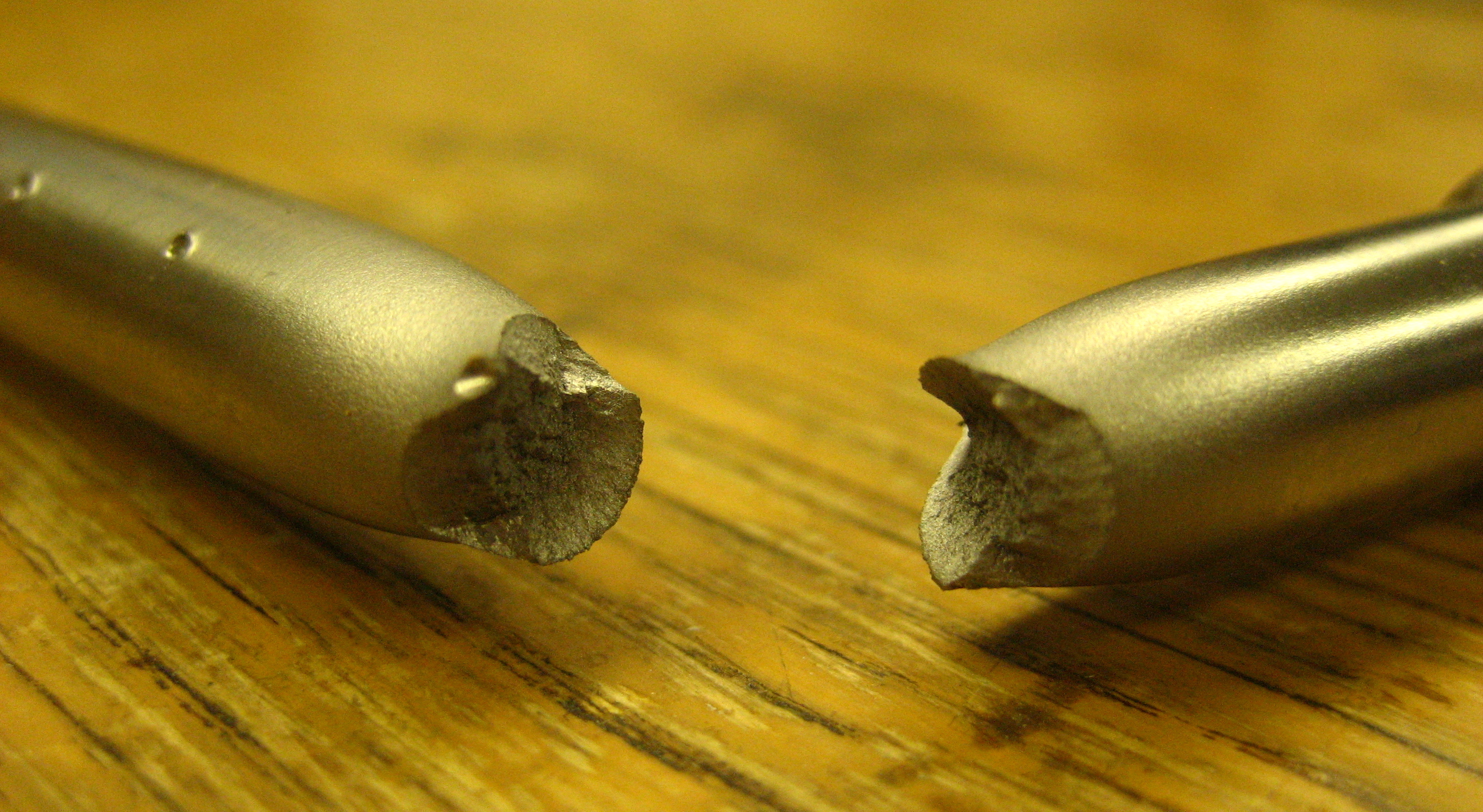
Fracture is the separation of an object or material into two or more pieces under the action of stress. The fracture of a solid usually occurs due to the development of certain displacement discontinuity surfaces within the solid. If a displacement develops perpendicular to the surface, it is called a normal tensile crack or simply a crack; if a displacement develops tangentially, it is called a shear crack, slip band or dislocation. Brittle fractures occur with no apparent deformation before fracture. Ductile fractures occur after visible deformation. Fracture strength, or breaking strength, is the stress when a specimen fails or fractures. The detailed understanding of how a fracture occurs and develops in materials is the object of fracture mechanics. Strength Fracture strength, also known as breaking strength, is the stress at which a specimen fails via fracture. This is usually determined for a given specimen by a tensile test, which charts the stress–strain cu ... [...More Info...] [...Related Items...] OR: [Wikipedia] [Google] [Baidu] |
Tibia
The tibia (; ), also known as the shinbone or shankbone, is the larger, stronger, and anterior (frontal) of the two bones in the leg below the knee in vertebrates (the other being the fibula, behind and to the outside of the tibia); it connects the knee with the ankle. The tibia is found on the medial side of the leg next to the fibula and closer to the median plane. The tibia is connected to the fibula by the interosseous membrane of leg, forming a type of fibrous joint called a syndesmosis with very little movement. The tibia is named for the flute ''tibia''. It is the second largest bone in the human body, after the femur. The leg bones are the strongest long bones as they support the rest of the body. Structure In human anatomy, the tibia is the second largest bone next to the femur. As in other vertebrates the tibia is one of two bones in the lower leg, the other being the fibula, and is a component of the knee and ankle joints. The ossification or formation of the bone ... [...More Info...] [...Related Items...] OR: [Wikipedia] [Google] [Baidu] |
Epiphysis
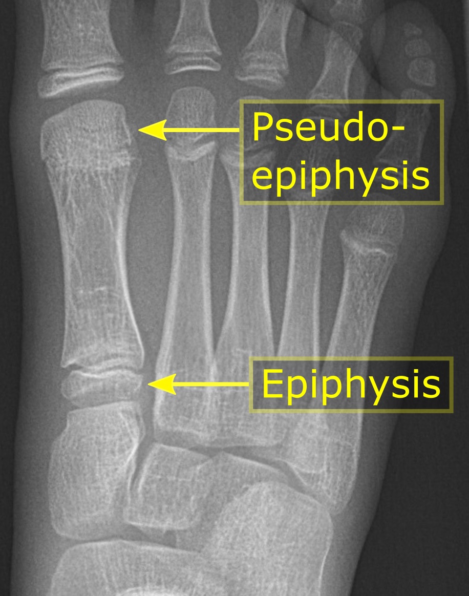
The epiphysis () is the rounded end of a long bone, at its joint with adjacent bone(s). Between the epiphysis and diaphysis (the long midsection of the long bone) lies the metaphysis, including the epiphyseal plate (growth plate). At the joint, the epiphysis is covered with articular cartilage; below that covering is a zone similar to the epiphyseal plate, known as subchondral bone. The epiphysis is filled with red bone marrow, which produces erythrocytes (red blood cells). Structure There are four types of epiphysis: # Pressure epiphysis: The region of the long bone that forms the joint is a pressure epiphysis (e.g. the head of the femur, part of the hip joint complex). Pressure epiphyses assist in transmitting the weight of the human body and are the regions of the bone that are under pressure during movement or locomotion. Another example of a pressure epiphysis is the head of the humerus which is part of the shoulder complex. condyles of femur and tibia also comes under ... [...More Info...] [...Related Items...] OR: [Wikipedia] [Google] [Baidu] |
Anterior Inferior Tibiofibular Ligament
Standard anatomical terms of location are used to unambiguously describe the anatomy of animals, including humans. The terms, typically derived from Latin or Greek roots, describe something in its standard anatomical position. This position provides a definition of what is at the front ("anterior"), behind ("posterior") and so on. As part of defining and describing terms, the body is described through the use of anatomical planes and anatomical axes. The meaning of terms that are used can change depending on whether an organism is bipedal or quadrupedal. Additionally, for some animals such as invertebrates, some terms may not have any meaning at all; for example, an animal that is radially symmetrical will have no anterior surface, but can still have a description that a part is close to the middle ("proximal") or further from the middle ("distal"). International organisations have determined vocabularies that are often used as standard vocabularies for subdisciplines of anat ... [...More Info...] [...Related Items...] OR: [Wikipedia] [Google] [Baidu] |
Abduction (kinesiology)
Motion, the process of movement, is described using specific anatomical terms. Motion includes movement of organs, joints, limbs, and specific sections of the body. The terminology used describes this motion according to its direction relative to the anatomical position of the body parts involved. Anatomists and others use a unified set of terms to describe most of the movements, although other, more specialized terms are necessary for describing unique movements such as those of the hands, feet, and eyes. In general, motion is classified according to the anatomical plane it occurs in. ''Flexion'' and ''extension'' are examples of ''angular'' motions, in which two axes of a joint are brought closer together or moved further apart. ''Rotational'' motion may occur at other joints, for example the shoulder, and are described as ''internal'' or ''external''. Other terms, such as ''elevation'' and ''depression'', describe movement above or below the horizontal plane. Many anatomic ... [...More Info...] [...Related Items...] OR: [Wikipedia] [Google] [Baidu] |
External Rotation
Motion, the process of movement, is described using specific anatomical terms. Motion includes movement of organs, joints, limbs, and specific sections of the body. The terminology used describes this motion according to its direction relative to the anatomical position of the body parts involved. Anatomists and others use a unified set of terms to describe most of the movements, although other, more specialized terms are necessary for describing unique movements such as those of the hands, feet, and eyes. In general, motion is classified according to the anatomical plane it occurs in. ''Flexion'' and ''extension'' are examples of ''angular'' motions, in which two axes of a joint are brought closer together or moved further apart. ''Rotational'' motion may occur at other joints, for example the shoulder, and are described as ''internal'' or ''external''. Other terms, such as ''elevation'' and ''depression'', describe movement above or below the horizontal plane. Many anatomica ... [...More Info...] [...Related Items...] OR: [Wikipedia] [Google] [Baidu] |
Anterior Tibiofibular Ligament
The anterior ligament of the lateral malleolus (anterior tibiofibular ligament or anterior inferior ligament) is a flat, trapezoidal band of fibers, broader below than above, which extends obliquely downward and lateralward between the adjacent margins of the tibia and fibula, on the front aspect of the syndesmosis. It is in relation, in front, with the fibularis tertius, the aponeurosis of the leg, and the integument; behind, with the interosseous ligament; and lies in contact with the cartilage Cartilage is a resilient and smooth type of connective tissue. In tetrapods, it covers and protects the ends of long bones at the joints as articular cartilage, and is a structural component of many body parts including the rib cage, the neck an ... covering the talus. References External links * - "Anterior inferior tibiofibular ligament" Additional images File:Slide2tat.JPG, Ankle joint. Deep dissection. Anterior view. File:Slide2coco.JPG, Dorsum of Foot. Ankle joint. Deep diss ... [...More Info...] [...Related Items...] OR: [Wikipedia] [Google] [Baidu] |
Open Reduction And Internal Fixation
Internal fixation is an operation in orthopedics that involves the surgical implementation of implants for the purpose of repairing a bone, a concept that dates to the mid-nineteenth century and was made applicable for routine treatment in the mid-twentieth century. An internal fixator may be made of stainless steel, titanium alloy, or cobalt-chrome alloy. or plastics. Types of internal fixators include: * Plate and screws * Kirschner wires * Intramedullary nails Open reduction Open Reduction Internal Fixation (ORIF) involves the implementation of implants to guide the healing process of a bone, as well as the open reduction, or setting, of the bone. ''Open reduction'' refers to open surgery to set bones, as is necessary for some fractures. ''Internal fixation'' refers to fixation of screws and/or plates, intramedullary rods and other devices to enable or facilitate healing. Rigid fixation prevents micro-motion across lines of fracture to enable healing and prevent infectio ... [...More Info...] [...Related Items...] OR: [Wikipedia] [Google] [Baidu] |


