|
Thrombophlebitis
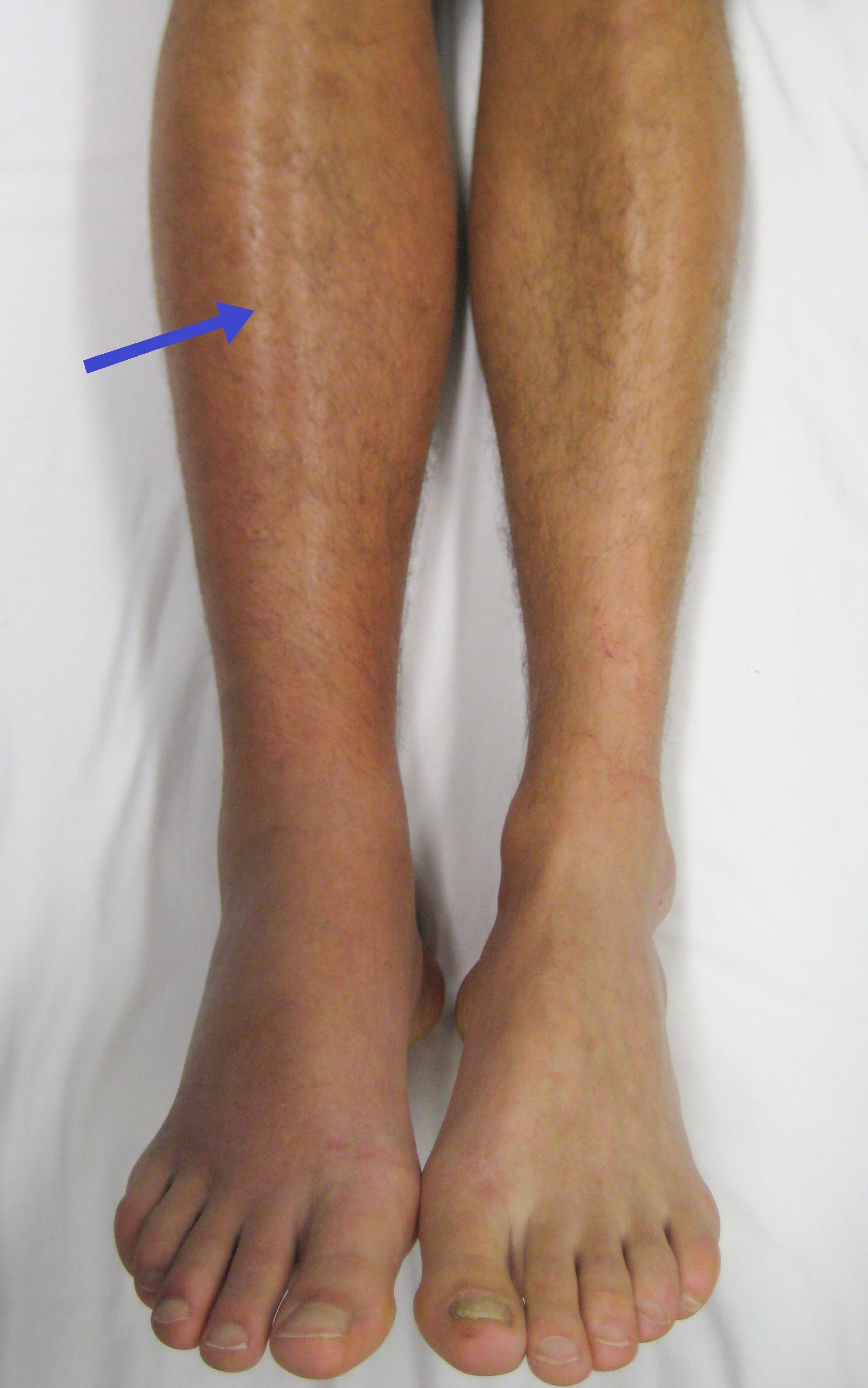
Thrombophlebitis is a phlebitis (inflammation of a vein) related to a thrombus (blood clot). When it occurs repeatedly in different locations, it is known as thrombophlebitis migrans (migratory thrombophlebitis). Signs and symptoms The following symptoms or signs are often associated with thrombophlebitis, although thrombophlebitis is not restricted to the veins of the legs. * Pain (area affected) * Skin redness/inflammation * Edema * Veins hard and cord-like * Tenderness Complications In terms of complications, one of the most serious occurs when the superficial blood clot is associated with a deep vein thrombosis; this can then dislodge, traveling through the heart and occluding the dense capillary network of the lungs This is a pulmonary embolism which can be life-threatening. Causes Thrombophlebitis causes include disorders related to increased tendency for blood clotting and reduced speed of blood in the veins such as prolonged immobility; prolonged traveling (sitting) may ... [...More Info...] [...Related Items...] OR: [Wikipedia] [Google] [Baidu] |
Great Saphenous Vein
The great saphenous vein (GSV, alternately "long saphenous vein"; ) is a large, subcutaneous, superficial vein of the leg. It is the longest vein in the body, running along the length of the lower limb, returning blood from the foot, leg and thigh to the deep femoral vein at the femoral triangle. Structure The great saphenous vein originates from where the dorsal vein of the big toe (the hallux) merges with the dorsal venous arch of the foot. After passing in front of the medial malleolus (where it often can be visualized and palpated), it runs up the medial side of the leg. At the knee, it runs over the posterior border of the medial epicondyle of the femur bone. In the proximal anterior thigh inferolateral to the pubic tubercle, the great saphenous vein dives down deep through the cribriform fascia of the saphenous opening to join the femoral vein. It forms an arch, the saphenous arch, to join the common femoral vein in the region of the femoral triangle at the sapheno-femoral ... [...More Info...] [...Related Items...] OR: [Wikipedia] [Google] [Baidu] |
Vein
Veins are blood vessels in humans and most other animals that carry blood towards the heart. Most veins carry deoxygenated blood from the tissues back to the heart; exceptions are the pulmonary and umbilical veins, both of which carry oxygenated blood to the heart. In contrast to veins, arteries carry blood away from the heart. Veins are less muscular than arteries and are often closer to the skin. There are valves (called ''pocket valves'') in most veins to prevent backflow. Structure Veins are present throughout the body as tubes that carry blood back to the heart. Veins are classified in a number of ways, including superficial vs. deep, pulmonary vs. systemic, and large vs. small. * Superficial veins are those closer to the surface of the body, and have no corresponding arteries. *Deep veins are deeper in the body and have corresponding arteries. *Perforator veins drain from the superficial to the deep veins. These are usually referred to in the lower limbs and feet. *Communic ... [...More Info...] [...Related Items...] OR: [Wikipedia] [Google] [Baidu] |
Thrombus
A thrombus (plural thrombi), colloquially called a blood clot, is the final product of the blood coagulation step in hemostasis. There are two components to a thrombus: aggregated platelets and red blood cells that form a plug, and a mesh of cross-linked fibrin protein. The substance making up a thrombus is sometimes called cruor. A thrombus is a healthy response to injury intended to stop and prevent further bleeding, but can be harmful in thrombosis, when a clot obstructs blood flow through healthy blood vessels in the circulatory system. In the microcirculation consisting of the very small and smallest blood vessels the capillaries, tiny thrombi known as microclots can obstruct the flow of blood in the capillaries. This can cause a number of problems particularly affecting the alveoli in the lungs of the respiratory system resulting from reduced oxygen supply. Microclots have been found to be a characteristic feature in severe cases of COVID-19, and in long COVID. Mural th ... [...More Info...] [...Related Items...] OR: [Wikipedia] [Google] [Baidu] |
Protein S Deficiency
Protein S deficiency is a disorder associated with increased risk of venous thrombosis. Protein S, a vitamin K-dependent physiological anticoagulant, acts as a nonenzymatic cofactor to activate protein C in the degradation of factor Va and factor VIIIa. Decreased (antigen) levels or impaired function of protein S leads to decreased degradation of factor Va and factor VIIIa and an increased propensity to venous thrombosis. Protein S circulates in human plasma in two forms: approximately 60 percent is bound to complement component C4b β-chain while the remaining 40 percent is free, only free protein S has activated protein C cofactor activity Signs and symptoms Among the possible presentation of protein S deficiency are: Cause In terms of the cause of protein S deficiency it can be in ''inherited'' via autosomal dominance. A mutation in the PROS1 gene triggers the condition. The cytogenetic location of the gene in question is chromosome 3, specifically 3q11.1 Protein S deficie ... [...More Info...] [...Related Items...] OR: [Wikipedia] [Google] [Baidu] |
Protein C Deficiency
Protein C deficiency is a rare genetic trait that predisposes to thrombotic disease. It was first described in 1981. The disease belongs to a group of genetic disorders known as thrombophilias. Protein C deficiency is associated with an increased incidence of venous thromboembolism (relative risk 8–10), whereas no association with arterial thrombotic disease has been found. Presentation Complications Protein C is vitamin K-dependent. Patients with Protein C deficiency are at an increased risk of developing skin necrosis while on warfarin. Protein C has a short half life (8 hour) compared with other vitamin K-dependent factors and therefore is rapidly depleted with warfarin initiation, resulting in a transient hypercoagulable state. Pathophysiology The main function of protein C is its anticoagulant property as an inhibitor of coagulation factors V and VIII. A deficiency results in a loss of the normal cleaving of Factors Va and VIIIa. There are two main types of protein C mut ... [...More Info...] [...Related Items...] OR: [Wikipedia] [Google] [Baidu] |
Factor V Leiden
Factor V Leiden (rs6025 or ''F5'' p.R506Q) is a variant (mutated form) of human factor V (one of several substances that helps blood clot), which causes an increase in blood clotting (hypercoagulability). Due to this mutation, protein C, an anticoagulant protein that normally inhibits the pro-clotting activity of factor V, is not able to bind normally to factor V, leading to a hypercoagulable state, i.e., an increased tendency for the patient to form abnormal and potentially harmful blood clots. Factor V Leiden is the most common hereditary hypercoagulability (prone to clotting) disorder amongst ethnic Europeans. It is named after the Dutch city of Leiden, where it was first identified in 1994 by Rogier Maria Bertina under the direction of (and in the laboratory of) Pieter Hendrick Reitsma. Despite the increased risk of venous thromboembolisms, people with one copy of this gene have not been found to have shorter lives than the general population. Signs and symptoms The symptoms of ... [...More Info...] [...Related Items...] OR: [Wikipedia] [Google] [Baidu] |
Vasculitis
Vasculitis is a group of disorders that destroy blood vessels by inflammation. Both arteries and veins are affected. Lymphangitis (inflammation of lymphatic vessels) is sometimes considered a type of vasculitis. Vasculitis is primarily caused by leukocyte migration and resultant damage. Although both occur in vasculitis, inflammation of veins (phlebitis) or arteries (arteritis) on their own are separate entities. Signs and symptoms Possible signs and symptoms include: * General symptoms: Fever, unintentional weight loss * Skin: Palpable purpura, livedo reticularis * Muscles and joints: Muscle pain or inflammation, joint pain or joint swelling * Nervous system: Mononeuritis multiplex, headache, stroke, tinnitus, reduced visual acuity, acute visual loss * Heart and arteries: Heart attack, high blood pressure, gangrene * Respiratory tract: Nosebleeds, bloody cough, lung infiltrates * GI tract: Abdominal pain, bloody stool, perforations (hole in the GI tract) * Kidneys: Inflamma ... [...More Info...] [...Related Items...] OR: [Wikipedia] [Google] [Baidu] |
Behçet's Disease
Behçet's disease (BD) is a type of inflammatory disorder which affects multiple parts of the body. The most common symptoms include painful sores on the mucous membranes of the mouth and other parts of the body, inflammation of parts of the eye, and arthritis. The sores can last from a few days, up to a week or more. Less commonly there may be inflammation of the brain or spinal cord, blood clots, aneurysms, or blindness. Often, the symptoms come and go. The cause is unknown. It is believed to be partly genetic. Behçet's is not contagious. Diagnosis is based on at least three episodes of mouth sores in a year together with at least two of the following: genital sores, eye inflammation, skin sores, a positive skin prick test. There is no cure. Treatments may include immunosuppressive medication such as corticosteroids and lifestyle changes. Lidocaine mouthwash may help with the pain. Colchicine may decrease the frequency of attacks. While rare in the United States an ... [...More Info...] [...Related Items...] OR: [Wikipedia] [Google] [Baidu] |
Pulse
In medicine, a pulse represents the tactile arterial palpation of the cardiac cycle (heartbeat) by trained fingertips. The pulse may be palpated in any place that allows an artery to be compressed near the surface of the body, such as at the neck ( carotid artery), wrist (radial artery), at the groin (femoral artery), behind the knee (popliteal artery), near the ankle joint (posterior tibial artery), and on foot (dorsalis pedis artery). Pulse (or the count of arterial pulse per minute) is equivalent to measuring the heart rate. The heart rate can also be measured by listening to the heart beat by auscultation, traditionally using a stethoscope and counting it for a minute. The radial pulse is commonly measured using three fingers. This has a reason: the finger closest to the heart is used to occlude the pulse pressure, the middle finger is used get a crude estimate of the blood pressure, and the finger most distal to the heart (usually the ring finger) is used to nullify the eff ... [...More Info...] [...Related Items...] OR: [Wikipedia] [Google] [Baidu] |
Trousseau Sign Of Malignancy
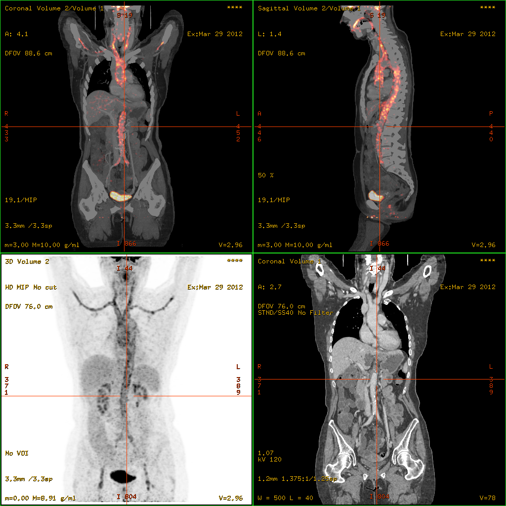
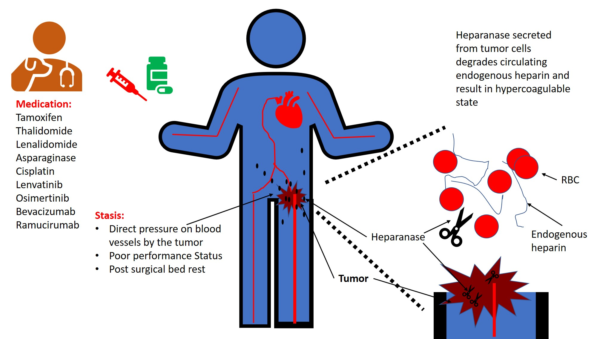
The Trousseau sign of malignancy or Trousseau's syndrome is a medical sign involving episodes of vessel inflammation due to blood clot (thrombophlebitis) which are recurrent or appearing in different locations over time (thrombophlebitis migrans or migratory thrombophlebitis). The location of the clot is tender and the clot can be felt as a nodule under the skin. Trousseau's syndrome is a rare variant of venous thrombosis that is characterized by recurrent, migratory thrombosis in superficial veins and in uncommon sites, such as the chest wall and arms. This syndrome is particularly associated with pancreatic, gastric and lung cancer and Trousseau's syndrome can be an early sign of cancer sometimes appearing months to years before the tumor would be otherwise detected. Heparin therapy is recommended to prevent future clots. The Trousseau sign of malignancy should not be confused with the Trousseau sign of latent tetany caused by low levels of calcium in the blood. History Armand ... [...More Info...] [...Related Items...] OR: [Wikipedia] [Google] [Baidu] |
Estrogen Replacement Therapy
Hormone replacement therapy (HRT), also known as menopausal hormone therapy or postmenopausal hormone therapy, is a form of hormone therapy used to treat symptoms associated with female menopause. These symptoms can include hot flashes, vaginal atrophy, accelerated skin aging, vaginal dryness, decreased muscle mass, sexual dysfunction, and bone loss or osteoporosis. They are in large part related to the diminished levels of sex hormones that occur during menopause. Estrogens and progestogens are the main hormone drugs used in HRT. Progesterone is the main female sex hormone that occurs naturally and is also manufactured into a drug that is used in menopausal hormone therapy. Although both classes of hormones can have symptomatic benefit, progestogen is specifically added to estrogen regimens, unless the uterus has been removed, to avoid the increased risk of endometrial cancer. Unopposed estrogen therapy promotes endometrial hyperplasia and increases the risk of cancer, while p ... [...More Info...] [...Related Items...] OR: [Wikipedia] [Google] [Baidu] |


