|
Tenon's Capsule
Tenon's capsule (), also known as the Tenon capsule, fascial sheath of the eyeball () or the fascia bulbi, is a thin membrane which envelops the eyeball from the optic nerve to the corneal limbus, separating it from the orbital fat and forming a socket in which it moves. The inner surface of Tenon's capsule is smooth and is separated from the outer surface of the sclera by the periscleral lymph space. This lymph space is continuous with the subdural and subarachnoid cavities and is traversed by delicate bands of connective tissue which extend between the capsule and the sclera. The capsule is perforated behind by the ciliary vessels and nerves and fuses with the sheath of the optic nerve and with the sclera around the entrance of the optic nerve. In front it adheres to the conjunctiva, and both structures are attached to the ciliary region of the eyeball. The structure was named after Jacques-René Tenon (1724–1816), a French surgeon and pathologist. Structure Relations Te ... [...More Info...] [...Related Items...] OR: [Wikipedia] [Google] [Baidu] |
Orbit
In celestial mechanics, an orbit is the curved trajectory of an object such as the trajectory of a planet around a star, or of a natural satellite around a planet, or of an artificial satellite around an object or position in space such as a planet, moon, asteroid, or Lagrange point. Normally, orbit refers to a regularly repeating trajectory, although it may also refer to a non-repeating trajectory. To a close approximation, planets and satellites follow elliptic orbits, with the center of mass being orbited at a focal point of the ellipse, as described by Kepler's laws of planetary motion. For most situations, orbital motion is adequately approximated by Newtonian mechanics, which explains gravity as a force obeying an inverse-square law. However, Albert Einstein's general theory of relativity, which accounts for gravity as due to curvature of spacetime, with orbits following geodesics, provides a more accurate calculation and understanding of the exact mechanics of orbi ... [...More Info...] [...Related Items...] OR: [Wikipedia] [Google] [Baidu] |
Inferior Tarsus
The tarsi (tarsal plates) are two comparatively thick, elongated plates of dense connective tissue, about in length for the upper eyelid and 5 mm for the lower eyelid; one is found in each eyelid, and contributes to its form and support. They are located directly above the lid margins. The tarsus has a lower and upper part making up the palpebrae. Superior The ''superior tarsus'' (''tarsus superior''; superior tarsal plate), the larger, is of a semilunar form, about in breadth at the center, and gradually narrowing toward its extremities. It is adjoined by the superior tarsal muscle. To the anterior surface of this plate the aponeurosis of the levator palpebræ superioris is attached. Inferior The ''inferior tarsus'' (''tarsus inferior''; inferior tarsal plate) is smaller, is thin, is elliptical in form, and has a vertical diameter of about . The free or ciliary margins of these plates are thick and straight. Relations The attached or orbital margins are connected to the ... [...More Info...] [...Related Items...] OR: [Wikipedia] [Google] [Baidu] |
Retrobulbar
A retrobulbar block is a regional anesthetic nerve block in the retrobulbar space, the area located behind the globe of the eye. Injection of local anesthetic into this space constitutes the retrobulbar block. This injection provides akinesia of the extraocular muscles by blocking cranial nerves II, III, and VI, thereby preventing movement of the globe. Cranial nerve IV lies outside the muscle cone, and therefore is not affected by the local anesthesia. As a result, intorsion of the eye is still possible. It also provides sensory anesthesia of the conjunctiva, cornea and uvea by blocking the ciliary nerves. This block is most commonly employed for cataract surgery, but also provides anesthesia for other intraocular surgeries. Side effects and complications Complications associated with this block are either ocular or systemic. Local ocular complications include hematoma formation, optic nerve damage and perforation of the globe with possible blindness. Systemic complicati ... [...More Info...] [...Related Items...] OR: [Wikipedia] [Google] [Baidu] |
Local Anaesthetic
A local anesthetic (LA) is a medication that causes absence of pain sensation. In the context of surgery, a local anesthetic creates an absence of pain in a specific location of the body without a loss of consciousness, as opposed to a general anesthetic. When it is used on specific nerve pathways (local anesthetic nerve block), paralysis (loss of muscle power) also can be achieved. Examples Short Duration & Low Potency Procaine Chloroprocaine Medium Duration & Potency Lidocaine Prilocaine High Duration & Potency Tetracaine Bupivacaine Cinchocaine Ropivacaine Clinical LAs belong to one of two classes: aminoamide and aminoester local anesthetics. Synthetic LAs are structurally related to cocaine. They differ from cocaine mainly in that they have a very low abuse potential and do not produce hypertension or (with few exceptions) vasoconstriction. They are used in various techniques of local anesthesia such as: * Topical anesthesia (surface) * Topical administration o ... [...More Info...] [...Related Items...] OR: [Wikipedia] [Google] [Baidu] |
Extraocular Muscles
The extraocular muscles (extrinsic ocular muscles), are the seven extrinsic muscles of the human eye. Six of the extraocular muscles, the four recti muscles, and the superior and inferior oblique muscles, control movement of the eye and the other muscle, the levator palpebrae superioris, controls eyelid elevation. The actions of the six muscles responsible for eye movement depend on the position of the eye at the time of muscle contraction. Structure Since only a small part of the eye called the fovea provides sharp vision, the eye must move to follow a target. Eye movements must be precise and fast. This is seen in scenarios like reading, where the reader must shift gaze constantly. Although under voluntary control, most eye movement is accomplished without conscious effort. Precisely how the integration between voluntary and involuntary control of the eye occurs is a subject of continuing research."eye, human."Encyclopædia Britannica from Encyclopædia Britannica 2006 Ulti ... [...More Info...] [...Related Items...] OR: [Wikipedia] [Google] [Baidu] |
Lacrimal Gland
The lacrimal glands are paired exocrine glands, one for each eye, found in most terrestrial vertebrates and some marine mammals, that secrete the aqueous layer of the tear film. In humans, they are situated in the upper lateral region of each orbit, in the lacrimal fossa of the orbit formed by the frontal bone. Inflammation of the lacrimal glands is called dacryoadenitis. The lacrimal gland produces tears which are secreted by the lacrimal ducts, and flow over the ocular surface, and then into canals that connect to the lacrimal sac. From that sac, the tears drain through the lacrimal duct into the nose. Anatomists divide the gland into two sections, a palpebral lobe, or portion, and an orbital lobe or portion. The smaller ''palpebral lobe'' lies close to the eye, along the inner surface of the eyelid; if the upper eyelid is everted, the palpebral portion can be seen. The orbital lobe of the gland, contains fine interlobular ducts that connect the orbital lobe and the palpebra ... [...More Info...] [...Related Items...] OR: [Wikipedia] [Google] [Baidu] |
Zygomatic Bone
In the human skull, the zygomatic bone (from grc, ζῠγόν, zugón, yoke), also called cheekbone or malar bone, is a paired irregular bone which articulates with the maxilla, the temporal bone, the sphenoid bone and the frontal bone. It is situated at the upper and lateral part of the face and forms the prominence of the cheek, part of the lateral wall and floor of the orbit, and parts of the temporal fossa and the infratemporal fossa. It presents a malar and a temporal surface; four processes (the frontosphenoidal, orbital, maxillary, and temporal), and four borders. Etymology The term ''zygomatic'' derives from the Ancient Greek , ''zygoma'', meaning "yoke". The zygomatic bone is occasionally referred to as the zygoma, but this term may also refer to the zygomatic arch. Structure Surfaces The ''malar surface'' is convex and perforated near its center by a small aperture, the zygomaticofacial foramen, for the passage of the zygomaticofacial nerve and vessels; below ... [...More Info...] [...Related Items...] OR: [Wikipedia] [Google] [Baidu] |
Suspensory Ligament Of The Eye
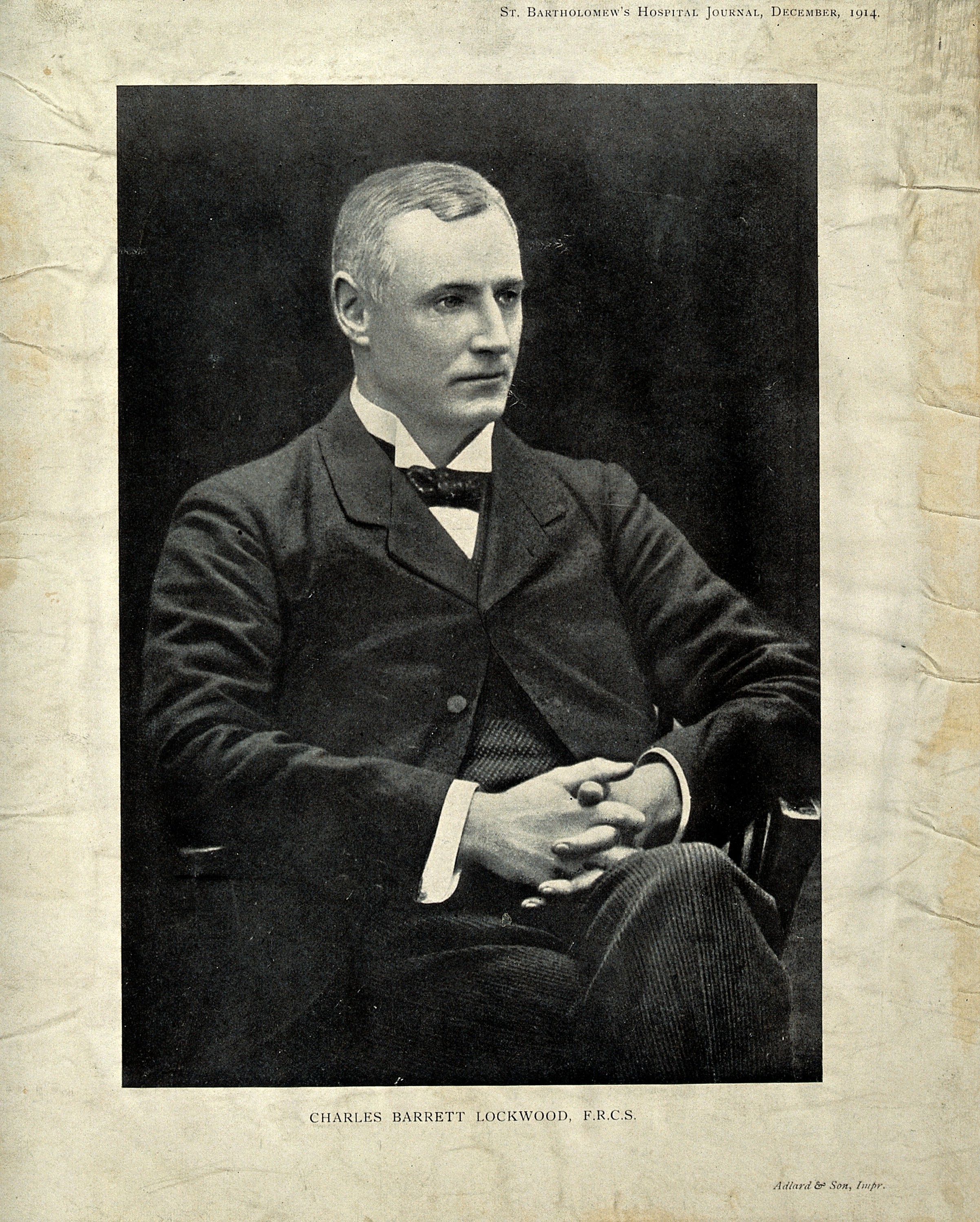
The suspensory ligament of eyeball (or Lockwood's ligament) forms a hammock stretching below the eyeball between the medial and lateral check ligaments and enclosing the inferior rectus and inferior oblique muscles of the eye. It is a thickening of Tenon's capsule, the dense connective tissue capsule surrounding the globe and separating it from orbital fat. This ligament is responsible for maintaining and supporting the position of the eyeball in its normal upward and forward position within the orbit, and prevents downward displacement of the eyeball. It can be considered a part of the bulbar sheath. It is named for Charles Barrett Lockwood Charles Barrett Lockwood (23 September 1856 – 8 November 1914) was a British surgeon and anatomist who practiced surgery at St. Bartholomew's Hospital in London. Lockwood was a member of the Royal College of Surgeons. Lockwood is remembere .... References Human eye anatomy {{Eye-stub ... [...More Info...] [...Related Items...] OR: [Wikipedia] [Google] [Baidu] |
Charles Barrett Lockwood
Charles Barrett Lockwood (23 September 1856 – 8 November 1914) was a British surgeon and anatomist who practiced surgery at St. Bartholomew's Hospital in London. Lockwood was a member of the Royal College of Surgeons. Lockwood is remembered for his surgical work with femoral and inguinal hernias. He developed an infra- inguinal approach for femoral hernia operations that is known today as the "low approach" or "Lockwood's operation". In 1893, he published an important book titled "Radical Cure of Femoral and Inguinal Hernia". He first conceived the idea of an Anatomical Society in 1887, acted as their first treasurer and was elected president of the society for 1901 to 1903. The " Lockwood's suspensory ligament" of the eye is named after him. This structure is the thickened area of contact between Tenon's capsule and the sheaths of the inferior rectus and inferior oblique muscles. This ligament is responsible for maintaining the position of the eyeball in its norma ... [...More Info...] [...Related Items...] OR: [Wikipedia] [Google] [Baidu] |
Check Ligaments
In anatomy, the alar ligaments are ligaments which connect the dens (a bony protrusion on the second cervical vertebra) to tubercles on the medial side of the occipital condyle. They are short, tough, fibrous cords that attach on the skull and on the axis, and function to check side-to-side movements of the head when it is turned. Because of their function, the alar ligaments are also known as the "check ligaments of the odontoid". Structure The alar ligaments are two strong, rounded cords of about 0.5 cm in diameter that run from the sides of the foramen magnum of the skull to the dens of the axis, the second cervical vertebra. They span almost horizontally, creating an angle between them of at least 140°. Development The alar ligaments, along with the transverse ligament of the atlas, derive from the axial component of the first cervical sclerotome. Function The function of the alar ligaments is to limit the amount of rotation of the head, and by their action on th ... [...More Info...] [...Related Items...] OR: [Wikipedia] [Google] [Baidu] |
Lacrimal Bone
The lacrimal bone is a small and fragile bone of the facial skeleton; it is roughly the size of the little fingernail. It is situated at the front part of the medial wall of the orbit. It has two surfaces and four borders. Several bony landmarks of the lacrimal bone function in the process of lacrimation or crying. Specifically, the lacrimal bone helps form the nasolacrimal canal necessary for tear translocation. A depression on the anterior inferior portion of the bone, the lacrimal fossa, houses the membranous lacrimal sac. Tears or lacrimal fluid, from the lacrimal glands, collect in this sac during excessive lacrimation. The fluid then flows through the nasolacrimal duct and into the nasopharynx. This drainage results in what is commonly referred to a runny nose during excessive crying or tear production. Injury or fracture of the lacrimal bone can result in posttraumatic obstruction of the lacrimal pathways. Structure Lateral or orbital surface The lateral or orbital surface i ... [...More Info...] [...Related Items...] OR: [Wikipedia] [Google] [Baidu] |


