|
Telocyte
Telocytes are a type of Interstitial cell, interstitial (Stromal cell, stromal) cells with very long (tens to hundreds of micrometres) and very thin prolongations (mostly below the resolving power of light microscopy) called telopodes. Rationale for the term ''telocyte'' Professor Laurențiu M. Popescu's group from Bucharest, Romania described a new type of cell (biology), cell. Popescu coined the terms telocytes (TC) for these cells, and ''telopodes'' (Tp) for their extremely long but thin prolongations in order to prevent further confusion with other interstitial (stromal) cells (e.g., fibroblast, fibroblast-like cells, myofibroblast, mesenchymal cells). Telopodes present an alternation of thin segments, ''podomeres'' (with caliber mostly under 200 nm, below the resolving power of light microscopy) and dilated segments, ''podoms'', which accommodate Mitochondrion, mitochondria, (rough) endoplasmic reticulum and caveolae - the so-called ''"Ca2+ uptake/releas ... [...More Info...] [...Related Items...] OR: [Wikipedia] [Google] [Baidu] |
Telocytes - Fig 6
Telocytes are a type of Interstitial cell, interstitial (Stromal cell, stromal) cells with very long (tens to hundreds of micrometres) and very thin prolongations (mostly below the resolving power of light microscopy) called telopodes. Rationale for the term ''telocyte'' Professor Laurențiu M. Popescu's group from Bucharest, Romania described a new type of cell (biology), cell. Popescu coined the terms telocytes (TC) for these cells, and ''telopodes'' (Tp) for their extremely long but thin prolongations in order to prevent further confusion with other interstitial (stromal) cells (e.g., fibroblast, fibroblast-like cells, myofibroblast, mesenchymal cells). Telopodes present an alternation of thin segments, ''podomeres'' (with caliber mostly under 200 nm, below the resolving power of light microscopy) and dilated segments, ''podoms'', which accommodate Mitochondrion, mitochondria, (rough) endoplasmic reticulum and caveolae - the so-called ''"Ca2+ uptake/releas ... [...More Info...] [...Related Items...] OR: [Wikipedia] [Google] [Baidu] |
Telocytes - Fig 1
Telocytes are a type of interstitial ( stromal) cells with very long (tens to hundreds of micrometres) and very thin prolongations (mostly below the resolving power of light microscopy) called telopodes. Rationale for the term ''telocyte'' Professor Laurențiu M. Popescu's group from Bucharest, Romania described a new type of cell. Popescu coined the terms telocytes (TC) for these cells, and ''telopodes'' (Tp) for their extremely long but thin prolongations in order to prevent further confusion with other interstitial (stromal) cells (e.g., fibroblast, fibroblast-like cells, myofibroblast, mesenchymal cells). Telopodes present an alternation of thin segments, ''podomeres'' (with caliber mostly under 200 nm, below the resolving power of light microscopy) and dilated segments, ''podoms'', which accommodate mitochondria, (rough) endoplasmic reticulum and caveolae - the so-called ''"Ca2+ uptake/release units"''. The concept of TC was promptly adopted by other lab ... [...More Info...] [...Related Items...] OR: [Wikipedia] [Google] [Baidu] |
Telocytes - Fig 2
Telocytes are a type of interstitial ( stromal) cells with very long (tens to hundreds of micrometres) and very thin prolongations (mostly below the resolving power of light microscopy) called telopodes. Rationale for the term ''telocyte'' Professor Laurențiu M. Popescu's group from Bucharest, Romania described a new type of cell. Popescu coined the terms telocytes (TC) for these cells, and ''telopodes'' (Tp) for their extremely long but thin prolongations in order to prevent further confusion with other interstitial (stromal) cells (e.g., fibroblast, fibroblast-like cells, myofibroblast, mesenchymal cells). Telopodes present an alternation of thin segments, ''podomeres'' (with caliber mostly under 200 nm, below the resolving power of light microscopy) and dilated segments, ''podoms'', which accommodate mitochondria, (rough) endoplasmic reticulum and caveolae - the so-called ''"Ca2+ uptake/release units"''. The concept of TC was promptly adopted by other lab ... [...More Info...] [...Related Items...] OR: [Wikipedia] [Google] [Baidu] |
Telocytes - Fig 3
Telocytes are a type of interstitial ( stromal) cells with very long (tens to hundreds of micrometres) and very thin prolongations (mostly below the resolving power of light microscopy) called telopodes. Rationale for the term ''telocyte'' Professor Laurențiu M. Popescu's group from Bucharest, Romania described a new type of cell. Popescu coined the terms telocytes (TC) for these cells, and ''telopodes'' (Tp) for their extremely long but thin prolongations in order to prevent further confusion with other interstitial (stromal) cells (e.g., fibroblast, fibroblast-like cells, myofibroblast, mesenchymal cells). Telopodes present an alternation of thin segments, ''podomeres'' (with caliber mostly under 200 nm, below the resolving power of light microscopy) and dilated segments, ''podoms'', which accommodate mitochondria, (rough) endoplasmic reticulum and caveolae - the so-called ''"Ca2+ uptake/release units"''. The concept of TC was promptly adopted by other lab ... [...More Info...] [...Related Items...] OR: [Wikipedia] [Google] [Baidu] |
Telocytes - Fig 4
Telocytes are a type of interstitial ( stromal) cells with very long (tens to hundreds of micrometres) and very thin prolongations (mostly below the resolving power of light microscopy) called telopodes. Rationale for the term ''telocyte'' Professor Laurențiu M. Popescu's group from Bucharest, Romania described a new type of cell. Popescu coined the terms telocytes (TC) for these cells, and ''telopodes'' (Tp) for their extremely long but thin prolongations in order to prevent further confusion with other interstitial (stromal) cells (e.g., fibroblast, fibroblast-like cells, myofibroblast, mesenchymal cells). Telopodes present an alternation of thin segments, ''podomeres'' (with caliber mostly under 200 nm, below the resolving power of light microscopy) and dilated segments, ''podoms'', which accommodate mitochondria, (rough) endoplasmic reticulum and caveolae - the so-called ''"Ca2+ uptake/release units"''. The concept of TC was promptly adopted by other lab ... [...More Info...] [...Related Items...] OR: [Wikipedia] [Google] [Baidu] |
Telocytes - Fig 7
Telocytes are a type of interstitial ( stromal) cells with very long (tens to hundreds of micrometres) and very thin prolongations (mostly below the resolving power of light microscopy) called telopodes. Rationale for the term ''telocyte'' Professor Laurențiu M. Popescu's group from Bucharest, Romania described a new type of cell. Popescu coined the terms telocytes (TC) for these cells, and ''telopodes'' (Tp) for their extremely long but thin prolongations in order to prevent further confusion with other interstitial (stromal) cells (e.g., fibroblast, fibroblast-like cells, myofibroblast, mesenchymal cells). Telopodes present an alternation of thin segments, ''podomeres'' (with caliber mostly under 200 nm, below the resolving power of light microscopy) and dilated segments, ''podoms'', which accommodate mitochondria, (rough) endoplasmic reticulum and caveolae - the so-called ''"Ca2+ uptake/release units"''. The concept of TC was promptly adopted by other lab ... [...More Info...] [...Related Items...] OR: [Wikipedia] [Google] [Baidu] |
Fibrosis
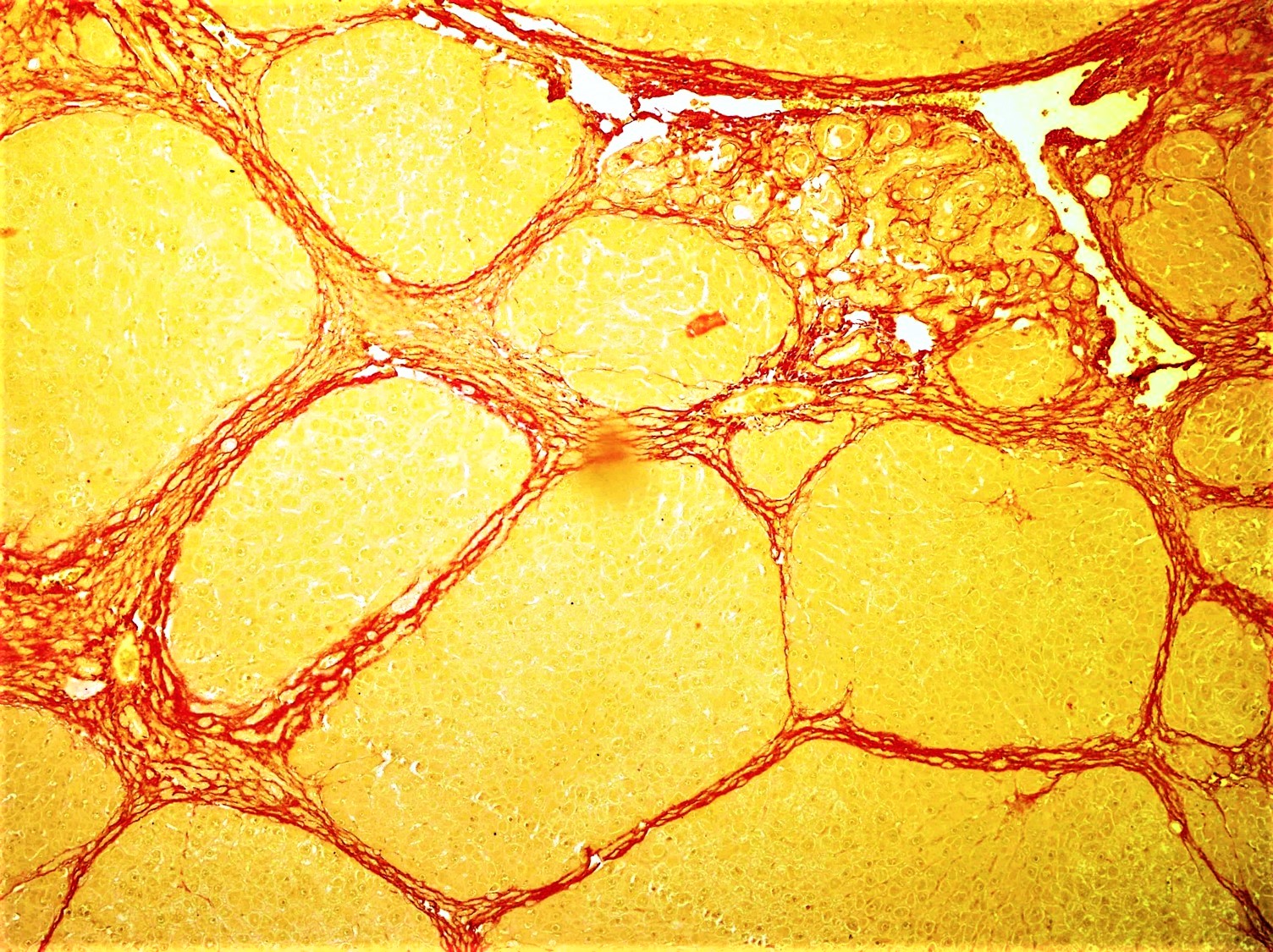
Fibrosis, also known as fibrotic scarring, is a pathological wound healing in which connective tissue replaces normal parenchymal tissue to the extent that it goes unchecked, leading to considerable tissue remodelling and the formation of permanent scar tissue. Repeated injuries, chronic inflammation and repair are susceptible to fibrosis where an accidental excessive accumulation of extracellular matrix components, such as the collagen is produced by fibroblasts, leading to the formation of a permanent fibrotic scar. In response to injury, this is called scarring, and if fibrosis arises from a single cell line, this is called a fibroma. Physiologically, fibrosis acts to deposit connective tissue, which can interfere with or totally inhibit the normal architecture and function of the underlying organ or tissue. Fibrosis can be used to describe the pathological state of excess deposition of fibrous tissue, as well as the process of connective tissue deposition in healing. Define ... [...More Info...] [...Related Items...] OR: [Wikipedia] [Google] [Baidu] |
Stroma (animal Tissue)
Stroma () is the part of a tissue or organ with a structural or connective role. It is made up of all the parts without specific functions of the organ - for example, connective tissue, blood vessels, ducts, etc. The other part, the parenchyma, consists of the cells that perform the function of the tissue or organ. There are multiple ways of classifying tissues: one classification scheme is based on tissue functions and another analyzes their cellular components. Stromal tissue falls into the "functional" class that contributes to the body's support and movement. The cells which make up stroma tissues serve as a matrix in which the other cells are embedded. Stroma is made of various types of stromal cells. Examples of stroma include: * stroma of iris * stroma of cornea * stroma of ovary * stroma of thyroid gland * stroma of thymus * stroma of bone marrow * lymph node stromal cell * multipotent stromal cell (mesenchymal stem cell) Structure Stromal connective tissues are foun ... [...More Info...] [...Related Items...] OR: [Wikipedia] [Google] [Baidu] |
Connective Tissue
Connective tissue is one of the four primary types of animal tissue, along with epithelial tissue, muscle tissue, and nervous tissue. It develops from the mesenchyme derived from the mesoderm the middle embryonic germ layer. Connective tissue is found in between other tissues everywhere in the body, including the nervous system. The three meninges, membranes that envelop the brain and spinal cord are composed of connective tissue. Most types of connective tissue consists of three main components: elastic and collagen fibers, ground substance, and cells. Blood, and lymph are classed as specialized fluid connective tissues that do not contain fiber. All are immersed in the body water. The cells of connective tissue include fibroblasts, adipocytes, macrophages, mast cells and leucocytes. The term "connective tissue" (in German, ''Bindegewebe'') was introduced in 1830 by Johannes Peter Müller. The tissue was already recognized as a distinct class in the 18th century. ... [...More Info...] [...Related Items...] OR: [Wikipedia] [Google] [Baidu] |
Collagen
Collagen () is the main structural protein in the extracellular matrix found in the body's various connective tissues. As the main component of connective tissue, it is the most abundant protein in mammals, making up from 25% to 35% of the whole-body protein content. Collagen consists of amino acids bound together to form a triple helix of elongated fibril known as a collagen helix. It is mostly found in connective tissue such as cartilage, bones, tendons, ligaments, and skin. Depending upon the degree of mineralization, collagen tissues may be rigid (bone) or compliant (tendon) or have a gradient from rigid to compliant (cartilage). Collagen is also abundant in corneas, blood vessels, the gut, intervertebral discs, and the dentin in teeth. In muscle tissue, it serves as a major component of the endomysium. Collagen constitutes one to two percent of muscle tissue and accounts for 6% of the weight of the skeletal muscle tissue. The fibroblast is the most common cell that crea ... [...More Info...] [...Related Items...] OR: [Wikipedia] [Google] [Baidu] |
Phenotype
In genetics, the phenotype () is the set of observable characteristics or traits of an organism. The term covers the organism's morphology or physical form and structure, its developmental processes, its biochemical and physiological properties, its behavior, and the products of behavior. An organism's phenotype results from two basic factors: the expression of an organism's genetic code, or its genotype, and the influence of environmental factors. Both factors may interact, further affecting phenotype. When two or more clearly different phenotypes exist in the same population of a species, the species is called polymorphic. A well-documented example of polymorphism is Labrador Retriever coloring; while the coat color depends on many genes, it is clearly seen in the environment as yellow, black, and brown. Richard Dawkins in 1978 and then again in his 1982 book ''The Extended Phenotype'' suggested that one can regard bird nests and other built structures such as cad ... [...More Info...] [...Related Items...] OR: [Wikipedia] [Google] [Baidu] |
Interstitial Cell
Interstitial cell refers to any cell that lies in the spaces between the functional cells of a tissue. Examples include: * Interstitial cell of Cajal (ICC) * Leydig cells, cells present in the male testes responsible for the production of androgen (male sex hormone) * A portion of the stroma of ovary * Certain cells in the pineal gland * Renal interstitial cells * neuroglial cells See also *List of human cell types derived from the germ layers This is a list of cells in humans derived from the three embryonic germ layers – ectoderm, mesoderm, and endoderm. Cells derived from ectoderm Surface ectoderm Skin * Trichocyte * Keratinocyte Anterior pituitary * Gonadotrope * Corticotro ... References *Sybil B Parker (ed). "Interstitial cell". McGraw Hill Dictionary of Scientific and Technical Terms. Fifth Edition. International Edition. 1994. Page 1041. {{DEFAULTSORT:Interstitial cells Cell biology Biology-related lists ... [...More Info...] [...Related Items...] OR: [Wikipedia] [Google] [Baidu] |


