|
Technicon
Siemens Healthineers AG (formerly Siemens Healthcare, Siemens Medical Solutions, Siemens Medical Systems) is a German medical device company. It is the parent company for several medical technology companies and is headquartered in Erlangen, Germany. The company dates its early beginnings in 1847 to a small family business in Berlin, co-founded by Werner von Siemens. Siemens Healthineers is connected to the larger corporation, Siemens AG. The name Siemens Medical Solutions was adopted in 2001, and the change to Siemens Healthcare was made in 2008. In 2015, Siemens named Bernd Montag as its new global CEO. In May 2016, the business operations of Siemens Healthcare GmbH were rebranded "Siemens Healthineers." Globally, the companies owned by Siemens Healthineers have 65,000 employees. History 19th century The history of Siemens Healthineers started in Berlin in the mid-19th century as a part of what is now known as Siemens AG. Siemens & Halske was founded by Werner von Siemens ... [...More Info...] [...Related Items...] OR: [Wikipedia] [Google] [Baidu] |
Aktiengesellschaft
(; abbreviated AG, ) is a German word for a corporation limited by Share (finance), share ownership (i.e. one which is owned by its shareholders) whose shares may be traded on a stock market. The term is used in Germany, Austria, Switzerland (where it is equivalent to a ''S.A. (corporation), société anonyme'' or a ''società per azioni''), and South Tyrol for companies incorporated there. It is also used in Luxembourg (as lb, Aktiëgesellschaft, label=none, ), although the equivalent French language term ''S.A. (corporation), société anonyme'' is more common. In the United Kingdom, the equivalent term is public limited company, "PLC" and in the United States while the terms Incorporation (business), "incorporated" or "corporation" are typically used, technically the more precise equivalent term is "joint-stock company" (though note for the British term only a minority of public limited companies have their shares listed on stock exchanges). Meaning of the word The German w ... [...More Info...] [...Related Items...] OR: [Wikipedia] [Google] [Baidu] |
X-ray
An X-ray, or, much less commonly, X-radiation, is a penetrating form of high-energy electromagnetic radiation. Most X-rays have a wavelength ranging from 10 picometers to 10 nanometers, corresponding to frequencies in the range 30 petahertz to 30 exahertz ( to ) and energies in the range 145 eV to 124 keV. X-ray wavelengths are shorter than those of UV rays and typically longer than those of gamma rays. In many languages, X-radiation is referred to as Röntgen radiation, after the German scientist Wilhelm Conrad Röntgen, who discovered it on November 8, 1895. He named it ''X-radiation'' to signify an unknown type of radiation.Novelline, Robert (1997). ''Squire's Fundamentals of Radiology''. Harvard University Press. 5th edition. . Spellings of ''X-ray(s)'' in English include the variants ''x-ray(s)'', ''xray(s)'', and ''X ray(s)''. The most familiar use of X-rays is checking for fractures (broken bones), but X-rays are also used in other ways. ... [...More Info...] [...Related Items...] OR: [Wikipedia] [Google] [Baidu] |
Linear Accelerator
A linear particle accelerator (often shortened to linac) is a type of particle accelerator that accelerates charged subatomic particles or ions to a high speed by subjecting them to a series of oscillating electric potentials along a linear beamline. The principles for such machines were proposed by Gustav Ising in 1924, while the first machine that worked was constructed by Rolf Widerøe in 1928 at the RWTH Aachen University. Linacs have many applications: they generate X-rays and high energy electrons for medicinal purposes in radiation therapy, serve as particle injectors for higher-energy accelerators, and are used directly to achieve the highest kinetic energy for light particles (electrons and positrons) for particle physics. The design of a linac depends on the type of particle that is being accelerated: electrons, protons or ions. Linacs range in size from a cathode ray tube (which is a type of linac) to the linac at the SLAC National Accelerator Laboratory in Menlo Park ... [...More Info...] [...Related Items...] OR: [Wikipedia] [Google] [Baidu] |
Time (magazine)
''Time'' (stylized in all caps) is an American news magazine based in New York City. For nearly a century, it was published Weekly newspaper, weekly, but starting in March 2020 it transitioned to every other week. It was first published in New York City on March 3, 1923, and for many years it was run by its influential co-founder, Henry Luce. A European edition (''Time Europe'', formerly known as ''Time Atlantic'') is published in London and also covers the Middle East, Africa, and, since 2003, Latin America. An Asian edition (''Time Asia'') is based in Hong Kong. The South Pacific edition, which covers Australia, New Zealand, and the Pacific Islands, is based in Sydney. Since 2018, ''Time'' has been published by Time USA, LLC, owned by Marc Benioff, who acquired it from Meredith Corporation. History ''Time'' has been based in New York City since its first issue published on March 3, 1923, by Briton Hadden and Henry Luce. It was the first weekly news magazine in the United St ... [...More Info...] [...Related Items...] OR: [Wikipedia] [Google] [Baidu] |
Computed Tomography
A computed tomography scan (CT scan; formerly called computed axial tomography scan or CAT scan) is a medical imaging technique used to obtain detailed internal images of the body. The personnel that perform CT scans are called radiographers or radiology technologists. CT scanners use a rotating X-ray tube and a row of detectors placed in a gantry to measure X-ray attenuations by different tissues inside the body. The multiple X-ray measurements taken from different angles are then processed on a computer using tomographic reconstruction algorithms to produce tomographic (cross-sectional) images (virtual "slices") of a body. CT scans can be used in patients with metallic implants or pacemakers, for whom magnetic resonance imaging (MRI) is contraindicated. Since its development in the 1970s, CT scanning has proven to be a versatile imaging technique. While CT is most prominently used in medical diagnosis, it can also be used to form images of non-living objects. The 1979 Nob ... [...More Info...] [...Related Items...] OR: [Wikipedia] [Google] [Baidu] |
Positron Emission Tomography
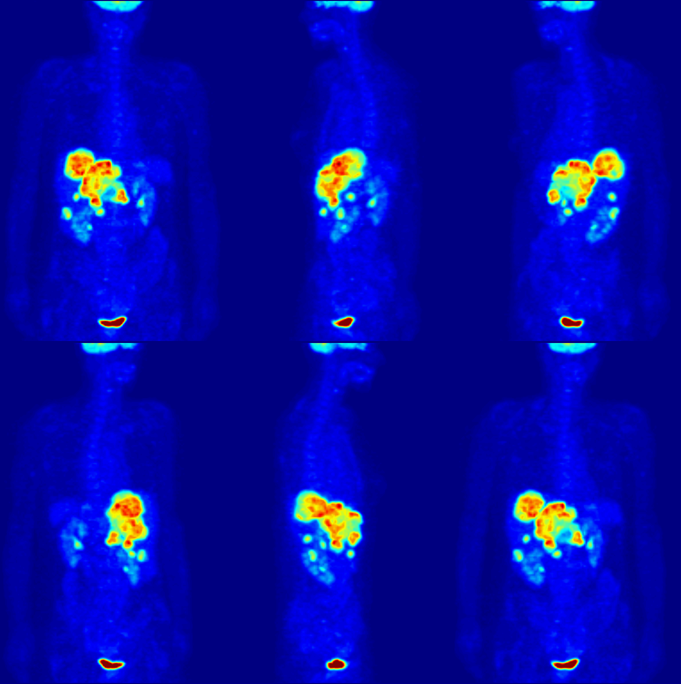
Positron emission tomography (PET) is a functional imaging technique that uses radioactive substances known as radiotracers to visualize and measure changes in Metabolism, metabolic processes, and in other physiological activities including blood flow, regional chemical composition, and absorption. Different tracers are used for various imaging purposes, depending on the target process within the body. For example, 18F-FDG, -FDG is commonly used to detect cancer, Sodium fluoride#Medical imaging, NaF is widely used for detecting bone formation, and Isotopes of oxygen#Oxygen-15, oxygen-15 is sometimes used to measure blood flow. PET is a common medical imaging, imaging technique, a Scintigraphy#Process, medical scintillography technique used in nuclear medicine. A radiopharmaceutical, radiopharmaceutical — a radioisotope attached to a drug — is injected into the body as a radioactive tracer, tracer. When the radiopharmaceutical undergoes beta plus decay, a positron is ... [...More Info...] [...Related Items...] OR: [Wikipedia] [Google] [Baidu] |
Magnetic Resonance Imaging
Magnetic resonance imaging (MRI) is a medical imaging technique used in radiology to form pictures of the anatomy and the physiological processes of the body. MRI scanners use strong magnetic fields, magnetic field gradients, and radio waves to generate images of the organs in the body. MRI does not involve X-rays or the use of ionizing radiation, which distinguishes it from CT and PET scans. MRI is a medical application of nuclear magnetic resonance (NMR) which can also be used for imaging in other NMR applications, such as NMR spectroscopy. MRI is widely used in hospitals and clinics for medical diagnosis, staging and follow-up of disease. Compared to CT, MRI provides better contrast in images of soft-tissues, e.g. in the brain or abdomen. However, it may be perceived as less comfortable by patients, due to the usually longer and louder measurements with the subject in a long, confining tube, though "Open" MRI designs mostly relieve this. Additionally, implants and oth ... [...More Info...] [...Related Items...] OR: [Wikipedia] [Google] [Baidu] |
Radiological Society Of North America
The Radiological Society of North America (RSNA) is a non-profit organization and an international society of radiologists, medical physicists and other medical imaging professionals representing 31 radiologic subspecialties from 145 countries around the world. Based in Oak Brook, Illinois, RSNA was established in 1915. RSNA's organizational mission is to promote excellence in patient care and health care delivery through education, research and technologic innovation. The Society hosts an annual conference in Chicago and develops educational resources such as courses, workshops and webinars. RSNA also publishes five peer-reviewed radiology journals, offers quality improvement tools, sponsors research to advance quantitative imaging biomarkers, and conducts outreach to enhance radiology education and patient care in low-income and middle-income countries. RSNA Annual Meeting RSNA hosts the world's largest annual medical imaging conference, a five-day event starting the last ... [...More Info...] [...Related Items...] OR: [Wikipedia] [Google] [Baidu] |
Medical Ultrasound
Medical ultrasound includes diagnostic techniques (mainly imaging techniques) using ultrasound, as well as therapeutic applications of ultrasound. In diagnosis, it is used to create an image of internal body structures such as tendons, muscles, joints, blood vessels, and internal organs, to measure some characteristics (e.g. distances and velocities) or to generate an informative audible sound. Its aim is usually to find a source of disease or to exclude pathology. The usage of ultrasound to produce visual images for medicine is called medical ultrasonography or simply sonography. The practice of examining pregnant women using ultrasound is called obstetric ultrasonography, and was an early development of clinical ultrasonography. Ultrasound is composed of sound waves with frequencies which are significantly higher than the range of human hearing (>20,000 Hz). Ultrasonic images, also known as sonograms, are created by sending pulses of ultrasound into tissue using a pr ... [...More Info...] [...Related Items...] OR: [Wikipedia] [Google] [Baidu] |
Åke Senning
Åke Senning (* 14 December 1915 in Rättvik, Sweden; † 21 July 2000 in Zurich, Switzerland) was a Swedish cardiac surgeon who worked at Zurich University Hospital from 1961 until his retirement in 1985. Biography Åke Senning was born to the Swedish veterinarian David Senning and the nurse Elly Senning, née Säfström. He finished his schooling in Uppsala with the baccalaureate. He actually wanted to become an engineer. However, as a nurse in World War 1, his mother persuaded him to study medicine. He subsequently completed the pre-clinical part of his studies in Uppsala, the clinical part and his state examination in Stockholm in 1948. His subsequent further training in Stockholm included general surgery, orthopaedics and thoracic and neurosurgery. Clarence Crafoord introduced him to the field of cardiac surgery in 1948. The influence of this eminent surgeon, who had a major impact on thoracic and cardiac surgery, sparked Senning's love of cardiac surgery and thus hel ... [...More Info...] [...Related Items...] OR: [Wikipedia] [Google] [Baidu] |
Cardiac Pacemaker
350px, Image showing the cardiac pacemaker or SA node, the primary pacemaker within the electrical_conduction_system_of_the_heart">SA_node,_the_primary_pacemaker_within_the_electrical_conduction_system_of_the_heart. The_muscle_contraction.html" "title="electrical conduction system of the heart.">electrical conduction system of the heart">SA node, the primary pacemaker within the electrical conduction system of the heart. The muscle contraction">contraction of cardiac muscle (heart muscle) in all animals is initiated by electrical impulses known as action potentials that in the heart are known as cardiac action potentials. The rate at which these impulses fire controls the rate of cardiac contraction, that is, the heart rate. The cells that create these rhythmic impulses, setting the pace for blood pumping, are called pacemaker cells, and they directly control the heart rate. They make up the cardiac pacemaker, that is, the natural pacemaker of the heart. In most humans, the h ... [...More Info...] [...Related Items...] OR: [Wikipedia] [Google] [Baidu] |
Echocardiography
An echocardiography, echocardiogram, cardiac echo or simply an echo, is an ultrasound of the heart. It is a type of medical imaging of the heart, using standard ultrasound or Doppler ultrasound. Echocardiography has become routinely used in the diagnosis, management, and follow-up of patients with any suspected or known heart diseases. It is one of the most widely used diagnostic imaging modalities in cardiology. It can provide a wealth of helpful information, including the size and shape of the heart (internal chamber size quantification), pumping capacity, location and extent of any tissue damage, and assessment of valves. An echocardiogram can also give physicians other estimates of heart function, such as a calculation of the cardiac output, ejection fraction, and diastolic function (how well the heart relaxes). Echocardiography is an important tool in assessing wall motion abnormality in patients with suspected cardiac disease. It is a tool which helps in reaching an ear ... [...More Info...] [...Related Items...] OR: [Wikipedia] [Google] [Baidu] |


