|
Talairach Coordinates
Talairach coordinates, also known as Talairach space, is a 3-dimensional coordinate system (known as an 'atlas') of the human brain, which is used to brain mapping, map the location of brain structures independent from individual differences in the size and overall shape of the brain. It is still common to use Talairach coordinates in Functional imaging, functional brain imaging studies and to target transcranial stimulation of brain regions. However, alternative methods such as the MNI Coordinate System (originated at the Montreal Neurological Institute and Hospital) have largely replaced Talairach for Stereotactic surgery, stereotaxy and other procedures. History The coordinate system was first created by neurosurgeons Jean Talairach and Gabor Szikla in their work on the ''Talairach Atlas'' in 1967, creating a standardized grid for neurosurgery. The grid was based on the idea that distances to lesions in the brain are proportional to overall brain size (i.e., the distance between ... [...More Info...] [...Related Items...] OR: [Wikipedia] [Google] [Baidu] |
Cingulate Region Of Human Brain
Cingulata, part of the superorder Xenarthra, is an order (biology), order of armour (zoology), armored Americas, New World placental mammals. Dasypodidae, Dasypodids and Chlamyphoridae, chlamyphorids, the armadillos, are the only surviving family (biology), families in the order. Two groups of cingulates much larger than extant armadillos (maximum body mass of 45 kg (100 lb) in the case of the giant armadillo) existed until recently: Pampatheriidae, pampatheriids, which reached weights of up to 200 kg (440 lb) and chlamyphorid glyptodonts, which attained masses of 2,000 kg (4,400 lb) or more. The cingulate order originated in South America during the Paleocene epoch about 66 to 56 million years ago, and due to the continent's former isolation remained confined to it during most of the Cenozoic. However, the formation of a Isthmus of Panama, land bridge allowed members of all three families to migrate to southern North America during the Pliocene or ea ... [...More Info...] [...Related Items...] OR: [Wikipedia] [Google] [Baidu] |
Positron Emission Tomography
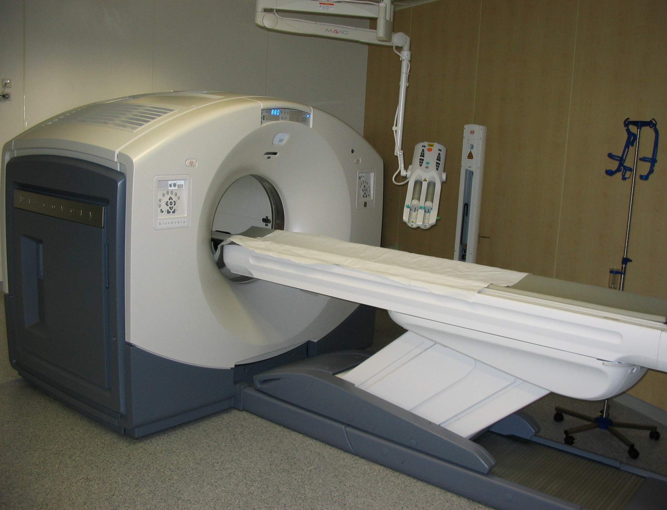
Positron emission tomography (PET) is a functional imaging technique that uses radioactive substances known as radiotracers to visualize and measure changes in Metabolism, metabolic processes, and in other physiological activities including blood flow, regional chemical composition, and absorption. Different tracers are used for various imaging purposes, depending on the target process within the body. For example, 18F-FDG, -FDG is commonly used to detect cancer, Sodium fluoride#Medical imaging, NaF is widely used for detecting bone formation, and Isotopes of oxygen#Oxygen-15, oxygen-15 is sometimes used to measure blood flow. PET is a common medical imaging, imaging technique, a Scintigraphy#Process, medical scintillography technique used in nuclear medicine. A radiopharmaceutical, radiopharmaceutical — a radioisotope attached to a drug — is injected into the body as a radioactive tracer, tracer. When the radiopharmaceutical undergoes beta plus decay, a positron is ... [...More Info...] [...Related Items...] OR: [Wikipedia] [Google] [Baidu] |
Brain
A brain is an organ that serves as the center of the nervous system in all vertebrate and most invertebrate animals. It is located in the head, usually close to the sensory organs for senses such as vision. It is the most complex organ in a vertebrate's body. In a human, the cerebral cortex contains approximately 14–16 billion neurons, and the estimated number of neurons in the cerebellum is 55–70 billion. Each neuron is connected by synapses to several thousand other neurons. These neurons typically communicate with one another by means of long fibers called axons, which carry trains of signal pulses called action potentials to distant parts of the brain or body targeting specific recipient cells. Physiologically, brains exert centralized control over a body's other organs. They act on the rest of the body both by generating patterns of muscle activity and by driving the secretion of chemicals called hormones. This centralized control allows rapid and coordinated respon ... [...More Info...] [...Related Items...] OR: [Wikipedia] [Google] [Baidu] |
List Of Neuroimaging Software
Neuroimaging software is used to study the structure and function of the brain. To see an NIH Blueprint for Neuroscience Research funded clearinghouse of many of these software applications, as well as hardware, etc. go to the NITRC web site. * 3D Slicer Extensible, free open source multi-purpose software for visualization and analysis. * Amira 3D visualization and analysis software * Analysis of Functional NeuroImages (AFNI) * Analyze developed by the Biomedical Imaging Resource (BIR) at Mayo Clinic. * Brain Image Analysis Package * CamBA * Caret Van Essen Lab, Washington University in St. Louis * CONN (functional connectivity toolbox) Diffusion Imaging in Python (DIPY)DL+DiReCT* EEGLAB * FMRIB Software Library (FSL) * FreeSurfer * Imarisbr>Imaris for Neuroscientists* ISAS (Ictal-Interictal SPECT Analysis by SPM) * LONI Pipeline, Laboratory of Neuro Imaging, USC * Mango * NITRC The Neuroimaging Informatics Tools and Resources Clearinghouse. An NIH funded database ... [...More Info...] [...Related Items...] OR: [Wikipedia] [Google] [Baidu] |
Statistical Parametric Mapping
Statistical parametric mapping (SPM) is a statistical technique for examining differences in brain activity recorded during functional neuroimaging experiments. It was created by Karl Friston. It may alternatively refer to software created by the Wellcome Department of Imaging Neuroscience at University College London to carry out such analyses. Approach Unit of measurement Functional neuroimaging is one type of 'brain scanning'. It involves the measurement of brain activity. The measurement technique depends on the imaging technology (e.g., fMRI and PET). The scanner produces a 'map' of the area that is represented as voxels. Each voxel represents the activity of a specific volume in three-dimensional space. The exact size of a voxel varies depending on the technology. fMRI voxels typically represent a volume of 27 mm3 in an equilateral cuboid. Experimental design Researchers examine brain activity linked to a specific mental process or processes. One approach involves aski ... [...More Info...] [...Related Items...] OR: [Wikipedia] [Google] [Baidu] |
Sulcus (neuroanatomy)
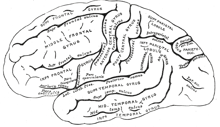
In neuroanatomy, a sulcus (Latin: "furrow", pl. ''sulci'') is a depression or groove in the cerebral cortex. It surrounds a gyrus (pl. gyri), creating the characteristic folded appearance of the brain in humans and other mammals. The larger sulci are usually called fissures. Structure Sulci, the grooves, and gyri, the folds or ridges, make up the folded surface of the cerebral cortex. Larger or deeper sulci are termed fissures, and in many cases the two terms are interchangeable. The folded cortex creates a larger surface area for the brain in humans and other mammals. When looking at the human brain, two-thirds of the surface are hidden in the grooves. The sulci and fissures are both grooves in the cortex, but they are differentiated by size. A sulcus is a shallower groove that surrounds a gyrus. A fissure is a large furrow that divides the brain into lobes and also into the two hemispheres as the longitudinal fissure. Importance of expanded surface area As the surfac ... [...More Info...] [...Related Items...] OR: [Wikipedia] [Google] [Baidu] |
Neuroimaging
Neuroimaging is the use of quantitative (computational) techniques to study the structure and function of the central nervous system, developed as an objective way of scientifically studying the healthy human brain in a non-invasive manner. Increasingly it is also being used for quantitative studies of brain disease and psychiatric illness. Neuroimaging is a highly multidisciplinary research field and is not a medical specialty. Neuroimaging differs from neuroradiology which is a medical specialty and uses brain imaging in a clinical setting. Neuroradiology is practiced by radiologists who are medical practitioners. Neuroradiology primarily focuses on identifying brain lesions, such as vascular disease, strokes, tumors and inflammatory disease. In contrast to neuroimaging, neuroradiology is qualitative (based on subjective impressions and extensive clinical training) but sometimes uses basic quantitative methods. Functional brain imaging techniques, such as functional magnet ... [...More Info...] [...Related Items...] OR: [Wikipedia] [Google] [Baidu] |
Cerebral Cortex
The cerebral cortex, also known as the cerebral mantle, is the outer layer of neural tissue of the cerebrum of the brain in humans and other mammals. The cerebral cortex mostly consists of the six-layered neocortex, with just 10% consisting of allocortex. It is separated into two cortices, by the longitudinal fissure that divides the cerebrum into the left and right cerebral hemispheres. The two hemispheres are joined beneath the cortex by the corpus callosum. The cerebral cortex is the largest site of neural integration in the central nervous system. It plays a key role in attention, perception, awareness, thought, memory, language, and consciousness. The cerebral cortex is part of the brain responsible for cognition. In most mammals, apart from small mammals that have small brains, the cerebral cortex is folded, providing a greater surface area in the confined volume of the cranium. Apart from minimising brain and cranial volume, cortical folding is crucial for the brain ... [...More Info...] [...Related Items...] OR: [Wikipedia] [Google] [Baidu] |
Korbinian Brodmann
Korbinian Brodmann (17 November 1868 – 22 August 1918) was a German neurologist who became famous for mapping the cerebral cortex and defining 52 distinct regions, known as Brodmann areas, based on their cytoarchitectonic (histological) characteristics. Life and career Brodmann was born in Liggersdorf, Province of Hohenzollern. He studied medicine in Munich, Würzburg, Berlin, and Freiburg, where he received his medical diploma in 1895. Subsequently he studied at the Medical School in the University of Lausanne in Switzerland, and then worked in the University Clinic in Munich. He received a doctor of medicine degree from the University of Leipzig in 1898, with a thesis on chronic ependymal sclerosis. From 1900 to 1901, Brodmann also worked in the Psychiatric Clinic at the University of Jena, with Ludwig Binswanger, and in the Municipal Mental Asylum in Frankfurt. There, he met Alois Alzheimer, who was influential in his decision to pursue basic neuroscience research. Fol ... [...More Info...] [...Related Items...] OR: [Wikipedia] [Google] [Baidu] |
Cytoarchitectonics
Cytoarchitecture (Greek '' κύτος''= "cell" + '' ἀρχιτεκτονική''= "architecture"), also known as cytoarchitectonics, is the study of the cellular composition of the central nervous system's tissues under the microscope. Cytoarchitectonics is one of the ways to parse the brain, by obtaining sections of the brain using a microtome and staining them with chemical agents which reveal where different neurons are located. The study of the parcellation of ''nerve fibers'' (primarily axons) into layers forms the subject of myeloarchitectonics ( History of the cerebral cytoarchitecture Defining cerebral cytoarchitecture began with the advent of —the science of slicing a ...[...More Info...] [...Related Items...] OR: [Wikipedia] [Google] [Baidu] |
Brodmann Areas
A Brodmann area is a region of the cerebral cortex, in the human or other primate brain, defined by its cytoarchitecture, or histological structure and organization of cells. History Brodmann areas were originally defined and numbered by the German anatomist Korbinian Brodmann based on the cytoarchitectural organization of neurons he observed in the cerebral cortex using the Nissl method of cell staining. Brodmann published his maps of cortical areas in humans, monkeys, and other species in 1909, along with many other findings and observations regarding the general cell types and laminar organization of the mammalian cortex. The same Brodmann area number in different species does not necessarily indicate homologous areas. A similar, but more detailed cortical map was published by Constantin von Economo and Georg N. Koskinas in 1925. Present importance Brodmann areas have been discussed, debated, refined, and renamed exhaustively for nearly a century and remain the most wid ... [...More Info...] [...Related Items...] OR: [Wikipedia] [Google] [Baidu] |
Magnetic Resonance Imaging
Magnetic resonance imaging (MRI) is a medical imaging technique used in radiology to form pictures of the anatomy and the physiological processes of the body. MRI scanners use strong magnetic fields, magnetic field gradients, and radio waves to generate images of the organs in the body. MRI does not involve X-rays or the use of ionizing radiation, which distinguishes it from CT and PET scans. MRI is a medical application of nuclear magnetic resonance (NMR) which can also be used for imaging in other NMR applications, such as NMR spectroscopy. MRI is widely used in hospitals and clinics for medical diagnosis, staging and follow-up of disease. Compared to CT, MRI provides better contrast in images of soft-tissues, e.g. in the brain or abdomen. However, it may be perceived as less comfortable by patients, due to the usually longer and louder measurements with the subject in a long, confining tube, though "Open" MRI designs mostly relieve this. Additionally, implants and oth ... [...More Info...] [...Related Items...] OR: [Wikipedia] [Google] [Baidu] |


