|
TAB1
Mitogen-activated protein kinase kinase kinase 7-interacting protein 1 is an enzyme that in humans is encoded by the ''TAB1'' gene. Function The protein encoded by this gene was identified as a regulator of the MAP kinase kinase kinase MAP3K7/TAK1, which is known to mediate various intracellular signaling pathways, such as those induced by TGF-beta, interleukin-1, and WNT-1. This protein interacts and thus activates TAK1 kinase. It has been shown that the C-terminal portion of this protein is sufficient for binding and activation of TAK1, while a portion of the N-terminus acts as a dominant-negative inhibitor of TGF beta, suggesting that this protein may function as a mediator between TGF beta receptors and TAK1. This protein can also interact with and activate the mitogen-activated protein kinase 14 (MAPK14/p38alpha), and thus represents an alternative activation pathway, in addition to the MAPKK pathways, which contributes to the biological responses of MAPK14 to various stimu ... [...More Info...] [...Related Items...] OR: [Wikipedia] [Google] [Baidu] |
MAP3K7
Mitogen-activated protein kinase kinase kinase 7 (MAP3K7), also known as TAK1, is an enzyme that in humans is encoded by the ''MAP3K7'' gene. Structure TAK1 is an evolutionarily conserved kinase in the MAP3 K family and clusters with the tyrosine-like and sterile kinase families. The protein structure of TAK1 contains an N (residues 1–104)- and C (residues 111–303)-terminus connected through the hinge region (Met 104-Ser 111). The ATP binding pocket is located in the hinge region of the kinase. Additionally, TAK1 has a catalytic lysine (Lys63) in the active site. Crystal structure of TAK1-ATP have shown that ATP forms two hydrogen bonds with residues Ala 107 and Glu 105. Further hydrogen bonding is observed to Asp 175, which is the leading residue of the DFG motif. This residue is thought to interact with Lys 63 through polar interactions and is catalytically important for phosphate transfer to substrate molecules. Critical for the TAK1-TAB1 complex is a helical loop arou ... [...More Info...] [...Related Items...] OR: [Wikipedia] [Google] [Baidu] |
MAPK14
Mitogen-activated protein kinase 14, also called p38-α, is an enzyme that in humans is encoded by the ''MAPK14'' gene. MAPK14 encodes p38α mitogen-activated protein kinase (MAPK) which is the prototypic member of the p38 MAPK family. p38 MAPKs are also known as stress-activated serine/threonine-specific kinases (SAPKs). In addition to MAPK14 for p38α MAPK, the p38 MAPK family has three additional members, including MAPK11, MAPK12 and MAPK13 which encodes p38β MAPK, p38γ MAPK and p38δ MAPK isoforms, respectively. p38α MAPK was originally identified as a tyrosine phosphorylated protein detected in activated immune cell macrophages with an essential role in inflammatory cytokine induction, such as Tumor Necrotic Factor α (TNFα). However, p38α MAPK mediated kinase activity has been implicated in many tissues beyond immune systems. p38α MAPK is mainly activated through MAPK kinase kinase cascades and exerts its biological function via downstream substrate phosphorylati ... [...More Info...] [...Related Items...] OR: [Wikipedia] [Google] [Baidu] |
Mitogen-activated Protein Kinase 14
Mitogen-activated protein kinase 14, also called p38-α, is an enzyme that in humans is encoded by the ''MAPK14'' gene. MAPK14 encodes p38α mitogen-activated protein kinase (MAPK) which is the prototypic member of the p38 MAPK family. p38 MAPKs are also known as stress-activated serine/threonine-specific kinases (SAPKs). In addition to MAPK14 for p38α MAPK, the p38 MAPK family has three additional members, including MAPK11, MAPK12 and MAPK13 which encodes p38β MAPK, p38γ MAPK and p38δ MAPK isoforms, respectively. p38α MAPK was originally identified as a tyrosine phosphorylated protein detected in activated immune cell macrophages with an essential role in inflammatory cytokine induction, such as Tumor Necrotic Factor α (TNFα). However, p38α MAPK mediated kinase activity has been implicated in many tissues beyond immune systems. p38α MAPK is mainly activated through MAPK kinase kinase cascades and exerts its biological function via downstream substrate phosphorylation ... [...More Info...] [...Related Items...] OR: [Wikipedia] [Google] [Baidu] |
MAP3K7IP2
Mitogen-activated protein kinase kinase kinase 7-interacting protein 2 is an enzyme that in humans is encoded by the ''MAP3K7IP2'' gene. The protein encoded by this gene is an activator of MAP3K7/ TAK1, which is required for the IL-1 induced activation of nuclear factor kappaB and MAPK8/JNK. This protein forms a kinase complex with TRAF6, MAP3K7 and TAB1, thus serves as an adaptor linking MAP3K7 and TRAF6. This protein, TAB1, and MAP3K7 also participate in the signal transduction induced by TNFSF11/RANKL through the activation of the receptor activator of NF-kappB ( TNFRSF11A/RANK), which may regulate the development and function of osteoclasts. Mutations in MAP3K7IP2 have been associated with human congenital heart defects. Interactions MAP3K7IP2 has been shown to interact with: * HDAC3, * TAB1, * MAP3K7IP3, * MAP3K7, * NFKB1, * NUMBL, * Nuclear receptor co-repressor 1, * TRAF2, and * TRAF6 TRAF6 is a TRAF human protein. Function The protein encoded by this ... [...More Info...] [...Related Items...] OR: [Wikipedia] [Google] [Baidu] |
XIAP
X-linked inhibitor of apoptosis protein (XIAP), also known as inhibitor of apoptosis protein 3 (IAP3) and baculoviral IAP repeat-containing protein 4 (BIRC4), is a protein that stops apoptotic cell death. In humans, this protein (XIAP) is produced by a gene named ''XIAP'' gene located on the X chromosome. XIAP is a member of the inhibitor of apoptosis family of proteins (IAP). IAPs were initially identified in baculoviruses, but XIAP is one of the homologous proteins found in mammals. It is so called because it was first discovered by a 273 base pair site on the X chromosome. The protein is also called human IAP-like Protein (hILP), because it is not as well conserved as the human IAPS: hIAP-1 and hIAP-2. XIAP is the most potent human IAP protein currently identified. Discovery Neuronal apoptosis inhibitor protein ( NAIP) was the first homolog to baculoviral IAPs that was identified in humans. With the sequencing data of NIAP, the gene sequence for a RING zinc-finger doma ... [...More Info...] [...Related Items...] OR: [Wikipedia] [Google] [Baidu] |
TRAF6
TRAF6 is a TRAF human protein. Function The protein encoded by this gene is a member of the TNF receptor associated factor (TRAF) protein family. TRAF proteins are associated with, and mediate signal transduction from members of the TNF receptor superfamily. This protein mediates the signaling not only from the members of the TNF receptor superfamily, but also from the members of the Toll/IL-1 family. Signals from receptors such as CD40, TNFSF11/TRANCE/RANKL and IL-1 have been shown to be mediated by this protein. This protein also interacts with various protein kinases including IRAK1/IRAK, SRC and PKCzeta, which provides a link between distinct signaling pathways. This protein functions as a signal transducer in the NF-kappaB pathway that activates IkappaB kinase (IKK) in response to proinflammatory cytokines. The interaction of this protein with UBE2N/UBC13, and UBE2V1/UEV1A, which are ubiquitin conjugating enzymes catalyzing the formation of polyubiquitin chains, has been ... [...More Info...] [...Related Items...] OR: [Wikipedia] [Google] [Baidu] |
Mothers Against Decapentaplegic Homolog 7
Mothers against decapentaplegic homolog 7 or SMAD7 is a protein that in humans is encoded by the ''SMAD7'' gene. SMAD7 is a protein that, as its name describes, is a homolog of the Drosophila gene: "Mothers against decapentaplegic". It belongs to the SMAD family of proteins, which belong to the TGFβ superfamily of ligands. Like many other TGFβ family members, SMAD7 is involved in cell signalling. It is a TGFβ type 1 receptor antagonist. It blocks TGFβ1 and activin associating with the receptor, blocking access to SMAD2. It is an inhibitory SMAD (I-SMAD) and is enhanced by SMURF2. Smad7 enhances muscle differentiation. Structure Smad proteins contain two conserved domains. The Mad Homology domain 1 (MH1 domain) is at the N-terminal and the Mad Homology domain 2 (MH2 domain) is at the C-terminal. Between them there is a linker region which is full of regulatory sites. The MH1 domain has DNA binding activity while the MH2 domain has transcriptional activity. The linker re ... [...More Info...] [...Related Items...] OR: [Wikipedia] [Google] [Baidu] |
MAP3K7IP3
Mitogen-activated protein kinase kinase kinase 7-interacting protein 3 is an enzyme that in humans is encoded by the ''MAP3K7IP3'' gene. Function The product of this gene functions in the NF-kappaB signal transduction pathway. The encoded protein, and the similar and functionally redundant protein MAP3K7IP2/TAB2, forms a ternary complex with the protein kinase MAP3K7/TAK1 and either TRAF2 or TRAF6 in response to stimulation with the pro-inflammatory cytokines TNF or IL-1. Subsequent MAP3K7/TAK1 kinase activity triggers a signaling cascade leading to activation of the NF-kappaB transcription factor. The human genome contains a related pseudogene. Alternatively spliced transcript variants have been described, but their biological validity has not been determined. Interactions MAP3K7IP3 has been shown to interact with MAP3K7IP2 Mitogen-activated protein kinase kinase kinase 7-interacting protein 2 is an enzyme that in humans is encoded by the ''MAP3K7IP2'' gene. The protein ... [...More Info...] [...Related Items...] OR: [Wikipedia] [Google] [Baidu] |
Wound Repair
Wound healing refers to a living organism's replacement of destroyed or damaged tissue by newly produced tissue. In undamaged skin, the epidermis (surface, epithelial layer) and dermis (deeper, connective layer) form a protective barrier against the external environment. When the barrier is broken, a regulated sequence of biochemical events is set into motion to repair the damage. This process is divided into predictable phases: blood clotting (hemostasis), inflammation, tissue growth (cell proliferation), and tissue remodeling (maturation and cell differentiation). Blood clotting may be considered to be part of the inflammation stage instead of a separate stage. The wound healing process is not only complex but fragile, and it is susceptible to interruption or failure leading to the formation of non-healing chronic wounds. Factors that contribute to non-healing chronic wounds are diabetes, venous or arterial disease, infection, and metabolic deficiencies of old age.Enoch, S. P ... [...More Info...] [...Related Items...] OR: [Wikipedia] [Google] [Baidu] |
Oncogenesis
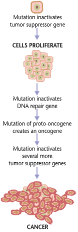
Carcinogenesis, also called oncogenesis or tumorigenesis, is the formation of a cancer, whereby normal cells are transformed into cancer cells. The process is characterized by changes at the cellular, genetic, and epigenetic levels and abnormal cell division. Cell division is a physiological process that occurs in almost all tissues and under a variety of circumstances. Normally, the balance between proliferation and programmed cell death, in the form of apoptosis, is maintained to ensure the integrity of tissues and organs. According to the prevailing accepted theory of carcinogenesis, the somatic mutation theory, mutations in DNA and epimutations that lead to cancer disrupt these orderly processes by interfering with the programming regulating the processes, upsetting the normal balance between proliferation and cell death. This results in uncontrolled cell division and the evolution of those cells by natural selection in the body. Only certain mutations lead to cancer w ... [...More Info...] [...Related Items...] OR: [Wikipedia] [Google] [Baidu] |
