|
Truncal Neural Crest
The trunk neural crest or truncal neural crest is one of the regions of neural crest in the embryo. The trunk neural crest lies between the vagal and sacral neural crest and gives rise to two groups of cells. One group migrates dorsolateral and populates the skin, forming pigment cells and the other migrates ventrolateral through the anterior sclerotome to become the epinephrine-producing cells of the adrenal gland and the neurons of the sympathetic nervous system. Some cells remain in the sclerotome to form the dorsal root ganglia :''Other Migration Locations:'' :*Proximal to the spinal cord and line up symmetrically to form the dorsal root ganglia. :*Into the skin to form melanocytes. :* Chromaffin cells of the adrenal medulla. :*Near the vertebral column and become sympathetic chain ganglia The sympathetic ganglia, or paravertebral ganglia are autonomic ganglia, of the sympathetic nervous system. Ganglia are 20,000 to 30,000 afferent and efferent nerve cell bodies that r ... [...More Info...] [...Related Items...] OR: [Wikipedia] [Google] [Baidu] |
Neural Crest
Neural crest cells are a temporary group of cells unique to vertebrates that arise from the embryonic ectoderm germ layer, and in turn give rise to a diverse cell lineage—including melanocytes, craniofacial cartilage and bone, smooth muscle, peripheral and enteric neurons and glia. After gastrulation, neural crest cells are specified at the border of the neural plate and the non-neural ectoderm. During neurulation, the borders of the neural plate, also known as the neural folds, converge at the dorsal midline to form the neural tube. Subsequently, neural crest cells from the roof plate of the neural tube undergo an epithelial to mesenchymal transition, delaminating from the neuroepithelium and migrating through the periphery where they differentiate into varied cell types. The emergence of neural crest was important in vertebrate evolution because many of its structural derivatives are defining features of the vertebrate clade. Underlying the development of neural crest is ... [...More Info...] [...Related Items...] OR: [Wikipedia] [Google] [Baidu] |
Embryo
An embryo is an initial stage of development of a multicellular organism. In organisms that reproduce sexually, embryonic development is the part of the life cycle that begins just after fertilization of the female egg cell by the male sperm cell. The resulting fusion of these two cells produces a single-celled zygote that undergoes many cell divisions that produce cells known as blastomeres. The blastomeres are arranged as a solid ball that when reaching a certain size, called a morula, takes in fluid to create a cavity called a blastocoel. The structure is then termed a blastula, or a blastocyst in mammals. The mammalian blastocyst hatches before implantating into the endometrial lining of the womb. Once implanted the embryo will continue its development through the next stages of gastrulation, neurulation, and organogenesis. Gastrulation is the formation of the three germ layers that will form all of the different parts of the body. Neurulation forms the nervous ... [...More Info...] [...Related Items...] OR: [Wikipedia] [Google] [Baidu] |
Pigment Cells
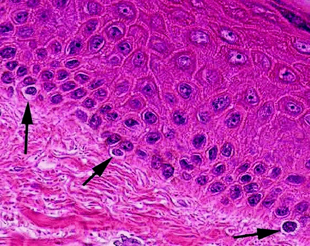
Melanocytes are melanin-producing neural crest-derived cells located in the bottom layer (the stratum basale) of the skin's epidermis, the middle layer of the eye (the uvea), the inner ear, vaginal epithelium, meninges, bones, and heart. Melanin is a dark pigment primarily responsible for skin color. Once synthesized, melanin is contained in special organelles called melanosomes which can be transported to nearby keratinocytes to induce pigmentation. Thus darker skin tones have more melanosomes present than lighter skin tones. Functionally, melanin serves as protection against UV radiation. Melanocytes also have a role in the immune system. Function Through a process called melanogenesis, melanocytes produce melanin, which is a pigment found in the skin, eyes, hair, nasal cavity, and inner ear. This melanogenesis leads to a long-lasting pigmentation, which is in contrast to the pigmentation that originates from oxidation of already-existing melanin. There are both basa ... [...More Info...] [...Related Items...] OR: [Wikipedia] [Google] [Baidu] |
Sclerotome
The somites (outdated term: primitive segments) are a set of bilaterally paired blocks of paraxial mesoderm that form in the embryonic stage of somitogenesis, along the head-to-tail axis in segmented animals. In vertebrates, somites subdivide into the dermatomes, myotomes, sclerotomes and syndetomes that give rise to the vertebrae of the vertebral column, rib cage, part of the occipital bone, skeletal muscle, cartilage, tendons, and skin (of the back). The word ''somite'' is sometimes also used in place of the word '' metamere''. In this definition, the somite is a homologously-paired structure in an animal body plan, such as is visible in annelids and arthropods. Development The mesoderm forms at the same time as the other two germ layers, the ectoderm and endoderm. The mesoderm at either side of the neural tube is called paraxial mesoderm. It is distinct from the mesoderm underneath the neural tube which is called the chordamesoderm that becomes the notochord. The pa ... [...More Info...] [...Related Items...] OR: [Wikipedia] [Google] [Baidu] |
Adrenal Gland
The adrenal glands (also known as suprarenal glands) are endocrine glands that produce a variety of hormones including adrenaline and the steroids aldosterone and cortisol. They are found above the kidneys. Each gland has an outer cortex which produces steroid hormones and an inner medulla. The adrenal cortex itself is divided into three main zones: the zona glomerulosa, the zona fasciculata and the zona reticularis. The adrenal cortex produces three main types of steroid hormones: mineralocorticoids, glucocorticoids, and androgens. Mineralocorticoids (such as aldosterone) produced in the zona glomerulosa help in the regulation of blood pressure and electrolyte balance. The glucocorticoids cortisol and cortisone are synthesized in the zona fasciculata; their functions include the regulation of metabolism and immune system suppression. The innermost layer of the cortex, the zona reticularis, produces androgens that are converted to fully functional sex hormones in the gonads ... [...More Info...] [...Related Items...] OR: [Wikipedia] [Google] [Baidu] |
Sympathetic Nervous System
The sympathetic nervous system (SNS) is one of the three divisions of the autonomic nervous system, the others being the parasympathetic nervous system and the enteric nervous system. The enteric nervous system is sometimes considered part of the autonomic nervous system, and sometimes considered an independent system. The autonomic nervous system functions to regulate the body's unconscious actions. The sympathetic nervous system's primary process is to stimulate the body's fight or flight response. It is, however, constantly active at a basic level to maintain homeostasis. The sympathetic nervous system is described as being antagonistic to the parasympathetic nervous system which stimulates the body to "feed and breed" and to (then) "rest-and-digest". Structure There are two kinds of neurons involved in the transmission of any signal through the sympathetic system: pre-ganglionic and post-ganglionic. The shorter preganglionic neurons originate in the thoracolumbar division o ... [...More Info...] [...Related Items...] OR: [Wikipedia] [Google] [Baidu] |
Dorsal Root Ganglia
A dorsal root ganglion (or spinal ganglion; also known as a posterior root ganglion) is a cluster of neurons (a ganglion) in a dorsal root of a spinal nerve. The cell bodies of sensory neurons known as first-order neurons are located in the dorsal root ganglia. The axons of dorsal root ganglion neurons are known as afferents. In the peripheral nervous system, afferents refer to the axons that relay sensory information into the central nervous system (i.e. the brain and the spinal cord). Structure The neurons comprising the dorsal root ganglion are of the pseudo-unipolar type, meaning they have a cell body (soma) with two branches that act as a single axon, often referred to as a ''distal process'' and a ''proximal process''. Unlike the majority of neurons found in the central nervous system, an action potential in posterior root ganglion neuron may initiate in the ''distal process'' in the periphery, bypass the cell body, and continue to propagate along the ''proximal pro ... [...More Info...] [...Related Items...] OR: [Wikipedia] [Google] [Baidu] |
Melanocytes
Melanocytes are melanin-producing neural crest-derived cells located in the bottom layer (the stratum basale) of the skin's epidermis, the middle layer of the eye (the uvea), the inner ear, vaginal epithelium, meninges, bones, and heart. Melanin is a dark pigment primarily responsible for skin color. Once synthesized, melanin is contained in special organelles called melanosomes which can be transported to nearby keratinocytes to induce pigmentation. Thus darker skin tones have more melanosomes present than lighter skin tones. Functionally, melanin serves as protection against UV radiation. Melanocytes also have a role in the immune system. Function Through a process called melanogenesis, melanocytes produce melanin, which is a pigment found in the skin, eyes, hair, nasal cavity, and inner ear. This melanogenesis leads to a long-lasting pigmentation, which is in contrast to the pigmentation that originates from oxidation of already-existing melanin. There are both ... [...More Info...] [...Related Items...] OR: [Wikipedia] [Google] [Baidu] |
Chromaffin Cell
Chromaffin cells, also called pheochromocytes (or phaeochromocytes), are neuroendocrine cells found mostly in the medulla of the adrenal glands in mammals. These cells serve a variety of functions such as serving as a response to stress, monitoring carbon dioxide and oxygen concentrations in the body, maintenance of respiration and the regulation of blood pressure. They are in close proximity to pre-synaptic sympathetic ganglia of the sympathetic nervous system, with which they communicate, and structurally they are similar to post-synaptic sympathetic neurons. In order to activate chromaffin cells, the splanchnic nerve of the sympathetic nervous system releases acetylcholine, which then binds to nicotinic acetylcholine receptors on the adrenal medulla. This causes the release of catecholamines. The chromaffin cells release catecholamines: ~80% of adrenaline ( epinephrine) and ~20% of noradrenaline (norepinephrine) into systemic circulation for systemic effects on multiple org ... [...More Info...] [...Related Items...] OR: [Wikipedia] [Google] [Baidu] |
Adrenal Medulla
The adrenal medulla ( la, medulla glandulae suprarenalis) is part of the adrenal gland. It is located at the center of the gland, being surrounded by the adrenal cortex. It is the innermost part of the adrenal gland, consisting of chromaffin cells that secrete catecholamines, including epinephrine (adrenaline), norepinephrine (noradrenaline), and a small amount of dopamine, in response to stimulation by sympathetic preganglionic neurons. Structure The adrenal medulla consists of irregularly shaped cells grouped around blood vessels. These cells are intimately connected with the sympathetic division of the autonomic nervous system (ANS). These adrenal medullary cells are modified postganglionic neurons, and preganglionic autonomic nerve fibers lead to them directly from the central nervous system. The adrenal medulla affects energy availability, heart rate, and basal metabolic rate. Recent research indicates that the adrenal medulla may receive input from higher-order cognitive ... [...More Info...] [...Related Items...] OR: [Wikipedia] [Google] [Baidu] |
Sympathetic Chain Ganglia
The sympathetic ganglia, or paravertebral ganglia are autonomic ganglia, of the sympathetic nervous system. Ganglia are 20,000 to 30,000 afferent and efferent nerve cell bodies that run along on either side of the spinal cord. Afferent nerve cell bodies bring information from the body to the brain and spinal cord, while efferent nerve cell bodies bring information from the brain and spinal cord to the rest of the body. The cell bodies create long sympathetic chains that are on either side of the spinal cord. They also form para- or pre-vertebral ganglia of gross anatomy. The efferent nerve cell bodies bring information from the brain to the body regarding perceptions of danger. This perception of danger can instigate the fight-or-flight response associated with the sympathetic nervous system. The fight-or-flight response is adaptive when there is a real and present danger which can be avoided or diminished through increased sympathetic activity. Sympathetic activity could be in ... [...More Info...] [...Related Items...] OR: [Wikipedia] [Google] [Baidu] |
.jpg)


