|
Transarterial Chemoembolization
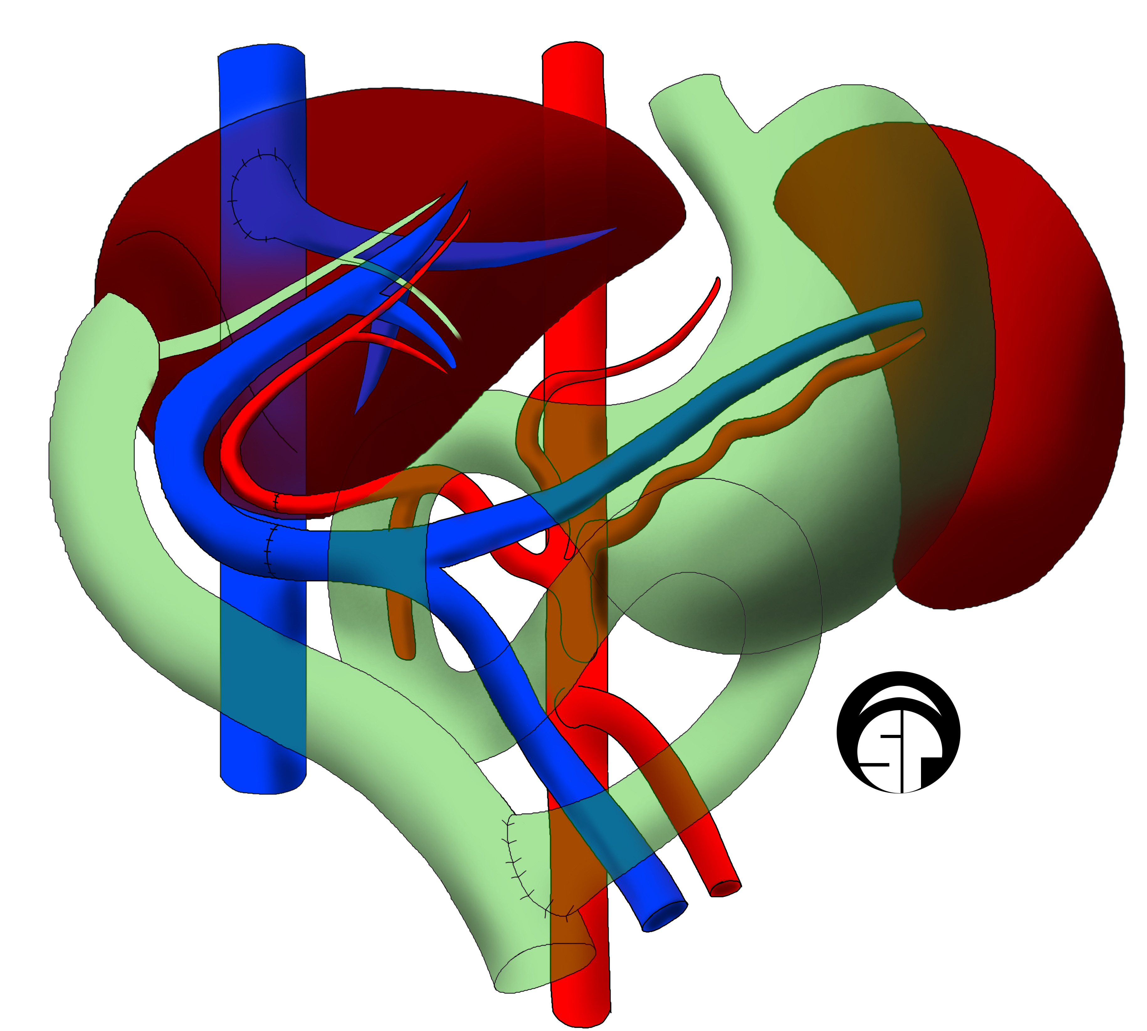
Transcatheter arterial chemoembolization (TACE) is a minimally invasive procedure performed in interventional radiology to restrict a tumor's blood supply. Small embolic particles coated with chemotherapeutic drugs are injected selectively through a catheter into an artery directly supplying the tumor. These particles both block the blood supply and induce cytotoxicity, attacking the tumor in several ways. The radiotherapeutic analogue (combining radiotherapy with embolization) is called radioembolization or selective internal radiation therapy (SIRT). Principles TACE derives its beneficial effect by two primary mechanisms. Most tumors within the liver are supplied by the proper hepatic artery, so arterial embolization preferentially interrupts the tumor's blood supply and stalls growth until neovascularization. Secondly, focused administration of chemotherapy allows for delivery of a higher dose to the tissue while simultaneously reducing systemic exposure, which is typically ... [...More Info...] [...Related Items...] OR: [Wikipedia] [Google] [Baidu] |
Interventional Radiology
Interventional radiology (IR) is a medical specialty that performs various minimally-invasive procedures using medical imaging guidance, such as x-ray fluoroscopy, computed tomography, magnetic resonance imaging, or ultrasound. IR performs both diagnostic and therapeutic procedures through very small incisions or body orifices. Diagnostic IR procedures are those intended to help make a diagnosis or guide further medical treatment, and include image-guided biopsy of a tumor or injection of an imaging contrast agent into a hollow structure, such as a blood vessel or a duct. By contrast, therapeutic IR procedures provide direct treatment—they include catheter-based medicine delivery, medical device placement (e.g., stents), and angioplasty of narrowed structures. The main benefits of interventional radiology techniques are that they can reach the deep structures of the body through a body orifice or tiny incision using small needles and wires. That decreases risks, pain, an ... [...More Info...] [...Related Items...] OR: [Wikipedia] [Google] [Baidu] |
Liver Transplantation
Liver transplantation or hepatic transplantation is the replacement of a diseased liver with the healthy liver from another person (allograft). Liver transplantation is a treatment option for end-stage liver disease and acute liver failure, although availability of donor organs is a major limitation. The most common technique is orthotopic transplantation, in which the native liver is removed and replaced by the donor organ in the same anatomic position as the original liver. The surgical procedure is complex, requiring careful harvest of the donor organ and meticulous implantation into the recipient. Liver transplantation is highly regulated, and only performed at designated transplant medical centers by highly trained transplant physicians and supporting medical team. The duration of the surgery ranges from 4 to 18 hours depending on outcome. Favorable outcomes require careful screening for eligible recipient, as well as a well-calibrated live or cadaveric donor match. Medic ... [...More Info...] [...Related Items...] OR: [Wikipedia] [Google] [Baidu] |
Abdominal Aorta
In human anatomy, the abdominal aorta is the largest artery in the abdominal cavity. As part of the aorta, it is a direct continuation of the descending aorta (of the thorax). Structure The abdominal aorta begins at the level of the thoracic diaphragm, diaphragm, crossing it via the aortic hiatus, technically behind the diaphragm, at the vertebral level of T12. It travels down the posterior wall of the abdomen, anterior to the vertebral column. It thus follows the curvature of the lumbar vertebrae, that is, convex anteriorly. The peak of this convexity is at the level of the third lumbar vertebra (L3). It runs parallel to the inferior vena cava, which is located just to the right of the abdominal aorta, and becomes smaller in diameter as it gives off branches. This is thought to be due to the large size of its principal branches. At the 11th rib, the diameter is 122mm long and 55mm wide and this is because of the constant pressure. The abdominal aorta is clinically divided int ... [...More Info...] [...Related Items...] OR: [Wikipedia] [Google] [Baidu] |
Common Femoral Artery
The femoral artery is a large artery in the thigh and the main arterial supply to the thigh and leg. The femoral artery gives off the deep femoral artery or profunda femoris artery and descends along the anteromedial part of the thigh in the femoral triangle. It enters and passes through the adductor canal, and becomes the popliteal artery as it passes through the adductor hiatus in the adductor magnus near the junction of the middle and distal thirds of the thigh. Structure The femoral artery enters the thigh from behind the inguinal ligament as the continuation of the external iliac artery. Here, it lies midway between the anterior superior iliac spine and the symphysis pubis (Mid-inguinal point). Segments In clinical parlance, the femoral artery has the following segments: *The common femoral artery (CFA) is the segment of the femoral artery between the inferior margin of the inguinal ligament and the branching point of the deep femoral artery/profunda femoris art ... [...More Info...] [...Related Items...] OR: [Wikipedia] [Google] [Baidu] |
Hepatic Artery
The common hepatic artery is a short blood vessel that supplies oxygenated blood to the liver, pylorus of the stomach, duodenum, pancreas, and gallbladder. It arises from the celiac artery and has the following branches: Additional images File:Common hepatic artery.jpg, Common hepatic artery and its branches including hepatic artery proper and right gastric artery (pyloric artery) References External links * - "Stomach, Spleen and Liver: Contents of the Hepatoduodenal ligament The hepatoduodenal ligament is the portion of the lesser omentum extending between the porta hepatis of the liver and the superior part of the duodenum. Running inside it are the following structures collectively known as the portal triad: * hep ..." * {{Authority control Arteries of the abdomen ... [...More Info...] [...Related Items...] OR: [Wikipedia] [Google] [Baidu] |
Seldinger Technique
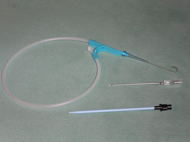
The Seldinger technique, also known as Seldinger wire technique, is a medical procedure to obtain safe access to blood vessels and other hollow organs. It is named after Sven Ivar Seldinger (1921–1998), a Swedish radiologist who introduced the procedure in 1953. Uses The Seldinger technique is used for angiography, insertion of chest drains and central venous catheters, insertion of PEG tubes using the push technique, insertion of the leads for an artificial pacemaker or implantable cardioverter-defibrillator, and numerous other interventional medical procedures. Complications The initial puncture is with a sharp instrument, and this may lead to hemorrhage or perforation of the organ in question. Infection is a possible complication, and hence asepsis is practiced during most Seldinger procedures. Loss of the guidewire into the cavity or blood vessel is a significant and generally preventable complication. Description The desired vessel or cavity is punctured with a shar ... [...More Info...] [...Related Items...] OR: [Wikipedia] [Google] [Baidu] |
Percutaneous
{{More citations needed, date=January 2021 In surgery, a percutaneous procedurei.e. Granger et al., 2012 is any medical procedure or method where access to inner organs or other tissue is done via needle-puncture of the skin, rather than by using an "open" approach where inner organs or tissue are exposed (typically with the use of a scalpel). The percutaneous approach is commonly used in vascular procedures such as angioplasty and stenting. This involves a needle catheter getting access to a blood vessel, followed by the introduction of a wire through the lumen (pathway) of the needle. It is over this wire that other catheters can be placed into the blood vessel. This technique is known as the modified Seldinger technique. More generally, "percutaneous", via its Latin roots means, 'by way of the skin'. An example would be percutaneous drug absorption from topical medications. More often, percutaneous is typically used in reference to placement of medical devices using a need ... [...More Info...] [...Related Items...] OR: [Wikipedia] [Google] [Baidu] |
Angiography
Angiography or arteriography is a medical imaging technique used to visualize the inside, or lumen, of blood vessels and organs of the body, with particular interest in the arteries, veins, and the heart chambers. Modern angiography is performed by injecting a radio-opaque contrast agent into the blood vessel and imaging using X-ray based techniques such as fluoroscopy. The word itself comes from the Greek words ἀγγεῖον ''angeion'' 'vessel' and γράφειν ''graphein'' 'to write, record'. The film or image of the blood vessels is called an ''angiograph'', or more commonly an ''angiogram''. Though the word can describe both an arteriogram and a venogram, in everyday usage the terms angiogram and arteriogram are often used synonymously, whereas the term venogram is used more precisely. The term angiography has been applied to radionuclide angiography and newer vascular imaging techniques such as CO2 angiography, CT angiography and MR angiography. The term ''isotope a ... [...More Info...] [...Related Items...] OR: [Wikipedia] [Google] [Baidu] |
Metastases
Metastasis is a pathogenic agent's spread from an initial or primary site to a different or secondary site within the host's body; the term is typically used when referring to metastasis by a cancerous tumor. The newly pathological sites, then, are metastases (mets). It is generally distinguished from cancer invasion, which is the direct extension and penetration by cancer cells into neighboring tissues. Cancer occurs after cells are genetically altered to proliferate rapidly and indefinitely. This uncontrolled proliferation by mitosis produces a primary heterogeneic tumour. The cells which constitute the tumor eventually undergo metaplasia, followed by dysplasia then anaplasia, resulting in a malignant phenotype. This malignancy allows for invasion into the circulation, followed by invasion to a second site for tumorigenesis. Some cancer cells known as circulating tumor cells acquire the ability to penetrate the walls of lymphatic or blood vessels, after which they are abl ... [...More Info...] [...Related Items...] OR: [Wikipedia] [Google] [Baidu] |
Colon Carcinoma
Colorectal cancer (CRC), also known as bowel cancer, colon cancer, or rectal cancer, is the development of cancer from the colon or rectum (parts of the large intestine). Signs and symptoms may include blood in the stool, a change in bowel movements, weight loss, and fatigue. Most colorectal cancers are due to old age and lifestyle factors, with only a small number of cases due to underlying genetic disorders. Risk factors include diet, obesity, smoking, and lack of physical activity. Dietary factors that increase the risk include red meat, processed meat, and alcohol. Another risk factor is inflammatory bowel disease, which includes Crohn's disease and ulcerative colitis. Some of the inherited genetic disorders that can cause colorectal cancer include familial adenomatous polyposis and hereditary non-polyposis colon cancer; however, these represent less than 5% of cases. It typically starts as a benign tumor, often in the form of a polyp, which over time becomes cancerous. ... [...More Info...] [...Related Items...] OR: [Wikipedia] [Google] [Baidu] |

.gif)


