|
Time-domain Diffuse Optics
Time-domain diffuse optics or time-resolved functional near-infrared spectroscopy is a branch of functional near-Infrared spectroscopy which deals with light propagation in diffusive media. There are three main approaches to diffuse optics namely continuous wave (CW), frequency domain (FD) and time-domain (TD). Biological tissue in the range of red to near-infrared wavelengths are transparent to light and can be used to probe deep layers of the tissue thus enabling various in vivo applications and clinical trials. Physical concepts In this approach, a narrow pulse of light (20 MHz) and finally, sufficient laser power (>1 mW) to achieve good signal to noise ratio. In the past bulky tunable Ti:sapphire Lasers were used. They provided a wide wavelength range of 400 nm, a narrow FWHM (< 1 ps) high average power (up to 1W) and high repetition (up to 100 MHz) frequency. However, they are bulky, expensive and take a long time for wavelength swapping. In recent year ... [...More Info...] [...Related Items...] OR: [Wikipedia] [Google] [Baidu] |
Functional Near-infrared Spectroscopy
Functional near-infrared spectroscopy (fNIRS) is an optical brain monitoring technique which uses near-infrared spectroscopy for the purpose of functional neuroimaging. Using fNIRS, brain activity is measured by using near-infrared light to estimate cortical hemodynamic activity which occur in response to neural activity. Alongside EEG, fNIRS is one of the most common non-invasive neuroimaging techniques which can be used in portable contexts. The signal is often compared with the BOLD signal measured by fMRI and is capable of measuring changes both in oxy- and deoxyhemoglobin concentration, but can only measure from regions near the cortical surface. fNIRS may also be referred to as Optical Topography (OT) and is sometimes referred to simply as NIRS. Description fNIRS estimates the concentration of hemoglobin from changes in absorption of near infrared light. As light moves or propagates through the head, it is alternately scattered or absorbed by the tissue through which it tra ... [...More Info...] [...Related Items...] OR: [Wikipedia] [Google] [Baidu] |
Photomultiplier Tube
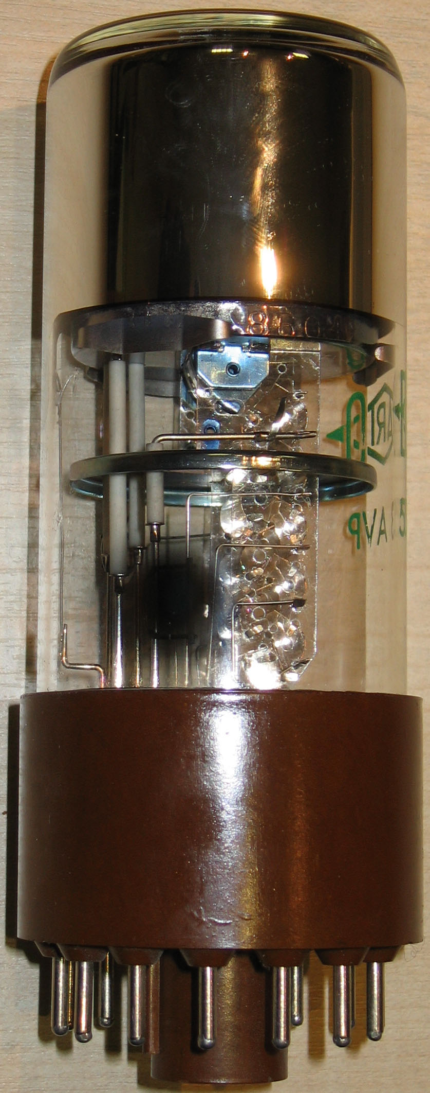
Photomultiplier tubes (photomultipliers or PMTs for short) are extremely sensitive detectors of light in the ultraviolet, visible, and near-infrared ranges of the electromagnetic spectrum. They are members of the class of vacuum tubes, more specifically vacuum phototubes. These detectors multiply the current produced by incident light by as much as 100 million times or 108 (i.e., 160 dB),Decibels are power ratios. Power is proportional to I2 (current squared). Thus a current gain of 108 produces a power gain of 1016, or 160 dB in multiple dynode stages, enabling (for example) individual photons to be detected when the incident flux of light is low. The combination of high gain, low noise, high frequency response or, equivalently, ultra-fast response, and large area of collection has maintained photomultipliers an essential place in low light level spectroscopy, confocal microscopy, Raman spectroscopy, fluorescence spectroscopy, nuclear and particle physics, astronomy, medical ... [...More Info...] [...Related Items...] OR: [Wikipedia] [Google] [Baidu] |
Functional Neuroimaging
Functional neuroimaging is the use of neuroimaging technology to measure an aspect of brain function, often with a view to understanding the relationship between activity in certain brain areas and specific mental functions. It is primarily used as a research tool in cognitive neuroscience, cognitive psychology, neuropsychology, and social neuroscience. Overview Common methods of functional neuroimaging include * Positron emission tomography (PET) * Functional magnetic resonance imaging (fMRI) * Electroencephalography (EEG) * Magnetoencephalography (MEG) * Functional near-infrared spectroscopy (fNIRS) * Single-photon emission computed tomography (SPECT) * Functional ultrasound imaging (fUS) PET, fMRI, fNIRS and fUS can measure localized changes in cerebral blood flow related to neural activity. These changes are referred to as ''activations''. Regions of the brain which are activated when a subject performs a particular task may play a role in the computational neuroscience, n ... [...More Info...] [...Related Items...] OR: [Wikipedia] [Google] [Baidu] |
Neuroimaging
Neuroimaging is the use of quantitative (computational) techniques to study the structure and function of the central nervous system, developed as an objective way of scientifically studying the healthy human brain in a non-invasive manner. Increasingly it is also being used for quantitative studies of brain disease and psychiatric illness. Neuroimaging is a highly multidisciplinary research field and is not a medical specialty. Neuroimaging differs from neuroradiology which is a medical specialty and uses brain imaging in a clinical setting. Neuroradiology is practiced by radiologists who are medical practitioners. Neuroradiology primarily focuses on identifying brain lesions, such as vascular disease, strokes, tumors and inflammatory disease. In contrast to neuroimaging, neuroradiology is qualitative (based on subjective impressions and extensive clinical training) but sometimes uses basic quantitative methods. Functional brain imaging techniques, such as functional magnet ... [...More Info...] [...Related Items...] OR: [Wikipedia] [Google] [Baidu] |
Diffuse Optical Imaging
Diffuse optical imaging (DOI) is a method of imaging using near-infrared spectroscopy (NIRS) or fluorescence-based methods. When used to create 3D volumetric models of the imaged material DOI is referred to as diffuse optical tomography, whereas 2D imaging methods are classified as diffuse optical imaging. The technique has many applications to neuroscience, sports medicine, wound monitoring, and cancer detection. Typically DOI techniques monitor changes in concentrations of oxygenated and deoxygenated hemoglobin and may additionally measure redox states of cytochromes. The technique may also be referred to as diffuse optical tomography (DOT), near infrared optical tomography (NIROT) or fluorescence diffuse optical tomography (FDOT), depending on the usage. In neuroscience, functional measurements made using NIR wavelengths, DOI techniques may classify as functional near infrared spectroscopy fNIRS. Physical mechanism Biological tissues can be considered strongly diffusive me ... [...More Info...] [...Related Items...] OR: [Wikipedia] [Google] [Baidu] |
Near-infrared Spectroscopy
Near-infrared spectroscopy (NIRS) is a spectroscopic method that uses the near-infrared region of the electromagnetic spectrum (from 780 nm to 2500 nm). Typical applications include medical and physiological diagnostics and research including blood sugar, pulse oximetry, functional neuroimaging, sports medicine, elite sports training, ergonomics, rehabilitation, neonatal research, brain computer interface, urology (bladder contraction), and neurology (neurovascular coupling). There are also applications in other areas as well such as pharmaceutical, food and agrochemical quality control, atmospheric chemistry, combustion research and astronomy. Theory Near-infrared spectroscopy is based on molecular overtone and combination vibrations. Such transitions are forbidden by the selection rules of quantum mechanics. As a result, the molar absorptivity in the near-IR region is typically quite small. (NIR absorption bands are typically 10–100 times weaker than the correspond ... [...More Info...] [...Related Items...] OR: [Wikipedia] [Google] [Baidu] |
Diffuse Optical Mammography
Diffuse optical mammography, or simply optical mammography, is an emerging imaging technique that enables the investigation of the breast composition through spectral analysis. It combines in a single non-invasive tool the capability to implement breast cancer risk assessment, lesion characterization, therapy monitoring and prediction of therapy outcome. It is an application of diffuse optics, which studies light propagation in strongly diffusive media, such as biological tissues, working in the red and near-infrared spectral range, between 600 and 1100 nm. Comparison with conventional imaging techniques Currently, the most common breast imaging techniques are X-ray mammography, ultrasounds, MRI and PET. X-ray mammography is widely spread for breast screening, thanks to its high spatial resolution and the short measurement time. However, it is not sensitive to the breast physiology, it is characterized by a limited efficiency in investigating dense breasts and it is h ... [...More Info...] [...Related Items...] OR: [Wikipedia] [Google] [Baidu] |
Time-to-digital Converter
In electronic instrumentation and signal processing, a time-to-digital converter (TDC) is a device for recognizing events and providing a digital representation of the time they occurred. For example, a TDC might output the time of arrival for each incoming pulse. Some applications wish to measure the time interval between two events rather than some notion of an absolute time. In electronics time-to-digital converters (TDCs) or time digitizers are devices commonly used to measure a time interval and convert it into digital (binary) output. In some cases interpolating TDCs are also called time counters (TCs). TDCs are used to determine the time interval between two signal pulses (known as start and stop pulse). Measurement is started and stopped when the rising or falling edge of a signal pulse crosses a set threshold. This pattern is seen in many physical experiments, like time-of-flight and lifetime measurements in atomic and high energy physics, experiments that involve laser ... [...More Info...] [...Related Items...] OR: [Wikipedia] [Google] [Baidu] |
Analog-to-digital Converter
In electronics, an analog-to-digital converter (ADC, A/D, or A-to-D) is a system that converts an analog signal, such as a sound picked up by a microphone or light entering a digital camera, into a digital signal. An ADC may also provide an isolated measurement such as an electronic device that converts an analog input voltage or current to a digital number representing the magnitude of the voltage or current. Typically the digital output is a two's complement binary number that is proportional to the input, but there are other possibilities. There are several ADC architectures. Due to the complexity and the need for precisely matched components, all but the most specialized ADCs are implemented as integrated circuits (ICs). These typically take the form of metal–oxide–semiconductor (MOS) mixed-signal integrated circuit chips that integrate both analog and digital circuits. A digital-to-analog converter (DAC) performs the reverse function; it converts a digital signa ... [...More Info...] [...Related Items...] OR: [Wikipedia] [Google] [Baidu] |
Time-correlated Single Photon Counting
Ultrafast laser spectroscopy is a spectroscopic technique that uses ultrashort pulse lasers for the study of dynamics on extremely short time scales ( attoseconds to nanoseconds). Different methods are used to examine the dynamics of charge carriers, atoms, and molecules. Many different procedures have been developed spanning different time scales and photon energy ranges; some common methods are listed below. Attosecond-to-picosecond spectroscopy Dynamics on the as to fs time scale are in general too fast to be measured electronically. Most measurements are done by employing a sequence of ultrashort light pulses to initiate a process and record its dynamics. The temporal width (duration) of the light pulses has to be on the same scale as the dynamics that are to be measured or even shorter. Light sources Titanium-sapphire laser Ti-sapphire lasers are tunable lasers that emit red and near-infrared light (700 nm- 1100 nm).Ti-sapphire laser oscillators use Ti doped-sapphi ... [...More Info...] [...Related Items...] OR: [Wikipedia] [Google] [Baidu] |
Silicon Photomultiplier
Silicon photomultipliers, often called "SiPM" in the literature, are solid-state single-photon-sensitive devices based on Single-photon avalanche diode (SPAD) implemented on common silicon substrate. The dimension of each single SPAD can vary from 10 to 100 micrometres, and their density can be up to 10000 per square millimeter. Every SPAD in SiPM operates in Geiger mode and is coupled with the others by a metal or polysilicon quenching resistor. Although the device works in digital/switching mode, most of SiPM are an analog device because all the microcells are read in parallel, making it possible to generate signals within a dynamic range from a single photon to 1000 photons for a device with just a square-millimeter area. More advanced readout schemes are utilized for the lidar applications. The supply voltage (''V''b) depends on APD technology used and typically varies between 20 V and 100 V, thus being from 15 to 75 times lower than the voltage required for a traditional pho ... [...More Info...] [...Related Items...] OR: [Wikipedia] [Google] [Baidu] |
Single-photon Avalanche Diode
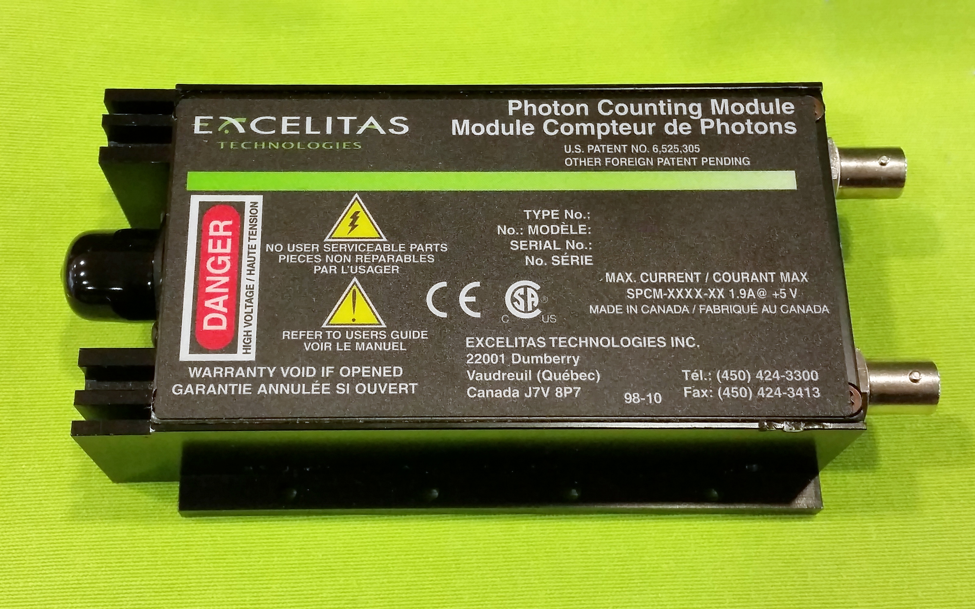
A single-photon avalanche diode (SPAD) is a solid-state photodetector within the same family as photodiodes and avalanche photodiodes (APDs), while also being fundamentally linked with basic diode behaviours. As with photodiodes and APDs, a SPAD is based around a semi-conductor p-n junction that can be illuminated with ionizing radiation such as gamma, x-rays, beta and alpha particles along with a wide portion of the electromagnetic spectrum from ultraviolet (UV) through the visible wavelengths and into the infrared (IR). In a photodiode, with a low reverse bias voltage, the leakage current changes linearly with absorption of photons, i.e. the liberation of current carriers (electrons and/or holes) due to the internal photoelectric effect. However, in a SPAD, the reverse bias is so high that a phenomenon called impact ionisation occurs which is able to cause an avalanche current to develop. Simply, a photo-generated carrier is accelerated by the electric field in the device to ... [...More Info...] [...Related Items...] OR: [Wikipedia] [Google] [Baidu] |


