|
Temporal Styloid Process
The temporal styloid process is a slender bony process of the temporal bone extending downward and forward from the undersurface of the temporal bone just below the ear. The styloid process gives attachments to several muscles, and ligaments. Structure The styloid process is a slender and pointed bony process of the temporal bone projecting anteroinferiorly from the inferior surface of the temporal bone just below the ear. Its length normally ranges from just under 3 cm to just over 4 cm. It is usually nearly straight, but may be curved in some individuals. Its ''proximal'' (''tympanohyal'') ''part'' is ensheathed by the tympanic part of the temporal bone ''(vaginal process), whereas'' its ''distal (stylohyal)'' ''part'' gives attachment to several structures. Attachments The styloid process gives attachments to several muscles, and ligaments. It serves as an anchor point for several muscles associated with the tongue and larynx. * stylohyoid ligament * stylomandi ... [...More Info...] [...Related Items...] OR: [Wikipedia] [Google] [Baidu] |
Temporal Bone
The temporal bones are situated at the sides and base of the skull, and lateral to the temporal lobes of the cerebral cortex. The temporal bones are overlaid by the sides of the head known as the temples, and house the structures of the ears. The lower seven cranial nerves and the major vessels to and from the brain traverse the temporal bone. Structure The temporal bone consists of four parts— the squamous, mastoid, petrous and tympanic parts. The squamous part is the largest and most superiorly positioned relative to the rest of the bone. The zygomatic process is a long, arched process projecting from the lower region of the squamous part and it articulates with the zygomatic bone. Posteroinferior to the squamous is the mastoid part. Fused with the squamous and mastoid parts and between the sphenoid and occipital bones lies the petrous part, which is shaped like a pyramid. The tympanic part is relatively small and lies inferior to the squamous part, anterior to t ... [...More Info...] [...Related Items...] OR: [Wikipedia] [Google] [Baidu] |
Stylohyoid Muscle
The stylohyoid muscle is a slender muscle, lying anterior and superior of the posterior belly of the digastric muscle. It is one of the suprahyoid muscles. It shares this muscle's innervation by the facial nerve, and functions to draw the hyoid bone backwards and elevate the tongue. Its origin is the styloid process of the temporal bone. It inserts on the body of the hyoid. Structure The stylohyoid muscle originates from the posterior and lateral surface of the styloid process of the temporal bone, near the base. Passing inferior and anterior, it inserts into the body of the hyoid bone, at its junction with the greater cornu, and just superior to the omohyoid muscle. It belongs to the group of suprahyoid muscles. It is perforated, near its insertion, by the intermediate tendon of the digastric muscle. The stylohyoid muscle has vascular supply from the lingual artery, a branch of the external carotid artery. Nerve supply A branch of the facial nerve (CN VII) innervates the s ... [...More Info...] [...Related Items...] OR: [Wikipedia] [Google] [Baidu] |
Cartilage
Cartilage is a resilient and smooth type of connective tissue. In tetrapods, it covers and protects the ends of long bones at the joints as articular cartilage, and is a structural component of many body parts including the rib cage, the neck and the bronchial tubes, and the intervertebral discs. In other taxa, such as chondrichthyans, but also in cyclostomes, it may constitute a much greater proportion of the skeleton. It is not as hard and rigid as bone, but it is much stiffer and much less flexible than muscle. The matrix of cartilage is made up of glycosaminoglycans, proteoglycans, collagen fibers and, sometimes, elastin. Because of its rigidity, cartilage often serves the purpose of holding tubes open in the body. Examples include the rings of the trachea, such as the cricoid cartilage and carina. Cartilage is composed of specialized cells called chondrocytes that produce a large amount of collagenous extracellular matrix, abundant ground substance that is rich in p ... [...More Info...] [...Related Items...] OR: [Wikipedia] [Google] [Baidu] |
Endochondral Ossification
Endochondral ossification is one of the two essential processes during fetal development of the mammalian skeletal system by which bone tissue is produced. Unlike intramembranous ossification, the other process by which bone tissue is produced, cartilage is present during endochondral ossification. Endochondral ossification is also an essential process during the rudimentary formation of long bones, the growth of the length of long bones, and the natural healing of bone fractures. Growth of the cartilage model The cartilage model will grow in length by continuous cell division of chondrocytes, which is accompanied by further secretion of extracellular matrix. This is called interstitial growth. The process of appositional growth occurs when the cartilage model also grows in thickness due to the addition of more extracellular matrix on the peripheral cartilage surface, which is accompanied by new chondroblasts that develop from the perichondrium. Primary center of ossifica ... [...More Info...] [...Related Items...] OR: [Wikipedia] [Google] [Baidu] |
Eagle Syndrome
Eagle syndrome (also termed stylohyoid syndrome, styloid syndrome, styloid-stylohyoid syndrome, or styloid–carotid artery syndrome) is a rare condition commonly characterized but not limited to sudden, sharp nerve-like pain in the jaw bone and joint, back of the throat, and base of the tongue, triggered by swallowing, moving the jaw, or turning the neck. Since the brain to body's nerve connections pass through the neck, many seemingly random symptoms can be triggered by impingement or entanglement. First described by American otorhinolaryngologist Watt Weems Eagle in 1937, the condition is caused by an elongated or misshapen styloid process (the slender, pointed piece of bone just below the ear) and/or calcification of the stylohyoid ligament, either of which interferes with the functioning of neighboring regions in the body, giving rise to pain. Signs and symptoms Possible symptoms include: Classic eagle syndrome is present on only one side, however, rarely, it may be presen ... [...More Info...] [...Related Items...] OR: [Wikipedia] [Google] [Baidu] |
Calcification
Calcification is the accumulation of calcium salts in a body tissue. It normally occurs in the formation of bone, but calcium can be deposited abnormally in soft tissue,Miller, J. D. Cardiovascular calcification: Orbicular origins. ''Nature Materials'' 12, 476-478 (2013). causing it to harden. Calcifications may be classified on whether there is mineral balance or not, and the location of the calcification. Calcification may also refer to the processes of normal mineral deposition in biological systems, such as the formation of stromatolites or mollusc shells (see Biomineralization). Signs and symptoms Calcification can manifest itself in many ways in the body depending on the location. In the pulpal structure of a tooth, calcification often presents asymptomatically, and is diagnosed as an incidental finding during radiographic interpretation. Individual teeth with calcified pulp will typically respond negatively to vitality testing; teeth with calcified pulp often lack ... [...More Info...] [...Related Items...] OR: [Wikipedia] [Google] [Baidu] |
Glossopharyngeal Nerve
The glossopharyngeal nerve (), also known as the ninth cranial nerve, cranial nerve IX, or simply CN IX, is a cranial nerve that exits the brainstem from the sides of the upper medulla, just anterior (closer to the nose) to the vagus nerve. Being a mixed nerve (sensorimotor), it carries afferent sensory and efferent motor information. The motor division of the glossopharyngeal nerve is derived from the basal plate of the embryonic medulla oblongata, whereas the sensory division originates from the cranial neural crest. Structure From the anterior portion of the medulla oblongata, the glossopharyngeal nerve passes laterally across or below the flocculus, and leaves the skull through the central part of the jugular foramen. From the superior and inferior ganglia in jugular foramen, it has its own sheath of dura mater. The inferior ganglion on the inferior surface of petrous part of temporal is related with a triangular depression into which the aqueduct of cochlea opens. On the ... [...More Info...] [...Related Items...] OR: [Wikipedia] [Google] [Baidu] |
Stylopharyngeus
The stylopharyngeus is a muscle in the head that stretches between the temporal styloid process and the pharynx. Structure The stylopharyngeus is a long, slender muscle, cylindrical above, flattened below. It arises from the medial side of the base of the temporal styloid process, passes downward along the side of the pharynx between the superior pharyngeal constrictor and the middle pharyngeal constrictor, and spreads out beneath the mucous membrane. Some of its fibers are lost in the constrictor muscles while others, joining the palatopharyngeus muscle, are inserted into the posterior border of the thyroid cartilage. The glossopharyngeal nerve runs on the lateral side of this muscle, and crosses over it to reach the tongue. Nerve supply The stylopharyngeus is the only muscle in the pharynx innervated by the glossopharyngeal nerve (CN IX) via branchial motor neurons with their cell bodies in the rostral part of the nucleus ambiguus. Development Embryological origin i ... [...More Info...] [...Related Items...] OR: [Wikipedia] [Google] [Baidu] |
Facial Nerve
The facial nerve, also known as the seventh cranial nerve, cranial nerve VII, or simply CN VII, is a cranial nerve that emerges from the pons of the brainstem, controls the muscles of facial expression, and functions in the conveyance of taste sensations from the anterior two-thirds of the tongue. The nerve typically travels from the pons through the facial canal in the temporal bone and exits the skull at the stylomastoid foramen. It arises from the brainstem from an area posterior to the cranial nerve VI (abducens nerve) and anterior to cranial nerve VIII (vestibulocochlear nerve). The facial nerve also supplies preganglionic parasympathetic fibers to several head and neck ganglia. The facial and intermediate nerves can be collectively referred to as the nervus intermediofacialis. The path of the facial nerve can be divided into six segments: # intracranial (cisternal) segment # meatal (canalicular) segment (within the internal auditory canal) # labyrinthine segmen ... [...More Info...] [...Related Items...] OR: [Wikipedia] [Google] [Baidu] |
Hypoglossal Nerve
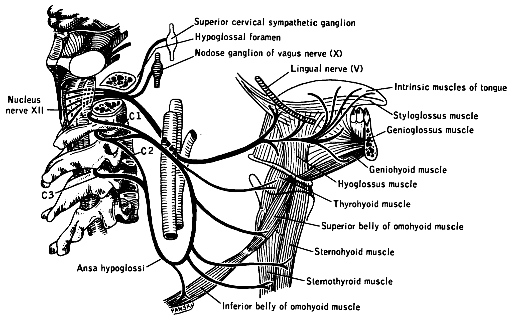
The hypoglossal nerve, also known as the twelfth cranial nerve, cranial nerve XII, or simply CN XII, is a cranial nerve that innervates all the extrinsic and intrinsic muscles of the tongue except for the palatoglossus, which is innervated by the vagus nerve. CN XII is a nerve with a solely motor function. The nerve arises from the hypoglossal nucleus in the medulla as a number of small rootlets, passes through the hypoglossal canal and down through the neck, and eventually passes up again over the tongue muscles it supplies into the tongue. The nerve is involved in controlling tongue movements required for speech and swallowing, including sticking out the tongue and moving it from side to side. Damage to the nerve or the neural pathways which control it can affect the ability of the tongue to move and its appearance, with the most common sources of damage being injury from trauma or surgery, and motor neuron disease. The first recorded description of the nerve is by Her ... [...More Info...] [...Related Items...] OR: [Wikipedia] [Google] [Baidu] |
Mandible
In anatomy, the mandible, lower jaw or jawbone is the largest, strongest and lowest bone in the human facial skeleton. It forms the lower jaw and holds the lower teeth in place. The mandible sits beneath the maxilla. It is the only movable bone of the skull (discounting the ossicles of the middle ear). It is connected to the temporal bones by the temporomandibular joints. The bone is formed in the fetus from a fusion of the left and right mandibular prominences, and the point where these sides join, the mandibular symphysis, is still visible as a faint ridge in the midline. Like other symphyses in the body, this is a midline articulation where the bones are joined by fibrocartilage, but this articulation fuses together in early childhood.Illustrated Anatomy of the Head and Neck, Fehrenbach and Herring, Elsevier, 2012, p. 59 The word "mandible" derives from the Latin word ''mandibula'', "jawbone" (literally "one used for chewing"), from '' mandere'' "to chew" and ''-bula'' ... [...More Info...] [...Related Items...] OR: [Wikipedia] [Google] [Baidu] |
Styloglossus
The styloglossus, the shortest and smallest of the three styloid muscles, arises from the anterior and lateral surfaces of the styloid process near its apex, and from the stylomandibular ligament. Passing inferiorly and anteriorly between the internal and external carotid arteries, it divides upon the side of the tongue near its dorsal surface, blending with the fibers of the longitudinalis inferior in front of the hyoglossus The hyoglossus, thin and quadrilateral, arises from the side of the body and from the whole length of the greater cornu of the hyoid bone, and passes almost vertically upward to enter the side of the tongue, between the styloglossus and the in ...; the other, oblique, overlaps the Hyoglossus and decussates with its fibers. Innervation The styloglossus is innervated by the hypoglossal nerve (CN XII) like all muscles of the tongue except palatoglossus which is innervated by the pharyngeal plexus of vagus nerve (CN X). Action The styloglossus draws u ... [...More Info...] [...Related Items...] OR: [Wikipedia] [Google] [Baidu] |


