|
Superior Sagittal Sinus
The superior sagittal sinus (also known as the superior longitudinal sinus), within the human head, is an unpaired area along the attached margin of the falx cerebri. It allows blood to drain from the lateral aspects of anterior cerebral hemispheres to the confluence of sinuses. Cerebrospinal fluid drains through arachnoid granulations into the superior sagittal sinus and is returned to venous circulation. Structure Commencing at the foramen cecum, through which it receives emissary veins from the nasal cavity, it runs from anterior to posterior, grooving the inner surface of the frontal, the adjacent margins of the two parietal lobes, and the superior division of the cruciate eminence of the occipital lobe. Near the internal occipital protuberance, it drains into the confluence of sinuses and deviates to either side (usually the right). At this point it is continued as the corresponding transverse sinus. The superior sagittal sinus is usually divided into three parts: anterior ... [...More Info...] [...Related Items...] OR: [Wikipedia] [Google] [Baidu] |
Superior Cerebral Veins
The superior cerebral veins, numbering eight to twelve, drain the superior, lateral, and medial surfaces of the hemispheres. They are predominantly found in the sulci between the gyri, but can also be found running across the gyri. Individually they drain into the superior sagittal sinus The superior sagittal sinus (also known as the superior longitudinal sinus), within the human head, is an unpaired area along the attached margin of the falx cerebri. It allows blood to drain from the lateral aspects of anterior cerebral hemispher .... The anterior veins run at near right angles to the sinus while the posterior and larger veins are directed at oblique angles, opening into the sinus in a direction opposed to the current (anterior to posterior) of the blood contained within it. Additional images File:Slide6Neo.JPG, Meninges and superficial cerebral veins. Deep dissection. Superior view. References External links Diagram Veins of the head and neck {{circulatory-stub ... [...More Info...] [...Related Items...] OR: [Wikipedia] [Google] [Baidu] |
Occipital Lobe
The occipital lobe is one of the four major lobes of the cerebral cortex in the brain of mammals. The name derives from its position at the back of the head, from the Latin ''ob'', "behind", and ''caput'', "head". The occipital lobe is the visual processing center of the mammalian brain containing most of the anatomical region of the visual cortex. The primary visual cortex is Brodmann area 17, commonly called V1 (visual one). Human V1 is located on the medial side of the occipital lobe within the calcarine sulcus; the full extent of V1 often continues onto the occipital pole. V1 is often also called striate cortex because it can be identified by a large stripe of myelin, the Stria of Gennari. Visually driven regions outside V1 are called extrastriate cortex. There are many extrastriate regions, and these are specialized for different visual tasks, such as visuospatial processing, color differentiation, and motion perception. Bilateral lesions of the occipital lobe can lead ... [...More Info...] [...Related Items...] OR: [Wikipedia] [Google] [Baidu] |
Parietal Foramina
A parietal foramen is an opening in the skull for the parietal emissary vein, which drains into the superior sagittal sinus. Occasionally, a small branch of the occipital artery can also pass through it. It is located at the back part of the parietal bone, close to the upper or sagittal border. It is not always present, and its size varies considerably. Parietal foramina tend to be symmetrical, with the same number on each side. See also * Foramina of skull This article lists foramina that occur in the human body. __TOC__ Skull The human skull has numerous openings (foramina), through which cranial nerves, arteries, veins, and other structures pass. These foramina vary in size and number, with ... References External links Foramina of the skull {{musculoskeletal-stub ... [...More Info...] [...Related Items...] OR: [Wikipedia] [Google] [Baidu] |
Pericranium
The periosteum is a membrane that covers the outer surface of all bones, except at the articular surfaces (i.e. the parts within a joint space) of long bones. Endosteum lines the inner surface of the medullary cavity of all long bones. Structure The periosteum consists of an outer fibrous layer, and an inner cambium layer (or osteogenic layer). The fibrous layer is of dense irregular connective tissue, containing fibroblasts, while the cambium layer is highly cellular containing progenitor cells that develop into osteoblasts. These osteoblasts are responsible for increasing the width of a long bone and the overall size of the other bone types. After a bone fracture, the progenitor cells develop into osteoblasts and chondroblasts, which are essential to the healing process. The outer fibrous layer and the inner cambium layer is differentiated under electron micrography. As opposed to osseous tissue, the periosteum has nociceptors, sensory neurons that make it very sensitive to ... [...More Info...] [...Related Items...] OR: [Wikipedia] [Google] [Baidu] |
Dura Mater
In neuroanatomy, dura mater is a thick membrane made of dense irregular connective tissue that surrounds the brain and spinal cord. It is the outermost of the three layers of membrane called the meninges that protect the central nervous system. The other two meningeal layers are the arachnoid mater and the pia mater. It envelops the arachnoid mater, which is responsible for keeping in the cerebrospinal fluid. It is derived primarily from the neural crest cell population, with postnatal contributions of the paraxial mesoderm. Structure The dura mater has several functions and layers. The dura mater is a membrane that envelops the arachnoid mater. It surrounds and supports the dural sinuses (also called dural venous sinuses, cerebral sinuses, or cranial sinuses) and carries blood from the brain toward the heart. Cranial dura mater has two layers called ''lamellae'', a superficial layer (also called the periosteal layer), which serves as the skull's inner periosteum, called the ... [...More Info...] [...Related Items...] OR: [Wikipedia] [Google] [Baidu] |
Diploë
Diploë ( or ) is the spongy cancellous bone separating the inner and outer layers of the cortical bone of the skull. In the cranial bones, the layers of compact cortical tissue are familiarly known as the tables of the skull; the outer one is thick and tough; the inner is thin, dense, and brittle, and hence is termed the ''vitreous table''. The intervening cancellous tissue is called the diploë. In certain regions of the skull The skull is a bone protective cavity for the brain. The skull is composed of four types of bone i.e., cranial bones, facial bones, ear ossicles and hyoid bone. However two parts are more prominent: the cranium and the mandible. In humans, th ... this becomes absorbed so as to leave spaces filled with liquid between the two tables. Etymology From Ancient Greek διπλόη (''diplóē'', “literally, a fold”), noun use of feminine of διπλόος (''diplóos'', “double”) References External links Skull {{musculoskeletal- ... [...More Info...] [...Related Items...] OR: [Wikipedia] [Google] [Baidu] |
Transverse Sinus
The transverse sinuses (left and right lateral sinuses), within the human head, are two areas beneath the brain which allow blood to drain from the back of the head. They run laterally in a groove along the interior surface of the occipital bone. They drain from the confluence of sinuses (by the internal occipital protuberance) to the sigmoid sinuses, which ultimately connect to the internal jugular vein. ''See diagram (at right)'': labeled under the brain as "" (for Latin: ''sinus transversus''). Structure The transverse sinuses are of large size and begin at the internal occipital protuberance; one, generally the right, being the direct continuation of the superior sagittal sinus, the other of the straight sinus. Each transverse sinus passes lateral and forward, describing a slight curve with its convexity upward, to the base of the petrous portion of the temporal bone, and lies, in this part of its course, in the attached margin of the tentorium cerebelli; it then leaves th ... [...More Info...] [...Related Items...] OR: [Wikipedia] [Google] [Baidu] |
Internal Occipital Protuberance
Along the internal surface of the occipital bone, at the point of intersection of the four divisions of the cruciform eminence, is the internal occipital protuberance. Running transversely on either side is a groove for the transverse sinus. Additional images See also * External occipital protuberance Near the middle of the squamous part of occipital bone is the external occipital protuberance, the highest point of which is referred to as the inion. The inion is the most prominent projection of the protuberance which is located at the posterioin ... References External links * Diagram at uni-mainz.de Bones of the head and neck {{musculoskeletal-stub ... [...More Info...] [...Related Items...] OR: [Wikipedia] [Google] [Baidu] |
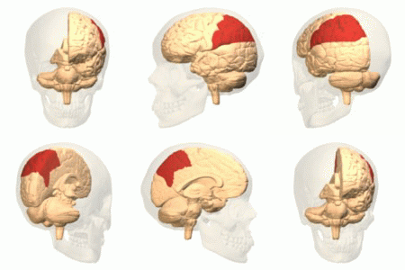
Parietal Lobes
The parietal lobe is one of the four major lobes of the cerebral cortex in the brain of mammals. The parietal lobe is positioned above the temporal lobe and behind the frontal lobe and central sulcus. The parietal lobe integrates sensory information among various modalities, including spatial sense and navigation (proprioception), the main sensory receptive area for the sense of touch in the somatosensory cortex which is just posterior to the central sulcus in the postcentral gyrus, and the dorsal stream of the visual system. The major sensory inputs from the skin (touch, temperature, and pain receptors), relay through the thalamus to the parietal lobe. Several areas of the parietal lobe are important in language processing. The somatosensory cortex can be illustrated as a distorted figure – the cortical homunculus (Latin: "little man") in which the body parts are rendered according to how much of the somatosensory cortex is devoted to them. The superior parietal lobule and in ... [...More Info...] [...Related Items...] OR: [Wikipedia] [Google] [Baidu] |
Confluence Of Sinuses
The confluence of sinuses (Latin: confluens sinuum), torcular Herophili, or torcula is the connecting point of the superior sagittal sinus, straight sinus, and occipital sinus. It is below the internal occipital protuberance of the skull. It drains venous blood from the brain into the transverse sinuses. It may be affected by arteriovenous fistulas, a thrombus, major trauma, or surgical damage, and may be imaged with many radiology techniques. Structure The confluence of sinuses is found deep to the internal occipital protuberance of the occipital bone of the skull. This puts it inferior to the occipital lobes of the brain, and posterosuperior to the cerebellum. It connects the ends of the superior sagittal sinus, the straight sinus, and the occipital sinus. Blood from it can drain into the left and right transverse sinuses. It is lined with endothelium, with some smooth muscle. Variation The confluence of sinuses shows significant variation. Most commonly, there is a conti ... [...More Info...] [...Related Items...] OR: [Wikipedia] [Google] [Baidu] |
Frontal Lobe
The frontal lobe is the largest of the four major lobes of the brain in mammals, and is located at the front of each cerebral hemisphere (in front of the parietal lobe and the temporal lobe). It is parted from the parietal lobe by a groove between tissues called the central sulcus and from the temporal lobe by a deeper groove called the lateral sulcus (Sylvian fissure). The most anterior rounded part of the frontal lobe (though not well-defined) is known as the frontal pole, one of the three poles of the cerebrum. The frontal lobe is covered by the frontal cortex. The frontal cortex includes the premotor cortex, and the primary motor cortex – parts of the motor cortex. The front part of the frontal cortex is covered by the prefrontal cortex. There are four principal gyri in the frontal lobe. The precentral gyrus is directly anterior to the central sulcus, running parallel to it and contains the primary motor cortex, which controls voluntary movements of specific body parts ... [...More Info...] [...Related Items...] OR: [Wikipedia] [Google] [Baidu] |
Emissary Veins
The emissary veins connect the extracranial venous system with the intracranial venous sinuses. They connect the veins outside the cranium to the venous sinuses inside the cranium. They drain from the scalp, through the skull, into the larger meningeal veins and dural venous sinuses. Emissary veins have an important role in selective cooling of the head. They also serve as routes where infections are carried into the cranial cavity from the extracranial veins to the intracranial veins. There are several types of emissary veins including posterior condyloid, mastoid, occipital and parietal emissary vein. Structure There are also emissary veins passing through the foramen ovale, jugular foramen, foramen lacerum, and hypoglossal canal. Function Because the emissary veins are valveless, they are an important part in selective brain cooling through bidirectional flow of cooler blood from the evaporating surface of the head. In general, blood flow is from external to internal but t ... [...More Info...] [...Related Items...] OR: [Wikipedia] [Google] [Baidu] |


