|
Superior Intercostal Vein
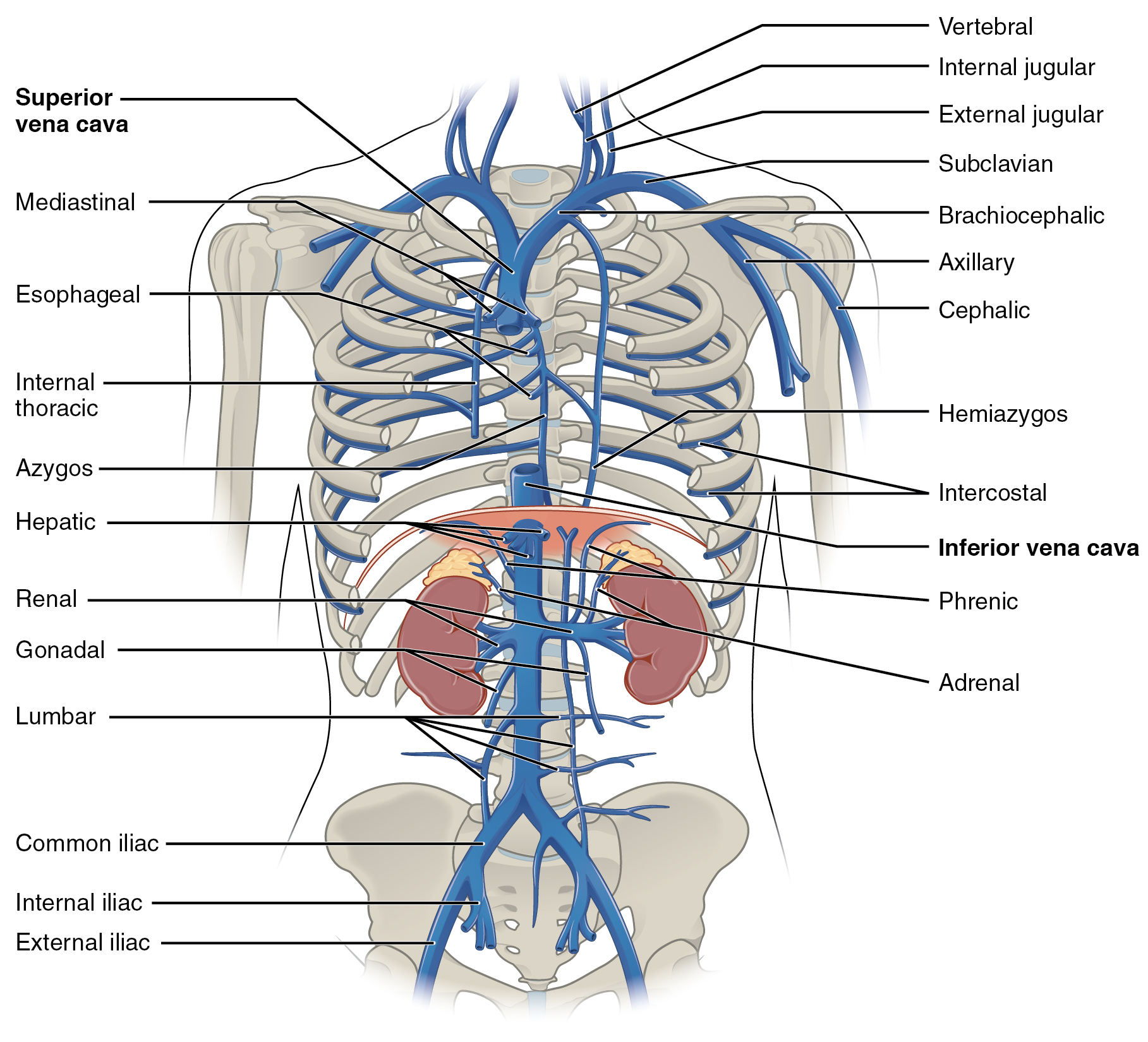
The superior intercostal veins are two veins that drain the 2nd, 3rd, and 4th intercostal spaces, one vein for each side of the body. Right superior intercostal vein The right superior intercostal vein drains the 2nd, 3rd, and 4th posterior intercostal veins on the right side of the body. It flows into the azygos vein. Left superior intercostal vein The left superior intercostal vein drains the 2nd and 3rd posterior intercostal veins on the left side of the body. It usually drains into the left brachiocephalic vein. It may also communicate with the accessory hemiazygos vein. As it passes posteriorly above the aortic arch, it crosses deep to the phrenic nerve and the pericardiacophrenic vessels and then superficial to the vagus nerve. See also * Supreme intercostal vein * Posterior intercostal veins The posterior intercostal veins are veins that drain the intercostal spaces posteriorly. They run with their corresponding posterior intercostal artery on the underside of the rib, ... [...More Info...] [...Related Items...] OR: [Wikipedia] [Google] [Baidu] |
Posterior Intercostal Veins
The posterior intercostal veins are veins that drain the intercostal spaces posteriorly. They run with their corresponding posterior intercostal artery on the underside of the rib, the vein superior to the artery. Each vein also gives off a dorsal branch that drains blood from the muscles of the back. There are eleven posterior intercostal veins on each side. Their patterns are variable, but they are commonly arranged as: * The 1st posterior intercostal vein, supreme intercostal vein, drains into the brachiocephalic vein or the vertebral vein. * The 2nd and 3rd (and often 4th) posterior intercostal veins drain into the superior intercostal vein. * The remaining posterior intercostal veins drain into the azygos vein on the right, or the hemiazygos and accessory hemiazygos vein The accessory hemiazygos vein, also called the superior hemiazygous vein, is a vein on the left side of the vertebral column that generally drains the fourth through eighth intercostal spaces on the left ... [...More Info...] [...Related Items...] OR: [Wikipedia] [Google] [Baidu] |
Azygos Vein
The azygos vein is a vein running up the right side of the thoracic vertebral column draining itself towards the superior vena cava. It connects the systems of superior vena cava and inferior vena cava and can provide an alternative path for blood to the right atrium when either of the venae cavae is blocked. Structure The azygos vein transports deoxygenated blood from the posterior walls of the thorax and abdomen into the superior vena cava. It is formed by the union of the ascending lumbar veins with the right subcostal veins at the level of the 12th thoracic vertebra, ascending to the right of the descending aorta and thoracic duct, passing behind the right crus of diaphragm, anterior to the vertebral bodies of T12 to T5 and right posterior intercostal arteries. At the level of T4 vertebrae, it arches over the root of the right lung from behind to the front to join the superior vena cava. The trachea and oesophagus is located medially to the arch of the azygous vein. The ... [...More Info...] [...Related Items...] OR: [Wikipedia] [Google] [Baidu] |
Brachiocephalic Vein
The left and right brachiocephalic veins (previously called innominate veins) are major veins in the upper chest, formed by the union of each corresponding internal jugular vein and subclavian vein. This is at the level of the sternoclavicular joint. The left brachiocephalic vein is nearly always longer than the right. These veins merge to form the superior vena cava, a great vessel, posterior to the junction of the first costal cartilage with the manubrium of the sternum. The brachiocephalic veins are the major veins returning blood to the superior vena cava. Tributaries The brachiocephalic vein is formed by the confluence of the subclavian and internal jugular veins. In addition it receives drainage from: * Left and right internal thoracic vein (Also called internal mammary veins): drain into the inferior border of their corresponding vein * Left and right inferior thyroid veins: drain into the superior aspect of their corresponding veins near the confluence * Left and righ ... [...More Info...] [...Related Items...] OR: [Wikipedia] [Google] [Baidu] |
Intercostal Arteries
The intercostal arteries are a group of arteries that supply the area between the ribs ("costae"), called the intercostal space. The highest intercostal artery (supreme intercostal artery or superior intercostal artery) is an artery in the human body that usually gives rise to the first and second posterior intercostal arteries, which supply blood to their corresponding intercostal space. It usually arises from the costocervical trunk, which is a branch of the subclavian artery. Some anatomists may contend that there is no supreme intercostal artery, only a supreme intercostal vein. The anterior intercostal branches of internal thoracic artery supply the upper five or six intercostal spaces. The internal thoracic artery (previously called as internal mammary artery) then divides into the superior epigastric artery and musculophrenic artery. The latter gives out the remaining anterior intercostal branches. Two in number in each space, these small vessels pass lateralward, one l ... [...More Info...] [...Related Items...] OR: [Wikipedia] [Google] [Baidu] |
Veins
Veins are blood vessels in humans and most other animals that carry blood towards the heart. Most veins carry deoxygenated blood from the tissues back to the heart; exceptions are the pulmonary and umbilical veins, both of which carry oxygenated blood to the heart. In contrast to veins, arteries carry blood away from the heart. Veins are less muscular than arteries and are often closer to the skin. There are valves (called ''pocket valves'') in most veins to prevent backflow. Structure Veins are present throughout the body as tubes that carry blood back to the heart. Veins are classified in a number of ways, including superficial vs. deep, pulmonary vs. systemic, and large vs. small. * Superficial veins are those closer to the surface of the body, and have no corresponding arteries. *Deep veins are deeper in the body and have corresponding arteries. *Perforator veins drain from the superficial to the deep veins. These are usually referred to in the lower limbs and feet. *Communic ... [...More Info...] [...Related Items...] OR: [Wikipedia] [Google] [Baidu] |
Intercostal Spaces
The intercostal space (ICS) is the anatomic space between two ribs (Lat. costa). Since there are 12 ribs on each side, there are 11 intercostal spaces, each numbered for the rib superior to it. Structures in intercostal space * several kinds of intercostal muscle * intercostal arteries and intercostal veins * intercostal lymph nodes * intercostal nerves Order of components Muscles There are 3 muscular layers in each intercostal space, consisting of the external intercostal muscle, the internal intercostal muscle, and the thinner innermost intercostal muscle. These muscles help to move the ribs during breathing. Neurovascular bundles Neurovascular bundles are located between the internal intercostal muscle and the innermost intercostal muscle. The neurovascular bundle has a strict order of vein-artery- nerve (VAN), from top to bottom. This neurovascular bundle runs high in the intercostal space, and the smaller collateral neurovascular bundle runs just superior ... [...More Info...] [...Related Items...] OR: [Wikipedia] [Google] [Baidu] |
Posterior Intercostal Veins
The posterior intercostal veins are veins that drain the intercostal spaces posteriorly. They run with their corresponding posterior intercostal artery on the underside of the rib, the vein superior to the artery. Each vein also gives off a dorsal branch that drains blood from the muscles of the back. There are eleven posterior intercostal veins on each side. Their patterns are variable, but they are commonly arranged as: * The 1st posterior intercostal vein, supreme intercostal vein, drains into the brachiocephalic vein or the vertebral vein. * The 2nd and 3rd (and often 4th) posterior intercostal veins drain into the superior intercostal vein. * The remaining posterior intercostal veins drain into the azygos vein on the right, or the hemiazygos and accessory hemiazygos vein The accessory hemiazygos vein, also called the superior hemiazygous vein, is a vein on the left side of the vertebral column that generally drains the fourth through eighth intercostal spaces on the left ... [...More Info...] [...Related Items...] OR: [Wikipedia] [Google] [Baidu] |
Azygos Vein
The azygos vein is a vein running up the right side of the thoracic vertebral column draining itself towards the superior vena cava. It connects the systems of superior vena cava and inferior vena cava and can provide an alternative path for blood to the right atrium when either of the venae cavae is blocked. Structure The azygos vein transports deoxygenated blood from the posterior walls of the thorax and abdomen into the superior vena cava. It is formed by the union of the ascending lumbar veins with the right subcostal veins at the level of the 12th thoracic vertebra, ascending to the right of the descending aorta and thoracic duct, passing behind the right crus of diaphragm, anterior to the vertebral bodies of T12 to T5 and right posterior intercostal arteries. At the level of T4 vertebrae, it arches over the root of the right lung from behind to the front to join the superior vena cava. The trachea and oesophagus is located medially to the arch of the azygous vein. The ... [...More Info...] [...Related Items...] OR: [Wikipedia] [Google] [Baidu] |
Accessory Hemiazygos Vein
The accessory hemiazygos vein, also called the superior hemiazygous vein, is a vein on the left side of the vertebral column that generally drains the fourth through eighth intercostal spaces on the left side of the body. Structure The accessory hemiazygos vein varies inversely in size with the left superior intercostal vein. It usually receives the posterior intercostal veins from the 4th, 5th, 6th, 7th, and 8th intercostal spaces between the left superior intercostal vein and highest tributary of the hemiazygos vein; the left bronchial vein sometimes opens into it. The vein usually crosses the body of the eighth thoracic vertebra to join the azygos vein The azygos vein is a vein running up the right side of the thoracic vertebral column draining itself towards the superior vena cava. It connects the systems of superior vena cava and inferior vena cava and can provide an alternative path for blo .... Alternatively, it ends in the hemiazygos vein. When this vein is small ... [...More Info...] [...Related Items...] OR: [Wikipedia] [Google] [Baidu] |
Aortic Arch
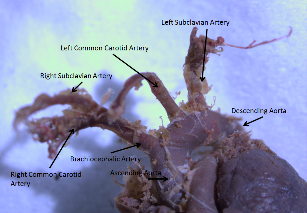
The aortic arch, arch of the aorta, or transverse aortic arch () is the part of the aorta between the ascending and descending aorta. The arch travels backward, so that it ultimately runs to the left of the trachea. Structure The aorta begins at the level of the upper border of the second/third sternocostal articulation of the right side, behind the ventricular outflow tract and pulmonary trunk. The right atrial appendage overlaps it. The first few centimeters of the ascending aorta and pulmonary trunk lies in the same pericardial sheath. and runs at first upward, arches over the pulmonary trunk, right pulmonary artery, and right main bronchus to lie behind the right second coastal cartilage. The right lung and sternum lies anterior to the aorta at this point. The aorta then passes posteriorly and to the left, anterior to the trachea, and arches over left main bronchus and left pulmonary artery, and reaches to the left side of the T4 vertebral body. Apart from T4 vertebral body ... [...More Info...] [...Related Items...] OR: [Wikipedia] [Google] [Baidu] |
Phrenic Nerve
The phrenic nerve is a mixed motor/sensory nerve which originates from the C3-C5 spinal nerves in the neck. The nerve is important for breathing because it provides exclusive motor control of the diaphragm, the primary muscle of respiration. In humans, the right and left phrenic nerves are primarily supplied by the C4 spinal nerve, but there is also contribution from the C3 and C5 spinal nerves. From its origin in the neck, the nerve travels downward into the chest to pass between the heart and lungs towards the diaphragm. In addition to motor fibers, the phrenic nerve contains sensory fibers, which receive input from the central tendon of the diaphragm and the mediastinal pleura, as well as some sympathetic nerve fibers. Although the nerve receives contributions from nerves roots of the cervical plexus and the brachial plexus, it is usually considered separate from either plexus. The name of the nerve comes from Ancient Greek ''phren'' 'diaphragm'. Structure The phrenic n ... [...More Info...] [...Related Items...] OR: [Wikipedia] [Google] [Baidu] |
Pericardiacophrenic
The pericardiacophrenic artery is a long slender branch of the internal thoracic artery. It anastomoses with the musculophrenic and superior phrenic arteries. Location The pericardiacophrenic artery branches from the internal thoracic artery. It accompanies the phrenic nerve between the pleura and pericardium, to the diaphragm. This is where both the artery and the phrenic nerve are distributed. Function The pericardiacophrenic arteries travel through the thoracic cavity, and are located within and supply the fibrous pericardium. Along with the musculophrenic arteries, they also provide arterial supply to the diaphragm. References External links * - "Pleural Cavities and Lungs: Structures Beneath the Left Mediastinal pleura The pulmonary pleurae (''sing.'' pleura) are the two opposing layers of serous membrane overlying the lungs and the inside of the surrounding chest walls. The inner pleura, called the visceral pleura, covers the surface of each lung and dips b ... [...More Info...] [...Related Items...] OR: [Wikipedia] [Google] [Baidu] |


