|
Submental Lymph Nodes
The submental glands (or suprahyoid) are situated between the anterior bellies of the digastric muscle and the hyoid bone. Their '' afferents'' drain the central portions of the lower lip and floor of the mouth and the apex of the tongue. Their '' efferents'' pass partly to the submandibular lymph nodes and partly to a gland of the deep cervical group situated on the internal jugular vein at the level of the cricoid cartilage. See also * Submental triangle References External links * () Image at umich.edu - must rolloverat Baylor College of Medicine Baylor College of Medicine (BCM) is a medical school and research center in Houston, Texas, within the Texas Medical Center, the world's largest medical center. BCM is composed of four academic components: the School of Medicine, the Graduate Sc ... Non-Hodgkin's Lymphoma , Symptoms and Types Lymphatics of the head and neck {{Portal bar, Anatomy ... [...More Info...] [...Related Items...] OR: [Wikipedia] [Google] [Baidu] |
Submandibular Lymph Nodes
The submandibular lymph nodes (submaxillary glands in older texts), three to six in number, are lymph nodes beneath the body of the mandible in the submandibular triangle, and rest on the superficial surface of the submandibular gland. One gland, the ''middle gland of Stahr'', which lies on the facial artery as it turns over the mandible, is the most constant of the series; small lymph glands are sometimes found on the deep surface of the submandibular gland. The ''afferents'' of the submandibular glands drain the medial canthus, the cheek, the side of the nose, the upper lip, the lateral part of the lower lip, the gums, and the anterior part of the margin of the tongue. Efferent lymph vessels from the facial and submental lymph nodes also enter the submandibular glands. Their efferent vessels pass to the superior deep cervical lymph nodes. Additional images File:illu_lymph_chain02.jpg, Deep Lymph Nodes References External links Archived Diagram via umich.edu - ro ... [...More Info...] [...Related Items...] OR: [Wikipedia] [Google] [Baidu] |
Lymphatic Vessel
The lymphatic vessels (or lymph vessels or lymphatics) are thin-walled vessels (tubes), structured like blood vessels, that carry lymph. As part of the lymphatic system, lymph vessels are complementary to the cardiovascular system. Lymph vessels are lined by endothelial cells, and have a thin layer of smooth muscle, and adventitia that binds the lymph vessels to the surrounding tissue. Lymph vessels are devoted to the propulsion of the lymph from the lymph capillaries, which are mainly concerned with the absorption of interstitial fluid from the tissues. Lymph capillaries are slightly bigger than their counterpart capillaries of the vascular system. Lymph vessels that carry lymph to a lymph node are called afferent lymph vessels, and those that carry it from a lymph node are called efferent lymph vessels, from where the lymph may travel to another lymph node, may be returned to a vein, or may travel to a larger lymph duct. Lymph ducts drain the lymph into one of the subclavia ... [...More Info...] [...Related Items...] OR: [Wikipedia] [Google] [Baidu] |
Submental Triangle
The submental triangle (or suprahyoid triangle) is a division of the anterior triangle of the neck. Boundaries It is limited to: * Lateral (away from the midline), formed by the anterior belly of the digastricus * Medial (towards the midline), formed by the midline of the neck between the mandible and the hyoid bone * Inferior (below), formed by the body of the hyoid bone *Floor is formed by the mylohyoideus *Roof is formed by Investing layer of deep cervical fascia Contents It contains one or two lymph glands, the submental lymph nodes The submental glands (or suprahyoid) are situated between the anterior bellies of the digastric muscle and the hyoid bone. Their '' afferents'' drain the central portions of the lower lip and floor of the mouth and the apex of the tongue. Their ... (three or four in number) and Submental veins and commencement of anterior jugular veins. (The contents of the triangle actually lie in the superficial fascia over the roof of submental tr ... [...More Info...] [...Related Items...] OR: [Wikipedia] [Google] [Baidu] |
Cricoid Cartilage
The cricoid cartilage , or simply cricoid (from the Greek ''krikoeides'' meaning "ring-shaped") or cricoid ring, is the only complete ring of cartilage around the trachea. It forms the back part of the voice box and functions as an attachment site for muscles, cartilages, and ligaments involved in opening and closing the airway and in producing speech. Structure The cricoid cartilage sits just inferior to the thyroid cartilage in the neck, at the level of the C6 vertebra, and is joined to it medially by the median cricothyroid ligament and postero-laterally by the cricothyroid joints. Inferior to it are the rings of cartilage around the trachea (which are not continuous – rather they are C-shaped with a gap posteriorly). The cricoid is joined to the first tracheal ring by the cricotracheal ligament, and this can be felt as a more yielding area between the firm thyroid cartilage and firmer cricoid. It is also anatomically related to the thyroid gland; although the ... [...More Info...] [...Related Items...] OR: [Wikipedia] [Google] [Baidu] |
Internal Jugular Vein
The internal jugular vein is a paired jugular vein that collects blood from the brain and the superficial parts of the face and neck. This vein runs in the carotid sheath with the common carotid artery and vagus nerve. It begins in the posterior compartment of the jugular foramen, at the base of the skull. It is somewhat dilated at its origin, which is called the ''superior bulb''. This vein also has a common trunk into which drains the anterior branch of the retromandibular vein, the facial vein, and the lingual vein. It runs down the side of the neck in a vertical direction, being at one end lateral to the internal carotid artery, and then lateral to the common carotid artery, and at the root of the neck, it unites with the subclavian vein to form the brachiocephalic vein (innominate vein); a little above its termination is a second dilation, the ''inferior bulb''. Above, it lies upon the rectus capitis lateralis, behind the internal carotid artery and the nerves passing ... [...More Info...] [...Related Items...] OR: [Wikipedia] [Google] [Baidu] |
Deep Cervical Group
Cervical lymph nodes are lymph nodes found in the neck. Of the 800 lymph nodes in the human body, 300 are in the neck. Cervical lymph nodes are subject to a number of different pathological conditions including tumours, infection and inflammation. Classification There are approximately 300 lymph nodes in the neck, and they can be classified in a number of different ways. History The classification of the cervical lymph nodes is generally attributed to Henri Rouvière in his 1932 publication "Anatomie des Lymphatiques de l'Homme" Rouviere described the cervical lymph nodes as a collar which surrounded the upper aerodigestive tract, consisting of submental, facial, submandibular, parotid, mastoid, occipital and retropharyngeal nodes, together with two chains that run in the long axis of the neck, the anterior cervical and postero-lateral cervical groups. However, this system was based upon anatomical landmarks found in dissection, making it imperfectly suited to the need ... [...More Info...] [...Related Items...] OR: [Wikipedia] [Google] [Baidu] |
Submandibular Lymph Nodes
The submandibular lymph nodes (submaxillary glands in older texts), three to six in number, are lymph nodes beneath the body of the mandible in the submandibular triangle, and rest on the superficial surface of the submandibular gland. One gland, the ''middle gland of Stahr'', which lies on the facial artery as it turns over the mandible, is the most constant of the series; small lymph glands are sometimes found on the deep surface of the submandibular gland. The ''afferents'' of the submandibular glands drain the medial canthus, the cheek, the side of the nose, the upper lip, the lateral part of the lower lip, the gums, and the anterior part of the margin of the tongue. Efferent lymph vessels from the facial and submental lymph nodes also enter the submandibular glands. Their efferent vessels pass to the superior deep cervical lymph nodes. Additional images File:illu_lymph_chain02.jpg, Deep Lymph Nodes References External links Archived Diagram via umich.edu - ro ... [...More Info...] [...Related Items...] OR: [Wikipedia] [Google] [Baidu] |
Apex Of The Tongue
The tongue is a muscular organ in the mouth of a typical tetrapod. It manipulates food for mastication and swallowing as part of the digestive process, and is the primary organ of taste. The tongue's upper surface (dorsum) is covered by taste buds housed in numerous lingual papillae. It is sensitive and kept moist by saliva and is richly supplied with nerves and blood vessels. The tongue also serves as a natural means of cleaning the teeth. A major function of the tongue is the enabling of speech in humans and vocalization in other animals. The human tongue is divided into two parts, an oral part at the front and a pharyngeal part at the back. The left and right sides are also separated along most of its length by a vertical section of fibrous tissue (the lingual septum) that results in a groove, the median sulcus, on the tongue's surface. There are two groups of muscles of the tongue. The four intrinsic muscles alter the shape of the tongue and are not attached to bone. The f ... [...More Info...] [...Related Items...] OR: [Wikipedia] [Google] [Baidu] |
Mouth
In animal anatomy, the mouth, also known as the oral cavity, or in Latin cavum oris, is the opening through which many animals take in food and issue vocal sounds. It is also the cavity lying at the upper end of the alimentary canal, bounded on the outside by the lips and inside by the pharynx. In tetrapods, it contains the tongue and, except for some like birds, teeth. This cavity is also known as the buccal cavity, from the Latin ''bucca'' ("cheek"). Some animal phyla, including arthropods, molluscs and chordates, have a complete digestive system, with a mouth at one end and an anus at the other. Which end forms first in ontogeny is a criterion used to classify bilaterian animals into protostomes and deuterostomes. Development In the first multicellular animals, there was probably no mouth or gut and food particles were engulfed by the cells on the exterior surface by a process known as endocytosis. The particles became enclosed in vacuoles into which enzymes were secr ... [...More Info...] [...Related Items...] OR: [Wikipedia] [Google] [Baidu] |
Lower Lip
The lips are the visible body part at the mouth of many animals, including humans. Lips are soft, movable, and serve as the opening for food intake and in the articulation of sound and speech. Human lips are a tactile sensory organ, and can be an erogenous zone when used in kissing and other acts of intimacy. Structure The upper and lower lips are referred to as the "Labium superius oris" and "Labium inferius oris", respectively. The juncture where the lips meet the surrounding skin of the mouth area is the vermilion border, and the typically reddish area within the borders is called the vermilion zone. The vermilion border of the upper lip is known as the cupid's bow. The fleshy protuberance located in the center of the upper lip is a tubercle known by various terms including the procheilon (also spelled ''prochilon''), the "tuberculum labii superioris", and the "labial tubercle". The vertical groove extending from the procheilon to the nasal septum is called the philtr ... [...More Info...] [...Related Items...] OR: [Wikipedia] [Google] [Baidu] |
Hyoid Bone
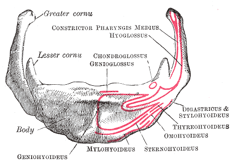
The hyoid bone (lingual bone or tongue-bone) () is a horseshoe-shaped bone situated in the anterior midline of the neck between the chin and the thyroid cartilage. At rest, it lies between the base of the mandible and the third cervical vertebra. Unlike other bones, the hyoid is only distantly articulated to other bones by muscles or ligaments. It is the only bone in the human body that is not connected to any other bones nearby. The hyoid is anchored by muscles from the anterior, posterior and inferior directions, and aids in tongue movement and swallowing. The hyoid bone provides attachment to the muscles of the floor of the mouth and the tongue above, the larynx below, and the epiglottis and pharynx behind. Its name is derived . Structure The hyoid bone is classed as an irregular bone and consists of a central part called the body, and two pairs of horns, the greater and lesser horns. Body The body of the hyoid bone is the central part of the hyoid bone. *At the fro ... [...More Info...] [...Related Items...] OR: [Wikipedia] [Google] [Baidu] |
Supraclavicular Lymph Nodes
Supraclavicular lymph nodes are lymph nodes found above the clavicle, that can be felt in the supraclavicular fossa. The supraclavicular lymph nodes on the left side are called Virchow's nodes.page 400 in: It leads to an appreciable mass that can be recognized clinically, called Troisier sign. Structure A Virchow's node is a left-sided supraclavicular lymph node.Clinical significance  Malignancies of the internal organs can reach an advanced stage before giving symptoms.
|


