|
Submandibular Triangle
The submandibular triangle (or submaxillary or digastric triangle) corresponds to the region of the neck immediately beneath the body of the mandible. Boundaries and coverings It is bounded: * ''above'', by the lower border of the body of the mandible, and a line drawn from its angle to the mastoid process; * ''below'', by the posterior belly of the Digastricus; in front, by the anterior belly of the Digastricus. It is covered by the integument, superficial fascia, Platysma, and deep fascia, ramifying in which are branches of the facial nerve and ascending filaments of the cutaneous cervical nerve. Its floor is formed by the Mylohyoideus anteriorly, and by the hyoglossus posteriorly. Triangles * Beclard Triangle * Lesser Triangle * Pirogoff Triangle Divisions It is divided into an anterior and a posterior part by the stylomandibular ligament. Anterior part The anterior part contains the submandibular gland, superficial to which is the anterior facial vein, while imbedded ... [...More Info...] [...Related Items...] OR: [Wikipedia] [Google] [Baidu] [Amazon] |
Human Mandible
In jawed vertebrates, the mandible (from the Latin ''mandibula'', 'for chewing'), lower jaw, or jawbone is a bone that makes up the lowerand typically more mobilecomponent of the mouth (the upper jaw being known as the maxilla). The jawbone is the skull's only movable, posable bone, sharing joints with the cranium's temporal bones. The mandible hosts the lower teeth (their depth delineated by the alveolar process). Many muscles attach to the bone, which also hosts nerves (some connecting to the teeth) and blood vessels. Amongst other functions, the jawbone is essential for chewing food. Owing to the Neolithic advent of agriculture (), human jaws evolved to be smaller. Although it is the strongest bone of the facial skeleton, the mandible tends to deform in old age; it is also subject to fracturing. Surgery allows for the removal of jawbone fragments (or its entirety) as well as regenerative methods. Additionally, the bone is of great forensic significance. Struct ... [...More Info...] [...Related Items...] OR: [Wikipedia] [Google] [Baidu] [Amazon] |
Submental Artery
The submental artery is the largest branch of the facial artery in the neck. It first runs forward under the mouth, then turns upward upon reaching the chin. Anatomy Origin The submental artery is the largest branch of the facial artery in the neck. It arises from the facial artery just as the facial artery splits the submandibular gland. Course and distribution The artery passes anterior-ward upon the mylohyoid muscle, coursing inferior to the body of the mandible and deep to the digastric muscle. Here, the artery supplies adjacent muscles and skin; it also forms anastomoses with the sublingual artery and with the mylohyoid branch of the inferior alveolar artery. Upon reaching the chin, artery turns superior-ward at the mandibular symphysis to pass over the mandible before dividing into a superficial branch and a deep branch; the two terminal branches are distributed to the chin and lower lip, and form anastomoses with the inferior labial and mental arteries. Distri ... [...More Info...] [...Related Items...] OR: [Wikipedia] [Google] [Baidu] [Amazon] |
Submandibular Space
The submandibular space is a fascial space of the head and neck (sometimes also termed fascial spaces or tissue spaces). It is a potential space, and is paired on either side, located on the superficial surface of the mylohyoid muscle between the anterior and posterior bellies of the digastric muscle. The space corresponds to the anatomic region termed the submandibular triangle, part of the anterior triangle of the neck. Location and structure Anatomic boundaries The anatomic boundaries of each submandibular space are: * the mylohyoid muscle superiorly, * the skin, superficial fascia, platysma muscle and superficial layer of the deep cervical fascia inferiorly and laterally, * the medial surface of the mandible anteriorly and laterally, * the hyoid bone posteriorly, * the anterior belly of the digastric muscle medially. Communications The communications of the submandibular space are: * medially and anteriorly to the submental space (located medial to the paired submandibular ... [...More Info...] [...Related Items...] OR: [Wikipedia] [Google] [Baidu] [Amazon] |
Anterior Triangle Of The Neck
The anterior triangle is a region of the neck. Structure The triangle is inverted with its apex inferior to its base which is under the chin. Investing fascia covers the roof of the triangle while visceral fascia covers the floor. Anatomy Muscles: * Suprahyoid muscles - Digastric (Ant and post belly), mylohyoid, geniohyoid and stylohyoid. * Infrahyoid muscles - Omohyoid, sternohyoid, sternothyroid, and thyrohyoid. Nerve supply 2 Bellies of digastric * Anterior: Mylohyoid nerve * Posterior: Facial nerve Stylohyoid: by the facial nerve, by a branch from that to the posterior belly of digastric. Mylohyoid: by its own nerve, a branch of the inferior alveolar (from the mandibular division of trigeminal nerve), which arises just before the parent nerve enters the mandibular foramen, pierces the sphenomandibular ligament, and runs forward on the inferior surface of the mylohyoid, supplying it and the anterior belly of the digastric. Geniohyoid: by a branch from the hypoglossa ... [...More Info...] [...Related Items...] OR: [Wikipedia] [Google] [Baidu] [Amazon] |
Hypoglossal Nerve
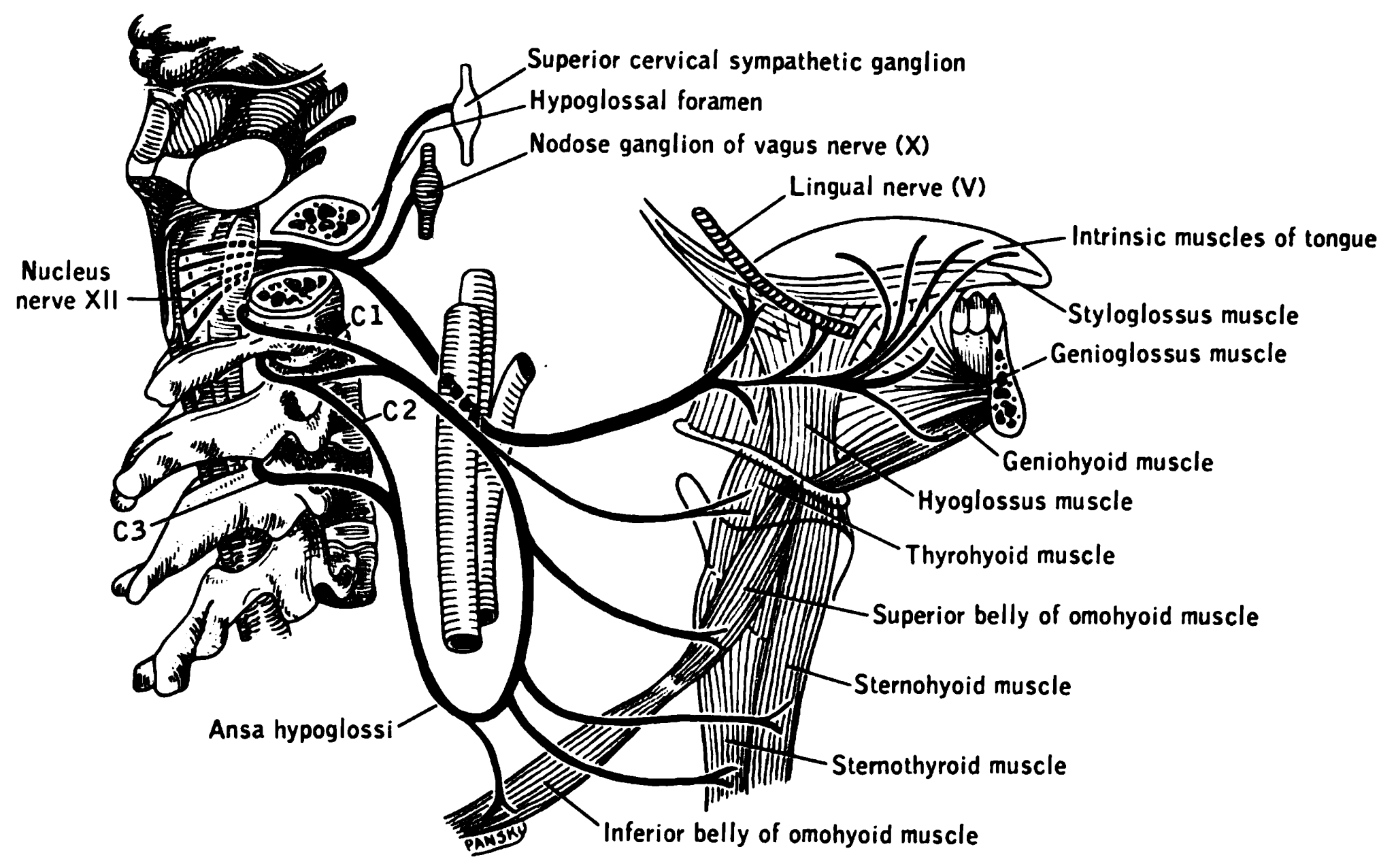
The hypoglossal nerve, also known as the twelfth cranial nerve, cranial nerve XII, or simply CN XII, is a cranial nerve that innervates all the extrinsic and intrinsic muscles of the tongue except for the palatoglossus, which is innervated by the vagus nerve. CN XII is a nerve with a sole motor function. The nerve arises from the hypoglossal nucleus in the medulla as a number of small rootlets, pass through the hypoglossal canal and down through the neck, and eventually passes up again over the tongue muscles it supplies into the tongue. The nerve is involved in controlling tongue movements required for speech and swallowing, including sticking out the tongue and moving it from side to side. Damage to the nerve or the neural pathways which control it can affect the ability of the tongue to move and its appearance, with the most common sources of damage being injury from trauma or surgery, and motor neuron disease. The first recorded description of the nerve was by Her ... [...More Info...] [...Related Items...] OR: [Wikipedia] [Google] [Baidu] [Amazon] |
Stylopharyngeus
The stylopharyngeus muscle is a muscle in the head. It originates from the temporal styloid process. Some of its fibres insert onto the thyroid cartilage, while others end by intermingling with proximal structures. It is innervated by the glossopharyngeal nerve (cranial nerve IX). It acts to elevate the larynx and pharynx, and dilate the pharynx, thus facilitating swallowing. Structure The stylopharyngeus is a long, slender, tapered pharyngeal muscle. It is cylindrical superiorly, and flattened inferiorly. It passes inferior-ward along the side of the pharynx between the superior pharyngeal constrictor (situated deep to the stylopharyngeus) and the middle pharyngeal constrictor (situated superficial to the stylopharyngeus), before spreads out beneath the mucous membrane. Origin It arises from (the medial side of the base of) the temporal styloid process. It is the only muscle of the pharynx not to originate in the pharyngeal wall. Insertion Some of its fibers are lost ... [...More Info...] [...Related Items...] OR: [Wikipedia] [Google] [Baidu] [Amazon] |
Styloglossus
The styloglossus muscle is a bilaterally paired muscle of the tongue. It originates at the styloid process of the temporal bone. It inserts onto the side of the tongue. It acts to elevate and retract the tongue. It is innervated by the hypoglossal nerve (cranial nerve XII). Anatomy The styloglossus muscle is the shortest and smallest of the three styloid muscles. Origin It arises from (the anterior and lateral surfaces of) the styloid process of the temporal bone near its apex, and from the stylomandibular ligament. Course and relations It passes anterioinferiorly from its origin to its insertion between the internal carotid artery and the external carotid artery, and between the superior pharyngeal constrictor muscle The superior pharyngeal constrictor muscle is a quadrilateral muscle of the pharynx. It is the uppermost and thinnest of the three pharyngeal constrictors. The muscle is divided into four parts according to its four distincts origins: a pterygop ... an ... [...More Info...] [...Related Items...] OR: [Wikipedia] [Google] [Baidu] [Amazon] |
Vagus Nerve
The vagus nerve, also known as the tenth cranial nerve (CN X), plays a crucial role in the autonomic nervous system, which is responsible for regulating involuntary functions within the human body. This nerve carries both sensory and motor fibers and serves as a major pathway that connects the brain to various organs, including the heart, lungs, and digestive tract. As a key part of the parasympathetic nervous system, the vagus nerve helps regulate essential involuntary functions like heart rate, breathing, and digestion. By controlling these processes, the vagus nerve contributes to the body's "rest and digest" response, helping to calm the body after stress, lower heart rate, improve digestion, and maintain homeostasis. The vagus nerve consists of two branches: the right and left vagus nerves. In the neck, the right vagus nerve contains approximately 105,000 fibers, while the left vagus nerve has about 87,000 fibers, according to one source. However, other sources report sl ... [...More Info...] [...Related Items...] OR: [Wikipedia] [Google] [Baidu] [Amazon] |
Internal Jugular Vein
The internal jugular vein is a paired jugular vein that collects blood from the brain and the superficial parts of the face and neck. This vein runs in the carotid sheath with the common carotid artery and vagus nerve. It begins in the posterior compartment of the jugular foramen, at the base of the skull. It is somewhat dilated at its origin, which is called the ''superior bulb''. This vein also has a common trunk into which drains the anterior branch of the retromandibular vein, the facial vein, and the lingual vein. It runs down the side of the neck in a vertical direction, being at one end lateral to the internal carotid artery, and then lateral to the common carotid artery, and at the root of the neck, it unites with the subclavian vein to form the brachiocephalic vein (innominate vein); a little above its termination is a second dilation, the ''inferior bulb''. Above, it lies upon the rectus capitis lateralis, behind the internal carotid artery and the nerves pa ... [...More Info...] [...Related Items...] OR: [Wikipedia] [Google] [Baidu] [Amazon] |
Internal Carotid
The internal carotid artery is an artery in the neck which supplies the anterior and middle cerebral circulation. In human anatomy, the internal and external carotid arise from the common carotid artery, where it bifurcates at cervical vertebrae C3 or C4. The internal carotid artery supplies the brain, including the eyes, while the external carotid nourishes other portions of the head, such as the face, scalp, skull, and meninges. Classification Terminologia Anatomica in 1998 subdivided the artery into four parts: "cervical", "petrous", "cavernous", and "cerebral". In clinical settings, however, usually the classification system of the internal carotid artery follows the 1996 recommendations by Bouthillier, describing seven anatomical segments of the internal carotid artery, each with a corresponding alphanumeric identifier: C1 cervical; C2 petrous; C3 lacerum; C4 cavernous; C5 clinoid; C6 ophthalmic; and C7 communicating. The Bouthillier nomenclature remains in widespread ... [...More Info...] [...Related Items...] OR: [Wikipedia] [Google] [Baidu] [Amazon] |
Internal Maxillary
Internal may refer to: *Internality as a concept in behavioural economics *Neijia, internal styles of Chinese martial arts *Neigong or "internal skills", a type of exercise in meditation associated with Daoism * ''Internal'' (album) by Safia, 2016 See also * *Internals (other) Internals usually refers to the internal parts of a machine, organism or other entity; or to the inner workings of a process. More specifically, internals may refer to: *the internal organs *the gastrointestinal tract The gastrointestinal tra ... * External (other) {{disambig ... [...More Info...] [...Related Items...] OR: [Wikipedia] [Google] [Baidu] [Amazon] |
Superficial Temporal
In human anatomy, the superficial temporal artery is a major artery of the head. It arises from the external carotid artery when it splits into the superficial temporal artery and maxillary artery. Its pulse can be felt above the zygomatic arch, above and in front of the tragus of the ear. Structure The superficial temporal artery is the smaller of two end branches that split superiorly from the external carotid. Based on its direction, the superficial temporal artery appears to be a continuation of the external carotid. It begins within the parotid gland, behind the neck of the mandible, and passes superficially over the posterior root of the zygomatic process of the temporal bone; about 5 cm above this process it divides into two branches: ''a. frontal'', and ''a. parietal''. Branches The parietal branch of the superficial temporal artery (posterior temporal) is a small artery in the head. It is larger than the frontal branch and curves upward and backward on the side ... [...More Info...] [...Related Items...] OR: [Wikipedia] [Google] [Baidu] [Amazon] |


