|
Submandibular Lymph Nodes
The submandibular lymph nodes (submaxillary glands in older texts), three to six in number, are lymph nodes beneath the body of the mandible in the submandibular triangle, and rest on the superficial surface of the submandibular gland. One gland, the ''middle gland of Stahr'', which lies on the facial artery as it turns over the mandible, is the most constant of the series; small lymph glands are sometimes found on the deep surface of the submandibular gland. The ''afferents'' of the submandibular glands drain the medial canthus, the cheek, the side of the nose, the upper lip, the lateral part of the lower lip, the gums, and the anterior part of the margin of the tongue. Efferent lymph vessel The lymphatic vessels (or lymph vessels or lymphatics) are thin-walled vessels (tubes), structured like blood vessels, that carry lymph. As part of the lymphatic system, lymph vessels are complementary to the cardiovascular system. Lymph vessel ...s from the facial and submental lymph ... [...More Info...] [...Related Items...] OR: [Wikipedia] [Google] [Baidu] |
Submental Lymph Nodes
The submental glands (or suprahyoid) are situated between the anterior bellies of the digastric muscle and the hyoid bone. Their '' afferents'' drain the central portions of the lower lip and floor of the mouth and the apex of the tongue. Their '' efferents'' pass partly to the submandibular lymph nodes and partly to a gland of the deep cervical group situated on the internal jugular vein at the level of the cricoid cartilage. See also * Submental triangle References External links * () Image at umich.edu - must rolloverat Baylor College of Medicine Baylor College of Medicine (BCM) is a medical school and research center in Houston, Texas, within the Texas Medical Center, the world's largest medical center. BCM is composed of four academic components: the School of Medicine, the Graduate S ... Non-Hodgkin's Lymphoma , Symptoms and Types Lymphatics of the head and neck {{Portal bar, Anatomy ... [...More Info...] [...Related Items...] OR: [Wikipedia] [Google] [Baidu] |
Facial Artery
The facial artery (external maxillary artery in older texts) is a branch of the external carotid artery that supplies structures of the superficial face. Structure The facial artery arises in the carotid triangle from the external carotid artery, a little above the lingual artery and, sheltered by the ramus of the mandible. It passes obliquely up beneath the digastric and stylohyoid muscles, over which it arches to enter a groove on the posterior surface of the submandibular gland. It then curves upward over the body of the mandible at the antero-inferior angle of the masseter; passes forward and upward across the cheek to the angle of the mouth, then ascends along the side of the nose, and ends at the medial commissure of the eye, under the name of the angular artery. The facial artery is remarkably tortuous. This is to accommodate itself to neck movements such as those of the pharynx in deglutition; and facial movements such as those of the mandible, lips, and cheeks. ... [...More Info...] [...Related Items...] OR: [Wikipedia] [Google] [Baidu] |
Superior Deep Cervical Lymph Nodes
The superior deep cervical lymph nodes are the deep cervical lymph nodes that are situated adjacent to the superior portion of the internal jugular vein. They drain either to the inferior deep cervical lymph nodes or into the jugular trunk. Most of these lymph nodes are situated deep to the sternocleidomastoid muscle The sternocleidomastoid muscle is one of the largest and most superficial cervical muscles. The primary actions of the muscle are rotation of the head to the opposite side and flexion of the neck. The sternocleidomastoid is innervated by the access ..., though some are not. Some are situated anterior and some posterior to the internal jugular vein. They are also situated adjacent to the accessory nerve (CN XI). Jugulodigastric group Superior deep cervical lymph nodes situated in a triangular region bounded by the posterior belly of the digastric muscle, the facial vein, and the internal jugular vein form a subgroup - the jugulodigastric group. The group consists ... [...More Info...] [...Related Items...] OR: [Wikipedia] [Google] [Baidu] |
Submental Lymph Nodes
The submental glands (or suprahyoid) are situated between the anterior bellies of the digastric muscle and the hyoid bone. Their '' afferents'' drain the central portions of the lower lip and floor of the mouth and the apex of the tongue. Their '' efferents'' pass partly to the submandibular lymph nodes and partly to a gland of the deep cervical group situated on the internal jugular vein at the level of the cricoid cartilage. See also * Submental triangle References External links * () Image at umich.edu - must rolloverat Baylor College of Medicine Baylor College of Medicine (BCM) is a medical school and research center in Houston, Texas, within the Texas Medical Center, the world's largest medical center. BCM is composed of four academic components: the School of Medicine, the Graduate S ... Non-Hodgkin's Lymphoma , Symptoms and Types Lymphatics of the head and neck {{Portal bar, Anatomy ... [...More Info...] [...Related Items...] OR: [Wikipedia] [Google] [Baidu] |
Efferent Lymph Vessel
The lymphatic vessels (or lymph vessels or lymphatics) are thin-walled vessels (tubes), structured like blood vessels, that carry lymph. As part of the lymphatic system, lymph vessels are complementary to the cardiovascular system. Lymph vessels are lined by endothelial cells, and have a thin layer of smooth muscle, and adventitia that binds the lymph vessels to the surrounding tissue. Lymph vessels are devoted to the propulsion of the lymph from the lymph capillaries, which are mainly concerned with the absorption of interstitial fluid from the tissues. Lymph capillaries are slightly bigger than their counterpart capillaries of the vascular system. Lymph vessels that carry lymph to a lymph node are called afferent lymph vessels, and those that carry it from a lymph node are called efferent lymph vessels, from where the lymph may travel to another lymph node, may be returned to a vein, or may travel to a larger lymph duct. Lymph ducts drain the lymph into one of the subclavian ... [...More Info...] [...Related Items...] OR: [Wikipedia] [Google] [Baidu] |
Tongue
The tongue is a muscular organ in the mouth of a typical tetrapod. It manipulates food for mastication and swallowing as part of the digestive process, and is the primary organ of taste. The tongue's upper surface (dorsum) is covered by taste buds housed in numerous lingual papillae. It is sensitive and kept moist by saliva and is richly supplied with nerves and blood vessels. The tongue also serves as a natural means of cleaning the teeth. A major function of the tongue is the enabling of speech in humans and vocalization in other animals. The human tongue is divided into two parts, an oral part at the front and a pharyngeal part at the back. The left and right sides are also separated along most of its length by a vertical section of fibrous tissue (the lingual septum) that results in a groove, the median sulcus, on the tongue's surface. There are two groups of muscles of the tongue. The four intrinsic muscles alter the shape of the tongue and are not attached to bone. ... [...More Info...] [...Related Items...] OR: [Wikipedia] [Google] [Baidu] |
Gums
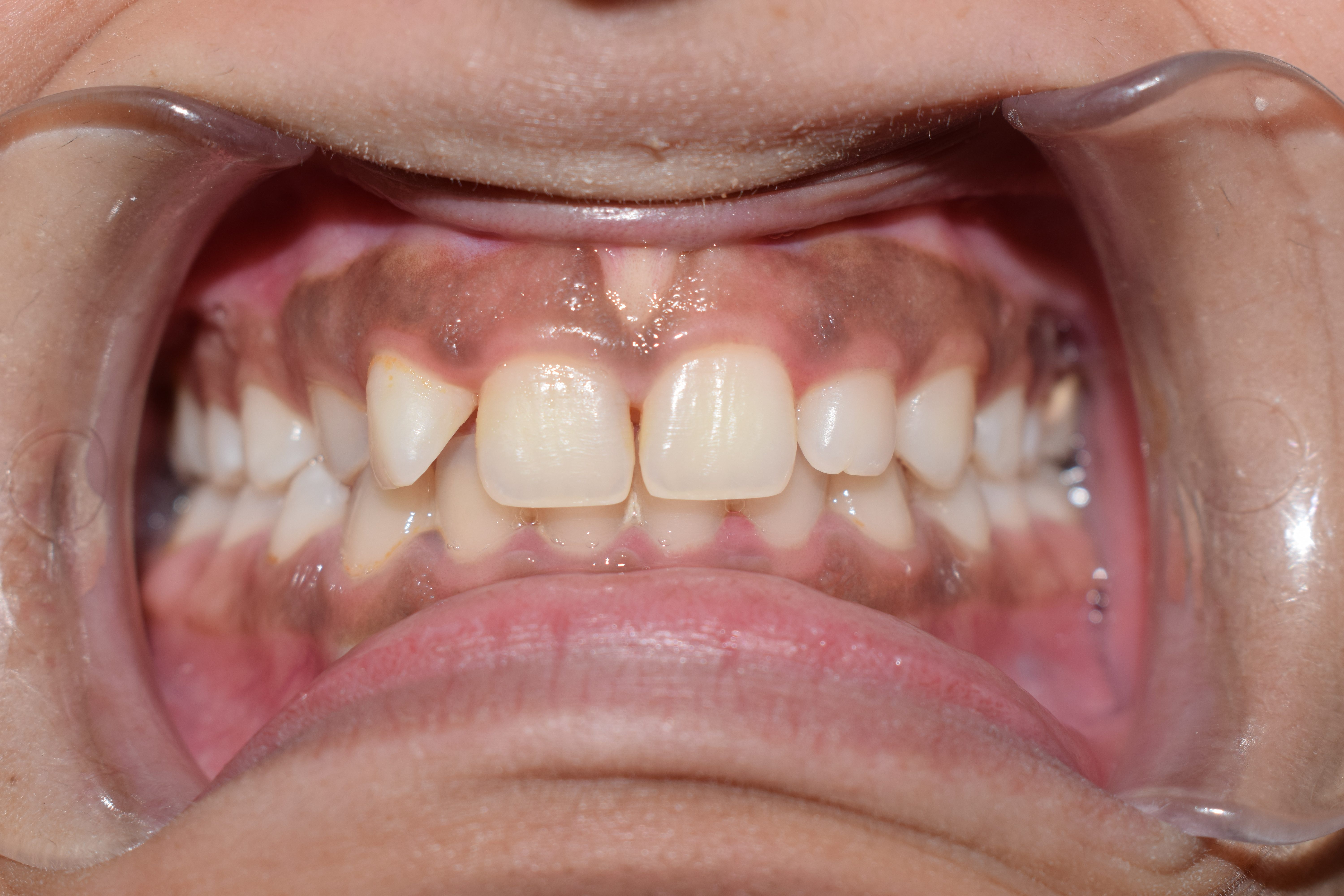
The gums or gingiva (plural: ''gingivae'') consist of the mucosal tissue that lies over the mandible and maxilla inside the mouth. Gum health and disease can have an effect on general health. Structure The gums are part of the soft tissue lining of the mouth. They surround the teeth and provide a seal around them. Unlike the soft tissue linings of the lips and cheeks, most of the gums are tightly bound to the underlying bone which helps resist the friction of food passing over them. Thus when healthy, it presents an effective barrier to the barrage of periodontal insults to deeper tissue. Healthy gums are usually coral pink in light skinned people, and may be naturally darker with melanin pigmentation. Changes in color, particularly increased redness, together with swelling and an increased tendency to bleed, suggest an inflammation that is possibly due to the accumulation of bacterial plaque. Overall, the clinical appearance of the tissue reflects the underlying histology, ... [...More Info...] [...Related Items...] OR: [Wikipedia] [Google] [Baidu] |
Lower Lip
The lips are the visible body part at the mouth of many animals, including humans. Lips are soft, movable, and serve as the opening for food intake and in the articulation of sound and speech. Human lips are a tactile sensory organ, and can be an erogenous zone when used in kissing and other acts of intimacy. Structure The upper and lower lips are referred to as the "Labium superius oris" and "Labium inferius oris", respectively. The juncture where the lips meet the surrounding skin of the mouth area is the vermilion border, and the typically reddish area within the borders is called the vermilion zone. The vermilion border of the upper lip is known as the cupid's bow. The fleshy protuberance located in the center of the upper lip is a tubercle known by various terms including the procheilon (also spelled ''prochilon''), the "tuberculum labii superioris", and the "labial tubercle". The vertical groove extending from the procheilon to the nasal septum is called the phi ... [...More Info...] [...Related Items...] OR: [Wikipedia] [Google] [Baidu] |
Upper Lip
The lips are the visible body part at the mouth of many animals, including humans. Lips are soft, movable, and serve as the opening for food intake and in the articulation of sound and speech. Human lips are a tactile sensory organ, and can be an erogenous zone when used in kissing and other acts of intimacy. Structure The upper and lower lips are referred to as the "Labium superius oris" and "Labium inferius oris", respectively. The juncture where the lips meet the surrounding skin of the mouth area is the vermilion border, and the typically reddish area within the borders is called the vermilion zone. The vermilion border of the upper lip is known as the cupid's bow. The fleshy protuberance located in the center of the upper lip is a tubercle known by various terms including the procheilon (also spelled ''prochilon''), the "tuberculum labii superioris", and the "labial tubercle". The vertical groove extending from the procheilon to the nasal septum is called the philt ... [...More Info...] [...Related Items...] OR: [Wikipedia] [Google] [Baidu] |
Human Nose
The human nose is the most protruding part of the face. It bears the nostrils and is the first organ of the respiratory system. It is also the principal organ in the olfactory system. The shape of the nose is determined by the nasal bones and the nasal cartilages, including the nasal septum which separates the nostrils and divides the nasal cavity into two. On average the nose of a male is larger than that of a female. The nose has an important function in breathing. The nasal mucosa lining the nasal cavity and the paranasal sinuses carries out the necessary conditioning of inhaled air by warming and moistening it. Nasal conchae, shell-like bones in the walls of the cavities, play a major part in this process. Filtering of the air by nasal hair in the nostrils prevents large particles from entering the lungs. Sneezing is a reflex to expel unwanted particles from the nose that irritate the mucosal lining. Sneezing can transmit infections, because aerosols are created ... [...More Info...] [...Related Items...] OR: [Wikipedia] [Google] [Baidu] |
Cheek
The cheeks ( la, buccae) constitute the area of the face below the eyes and between the nose and the left or right ear. "Buccal" means relating to the cheek. In humans, the region is innervated by the buccal nerve. The area between the inside of the cheek and the teeth and gums is called the vestibule or buccal pouch or buccal cavity and forms part of the mouth. In other animals the cheeks may also be referred to as jowls. Structure Humans Cheeks are fleshy in humans, the skin being suspended by the chin and the jaws, and forming the lateral wall of the human mouth, visibly touching the cheekbone below the eye. The inside of the cheek is lined with a mucous membrane (buccal mucosa, part of the oral mucosa). During mastication (chewing), the cheeks and tongue between them serve to keep the food between the teeth. Other animals The cheeks are covered externally by hairy skin, and internally by stratified squamous epithelium. This is mostly smooth, but may have caudally dir ... [...More Info...] [...Related Items...] OR: [Wikipedia] [Google] [Baidu] |
Canthus
The canthus (pl. canthi, palpebral commissures) is either corner of the eye where the upper and lower eyelids meet. More specifically, the inner and outer canthi are, respectively, the medial and lateral ends/angles of the palpebral fissure. The bicanthal plane is the transversal plane linking both canthi and defines the upper boundary of the midface. Etymology The word ' is the Latinized form of the Ancient Greek ('), meaning 'corner of the eye'. Population distribution The eyes of those of East Asian and some Southeast Asian people tend to have the inner canthus veiled by the epicanthus. In the Caucasian or double eyelid, the inner corner tends to be exposed completely. Commissures * The ''lateral palpebral commissure'' (commissura palpebrarum lateralis; external canthus) is more acute than the medial, and the eyelids here lie in close contact with the bulb of the eye. * The ''medial palpebral commissure'' (commissura palpebrarum medialis; internal canthus) is prolo ... [...More Info...] [...Related Items...] OR: [Wikipedia] [Google] [Baidu] |


