|
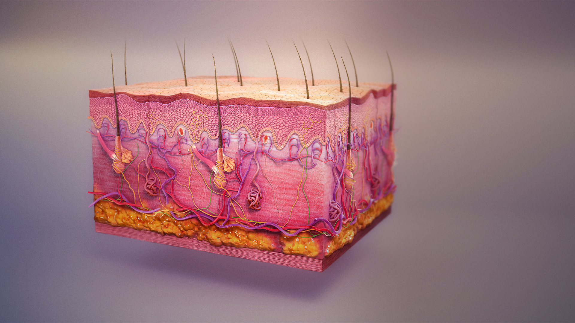
Subcutis
The subcutaneous tissue (), also called the hypodermis, hypoderm (), subcutis, superficial fascia, is the lowermost layer of the integumentary system in vertebrates. The types of cells found in the layer are fibroblasts, adipose cells, and macrophages. The subcutaneous tissue is derived from the mesoderm, but unlike the dermis, it is not derived from the mesoderm's dermatome region. It consists primarily of loose connective tissue, and contains larger blood vessels and nerves than those found in the dermis. It is a major site of fat storage in the body. In arthropods, a hypodermis can refer to an epidermal layer of cells that secretes the chitinous cuticle. The term also refers to a layer of cells lying immediately below the epidermis of plants. Structure * Fibrous bands anchoring the skin to the deep fascia * Collagen and elastin fibers attaching it to the dermis * Fat is absent from the eyelids, clitoris, penis, much of pinna, and scrotum * Blood vessels on route to t ... [...More Info...] [...Related Items...] OR: [Wikipedia] [Google] [Baidu] |
Integumentary
The integumentary system is the set of organs forming the outermost layer of an animal's body. It comprises the skin and its appendages, which act as a physical barrier between the external environment and the internal environment that it serves to protect and maintain the body of the animal. Mainly it is the body's outer skin. The integumentary system includes hair, scales, feathers, hooves, and nails. It has a variety of additional functions: it may serve to maintain water balance, protect the deeper tissues, excrete wastes, and regulate body temperature, and is the attachment site for sensory receptors which detect pain, sensation, pressure, and temperature. Structure Skin The skin is one of the largest organs of the body. In humans, it accounts for about 12 to 15 percent of total body weight and covers 1.5 to 2 m2 of surface area. The skin (integument) is a composite organ, made up of at least two major layers of tissue: the epidermis and the dermis. The epidermis ... [...More Info...] [...Related Items...] OR: [Wikipedia] [Google] [Baidu] |
Elastin
Elastin is a protein that in humans is encoded by the ''ELN'' gene. Elastin is a key component of the extracellular matrix in gnathostomes (jawed vertebrates). It is highly elastic and present in connective tissue allowing many tissues in the body to resume their shape after stretching or contracting. Elastin helps skin to return to its original position when it is poked or pinched. Elastin is also an important load-bearing tissue in the bodies of vertebrates and used in places where mechanical energy is required to be stored. Function The ''ELN'' gene encodes a protein that is one of the two components of elastic fibers. The encoded protein is rich in hydrophobic amino acids such as glycine and proline, which form mobile hydrophobic regions bounded by crosslinks between lysine residues. Multiple transcript variants encoding different isoforms have been found for this gene. Elastin's soluble precursor is tropoelastin. The characterization of disorder is consistent with ... [...More Info...] [...Related Items...] OR: [Wikipedia] [Google] [Baidu] |
Lobules
In anatomy, a lobe is a clear anatomical division or extension of an organ (as seen for example in the brain, lung, liver, or kidney) that can be determined without the use of a microscope at the gross anatomy level. This is in contrast to the much smaller lobule, which is a clear division only visible under the microscope. Interlobar ducts connect lobes and interlobular ducts connect lobules. Examples of lobes *The four main lobes of the brain **the frontal lobe **the parietal lobe **the occipital lobe **the temporal lobe *The three lobes of the human cerebellum **the flocculonodular lobe **the anterior lobe **the posterior lobe *The two lobes of the thymus *The two and three lobes of the lungs ** Left lung: superior and inferior ** Right lung: superior, middle, and inferior *The four lobes of the liver ** Left lobe of liver ** Right lobe of liver ** Quadrate lobe of liver ** Caudate lobe of liver *The renal lobes of the kidney * Earlobes Examples of lobules *the co ... [...More Info...] [...Related Items...] OR: [Wikipedia] [Google] [Baidu] |
Panniculus Adiposus
The panniculus adiposus is the fatty layer of the subcutaneous tissues, superficial to a deeper vestigial layer of muscle, the panniculus carnosus.McGrath, J.A.; Eady, R.A.; Pope, F.M. (2004). ''Rook's Textbook of Dermatology'' (Seventh Edition). Blackwell Publishing. Page 3.1. . It includes structures that are considered fascia A fascia (; plural fasciae or fascias; adjective fascial; from Latin: "band") is a band or sheet of connective tissue, primarily collagen, beneath the skin that attaches to, stabilizes, encloses, and separates muscles and other internal organ ... by some sources but not by others. Some examples include the fascia of Camper and the superficial cervical fascia. A group of disorders of inflammation of this layer is called panniculitis. References {{Authority control Skin anatomy ... [...More Info...] [...Related Items...] OR: [Wikipedia] [Google] [Baidu] |
Anchor Comment
An anchor is a device, normally made of metal , used to secure a vessel to the bed of a body of water to prevent the craft from drifting due to wind or current. The word derives from Latin ''ancora'', which itself comes from the Greek ἄγκυρα (ankȳra). Anchors can either be temporary or permanent. Permanent anchors are used in the creation of a mooring, and are rarely moved; a specialist service is normally needed to move or maintain them. Vessels carry one or more temporary anchors, which may be of different designs and weights. A sea anchor is a drag device, not in contact with the seabed, used to minimise drift of a vessel relative to the water. A drogue is a drag device used to slow or help steer a vessel running before a storm in a following or overtaking sea, or when crossing a bar in a breaking sea.. Overview Anchors achieve holding power either by "hooking" into the seabed, or mass, or a combination of the two. Permanent moorings use large mass ... [...More Info...] [...Related Items...] OR: [Wikipedia] [Google] [Baidu] |
Panniculus Carnosus
The panniculus carnosus is a part of the subcutaneous tissues in vertebrates. It is a layer of striated muscle deep to the panniculus adiposus.McGrath, J.A.; Eady, R.A.; Pope, F.M. (2004). ''Rook's Textbook of Dermatology'' (Seventh Edition). Blackwell Publishing. Page 3.1. . In humans the platysma muscle of the neck, palmaris brevis in the hand, and the dartos muscle in the scrotum are described as a discrete muscle of the panniculus carnosus. Some of the muscles of facial expression in the head are part of the panniculus carnosus. In other parts of the body, the layer is vestigial, and may be absent or may exist only as microscopic, disconnected fibers. In other animals, the panniculus carnosus is more extensive. A grazing animal may twitch the panniculus carnosus to frustrate the attempts of a bird to perch on its back. This is known as ''twitching the withers''. For another example, the panniculus carnosus in the echidna covers almost its entire body, enabling it to change ... [...More Info...] [...Related Items...] OR: [Wikipedia] [Google] [Baidu] |
Mast Cells
A mast cell (also known as a mastocyte or a labrocyte) is a resident cell of connective tissue that contains many granule (cell biology), granules rich in histamine and heparin. Specifically, it is a type of granulocyte derived from the CFU-GEMM, myeloid stem cell that is a part of the immune system, immune and neuroimmune system, neuroimmune systems. Mast cells were discovered by Paul Ehrlich in 1877. Although best known for their role in allergy and anaphylaxis, mast cells play an important protective role as well, being intimately involved in wound healing, angiogenesis, immune tolerance, defense against pathogens, and vascular permeability in brain tumours. The mast cell is very similar in both appearance and function to the Basophil granulocyte, basophil, another type of white blood cell. Although mast cells were once thought to be tissue-resident basophils, it has been shown that the two cells develop from different Haematopoiesis, hematopoietic lineages and thus cannot be t ... [...More Info...] [...Related Items...] OR: [Wikipedia] [Google] [Baidu] |
Pacinian Corpuscles
Pacinian corpuscle or lamellar corpuscle or Vater-Pacini corpuscle; is one of the four major types of mechanoreceptors (specialized nerve ending with adventitious tissue for mechanical sensation) found in mammalian skin. This type of mechanoreceptor is found in both glabrous (hairless) and hirsute (hairy) skins, viscera, joints and attached to periosteum of bone, primarily responsible for sensitivity to vibration. Few of them are also sensitive to quasi-static or low frequency pressure stimulus. Most of them respond only to sudden disturbances and are especially sensitive to vibration of few hundreds of Hz. The vibrational role may be used for detecting surface texture, e.g., rough vs. smooth. Most of the Pacinian corpuscles act as rapidly adapting mechanoreceptors. Groups of corpuscles respond to pressure changes, e.g. on grasping or releasing an object. Structure Pacinian corpuscles are larger and fewer in number than Meissner's corpuscle, Merkel cells and Ruffini's corpuscl ... [...More Info...] [...Related Items...] OR: [Wikipedia] [Google] [Baidu] |
Ruffini Ending
The Bulbous corpuscle or Ruffini ending or Ruffini corpuscle is a slowly adapting mechanoreceptor A mechanoreceptor, also called mechanoceptor, is a sensory receptor that responds to mechanical pressure or distortion. Mechanoreceptors are innervated by sensory neurons that convert mechanical pressure into electrical signals that, in animals, ... located in the cutaneous tissue between the dermal papillae and the hypodermis. It is named after Angelo Ruffini. Structure Ruffini corpuscles are enlarged dendritic endings with elongated capsules. Function This spindle-shaped receptor is sensitive to skin stretch, and contributes to the kinesthetic sense of and control of finger position and movement. They are at the highest density around the fingernails where they act in monitoring slippage of objects along the surface of the skin, allowing modulation of grip on an object. Ruffini corpuscles respond to sustained pressure and show very little adaptation. Ruffinian endings are ... [...More Info...] [...Related Items...] OR: [Wikipedia] [Google] [Baidu] |
Apocrine Sweat Glands
An apocrine sweat gland (; from Greek ''apo'' 'away' and ''krinein'' 'to separate') is composed of a coiled secretory portion located at the junction of the dermis and subcutaneous fat, from which a straight portion inserts and secretes into the infundibular portion of the hair follicle. In humans, apocrine sweat glands are found only in certain locations of the body: the axillae (armpits), areola and nipples of the breast, ear canal, eyelids, wings of the nostril, perineal region, and some parts of the external genitalia. Modified apocrine glands include the ciliary glands in the eyelids; the ceruminous glands, which produce ear wax; and the mammary glands, which produce milk. The rest of the body is covered by eccrine sweat glands. Most non-primate mammals, however, have apocrine sweat glands over the greater part of their body. Domestic animals such as dogs and cats have apocrine glands at each hair follicle and even in their urinary system, but eccrine glands only ... [...More Info...] [...Related Items...] OR: [Wikipedia] [Google] [Baidu] |


