|
Subcostal Artery
The subcostal arteries, so named because they lie below the last ribs, constitute the lowest pair of branches derived from the thoracic aorta, and are in series with the intercostal arteries. Each passes along the lower border of the twelfth rib behind the kidney and in front of the Quadratus lumborum muscle, and is accompanied by the twelfth thoracic nerve. It then pierces the posterior aponeurosis of the Transversus abdominis, and, passing forward between this muscle and the Internal Oblique, anastomoses with the superior epigastric, lower intercostal, and lumbar arteries. Each subcostal artery gives off a posterior branch which has a similar distribution to the posterior ramus of an intercostal artery. References External links * - "Branches of the ascending aorta, arch of the aorta, and the descending aorta In human anatomy, the descending aorta is part of the aorta, the largest artery in the body. The descending aorta begins at the aortic arch and runs down through ... [...More Info...] [...Related Items...] OR: [Wikipedia] [Google] [Baidu] |
Thoracic Aorta
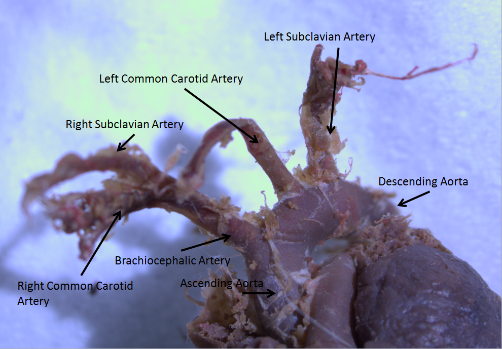
The descending thoracic aorta is a part of the aorta located in the thorax. It is a continuation of the aortic arch. It is located within the posterior mediastinal cavity, but frequently bulges into the left pleural cavity. The descending thoracic aorta begins at the lower border of the fourth thoracic vertebra and ends in front of the lower border of the twelfth thoracic vertebra, at the aortic hiatus in the diaphragm where it becomes the abdominal aorta. At its commencement, it is situated on the left of the vertebral column; it approaches the median line as it descends; and, at its termination, lies directly in front of the column. The descending thoracic aorta has a curved shape that faces forward, and has small branches. It has a radius of approximately 1.16 cm. Structure The descending thoracic aorta is part of the aorta, which has different parts named according to their structure or location. The descending thoracic aorta is a continuation of the descending aorta a ... [...More Info...] [...Related Items...] OR: [Wikipedia] [Google] [Baidu] |
Anastomoses
An anastomosis (, plural anastomoses) is a connection or opening between two things (especially cavities or passages) that are normally diverging or branching, such as between blood vessels, leaf veins, or streams. Such a connection may be normal (such as the foramen ovale in a fetus's heart) or abnormal (such as the patent foramen ovale in an adult's heart); it may be acquired (such as an arteriovenous fistula) or innate (such as the arteriovenous shunt of a metarteriole); and it may be natural (such as the aforementioned examples) or artificial (such as a surgical anastomosis). The reestablishment of an anastomosis that had become blocked is called a reanastomosis. Anastomoses that are abnormal, whether congenital or acquired, are often called fistulas. The term is used in medicine, biology, mycology, geology, and geography. Etymology Anastomosis: medical or Modern Latin, from Greek ἀναστόμωσις, anastomosis, "outlet, opening", Gr ana- "up, on, upon", stoma ... [...More Info...] [...Related Items...] OR: [Wikipedia] [Google] [Baidu] |
Aortic Arch
The aortic arch, arch of the aorta, or transverse aortic arch () is the part of the aorta between the ascending and descending aorta. The arch travels backward, so that it ultimately runs to the left of the trachea. Structure The aorta begins at the level of the upper border of the second/third sternocostal articulation of the right side, behind the ventricular outflow tract and pulmonary trunk. The right atrial appendage overlaps it. The first few centimeters of the ascending aorta and pulmonary trunk lies in the same pericardial sheath. and runs at first upward, arches over the pulmonary trunk, right pulmonary artery, and right main bronchus to lie behind the right second coastal cartilage. The right lung and sternum lies anterior to the aorta at this point. The aorta then passes posteriorly and to the left, anterior to the trachea, and arches over left main bronchus and left pulmonary artery, and reaches to the left side of the T4 vertebral body. Apart from T4 verte ... [...More Info...] [...Related Items...] OR: [Wikipedia] [Google] [Baidu] |
Ascending Aorta
The ascending aorta (AAo) is a portion of the aorta commencing at the upper part of the base of the left ventricle, on a level with the lower border of the third costal cartilage behind the left half of the sternum. Structure It passes obliquely upward, forward, and to the right, in the direction of the heart's axis, as high as the upper border of the second right costal cartilage, describing a slight curve in its course, and being situated, about behind the posterior surface of the sternum. The total length is about . Components The aortic root is the portion of the aorta beginning at the aortic annulus and extending to the sinotubular junction. It is sometimes regarded as a part of the ascending aorta, and sometimes regarded as a separate entity from the rest of the ascending aorta. Between each commissure of the aortic valve and opposite the cusps of the aortic valve, three small dilatations called the aortic sinuses. The sinotubular junction is the point in the ascend ... [...More Info...] [...Related Items...] OR: [Wikipedia] [Google] [Baidu] |
Posterior Ramus
Posterior may refer to: * Posterior (anatomy), the end of an organism opposite to its head ** Buttocks, as a euphemism * Posterior horn (other) * Posterior probability The posterior probability is a type of conditional probability that results from updating the prior probability with information summarized by the likelihood via an application of Bayes' rule. From an epistemological perspective, the posterior ..., the conditional probability that is assigned when the relevant evidence is taken into account * Posterior tense, a relative future tense {{disambiguation ... [...More Info...] [...Related Items...] OR: [Wikipedia] [Google] [Baidu] |
Lumbar Arteries
The lumbar arteries are arteries located in the lower back or lumbar region. The lumbar arteries are in parallel with the intercostals. They are usually four in number on either side, and arise from the back of the aorta, opposite the bodies of the upper four lumbar vertebrae. A fifth pair, small in size, is occasionally present: they arise from the middle sacral artery. They run lateralward and backward on the bodies of the lumbar vertebrae, behind the sympathetic trunk, to the intervals between the adjacent transverse processes, and are then continued into the abdominal wall. The arteries of the right side pass behind the inferior vena cava, and the upper two on each side run behind the corresponding crus of the diaphragm. The arteries of both sides pass beneath the tendinous arches which give origin to the psoas major, and are then continued behind this muscle and the lumbar plexus. They now cross the quadratus lumborum, the upper three arteries running behind, the l ... [...More Info...] [...Related Items...] OR: [Wikipedia] [Google] [Baidu] |
Lower Intercostal
The intercostal arteries are a group of arteries that supply the area between the ribs ("costae"), called the intercostal space. The highest intercostal artery (supreme intercostal artery or superior intercostal artery) is an artery in the human body that usually gives rise to the first and second posterior intercostal arteries, which supply blood to their corresponding intercostal space. It usually arises from the costocervical trunk, which is a branch of the subclavian artery. Some anatomists may contend that there is no supreme intercostal artery, only a supreme intercostal vein. The anterior intercostal branches of internal thoracic artery supply the upper five or six intercostal spaces. The internal thoracic artery (previously called as internal mammary artery) then divides into the superior epigastric artery and musculophrenic artery. The latter gives out the remaining anterior intercostal branches. Two in number in each space, these small vessels pass lateralward, o ... [...More Info...] [...Related Items...] OR: [Wikipedia] [Google] [Baidu] |
Superior Epigastric
In human anatomy, the superior epigastric artery is a blood vessel that carries oxygenated blood to the abdominal wall, and upper rectus abdominis muscle. It is a branch of the internal thoracic artery. It enters the rectus sheath to descend upon the inner surface of the rectus abdominis muscle. It anastomoses with the inferior epigastric artery. Structure Origin The superior epigastric artery arises from the internal thoracic artery (referred to as the internal mammary artery in the accompanying diagram). Course and relations The superior epigastric artery enters the rectus sheath to descend upon the deep surface of the rectus abdominis. Along its course, it is accompanied by a similarly named vein, the superior epigastric vein. Anastomoses It anastomoses with the inferior epigastric artery within the rectus abdominis muscle at the umbilicus. Distribution Where it anastomoses, the superior epigastric artery supplies the anterior part of the abdominal wall, uppe ... [...More Info...] [...Related Items...] OR: [Wikipedia] [Google] [Baidu] |
Internal Oblique
The abdominal internal oblique muscle, also internal oblique muscle or interior oblique, is an abdominal muscle in the abdominal wall that lies below the external oblique muscle and just above the transverse abdominal muscle. Structure Its fibers run perpendicular to the external oblique muscle, beginning in the thoracolumbar fascia of the lower back, the anterior 2/3 of the iliac crest (upper part of hip bone) and the lateral half of the inguinal ligament. The muscle fibers run from these points superomedially (up and towards midline) to the muscle's insertions on the inferior borders of the 10th through 12th ribs and the linea alba. In males, the cremaster muscle is also attached to the internal oblique. Nerve supply The internal oblique is supplied by the lower intercostal nerves, as well as the iliohypogastric nerve and the ilioinguinal nerve. Function The internal oblique performs two major functions. Firstly as an accessory muscle of respiration, it acts as an ant ... [...More Info...] [...Related Items...] OR: [Wikipedia] [Google] [Baidu] |
Subcostal Vein
The subcostal vein is a vein in the human body that runs along the bottom of the twelfth rib. It has the same essential qualities as the posterior intercostal veins The posterior intercostal veins are veins that drain the intercostal spaces posteriorly. They run with their corresponding posterior intercostal artery on the underside of the rib, the vein superior to the artery. Each vein also gives off a dors ..., except that it cannot be considered ''intercostal'' because it is not between two ribs. Each subcostal vein gives off a posterior (dorsal) branch which has a similar distribution to the posterior ramus of an intercostal artery. See also * Subcostal nerve * Subcostal artery External links * http://www.instantanatomy.net/thorax/vessels/vinsuperiormediastinum.html Veins of the torso {{circulatory-stub ... [...More Info...] [...Related Items...] OR: [Wikipedia] [Google] [Baidu] |
Transversus Abdominis
The transverse abdominal muscle (TVA), also known as the transverse abdominis, transversalis muscle and transversus abdominis muscle, is a muscle layer of the anterior and lateral (front and side) abdominal wall which is deep to (layered below) the internal oblique muscle. It is thought by most fitness instructors to be a significant component of the core. Structure The transverse abdominal, so called for the direction of its fibers, is the innermost of the flat muscles of the abdomen. It is positioned immediately inside of the internal oblique muscle. The transverse abdominal arises as fleshy fibers, from the lateral third of the inguinal ligament, from the anterior three-fourths of the inner lip of the iliac crest, from the inner surfaces of the cartilages of the lower six ribs, interdigitating with the diaphragm, and from the thoracolumbar fascia. It ends anteriorly in a broad aponeurosis (the Spigelian fascia), the lower fibers of which curve inferomedially (medially and ... [...More Info...] [...Related Items...] OR: [Wikipedia] [Google] [Baidu] |
Twelfth Thoracic Nerve
The subcostal nerve (anterior division of the twelfth thoracic nerve) is larger than the others. It runs along the lower border of the twelfth rib, often gives a communicating branch to the first lumbar nerve, and passes under the lateral lumbocostal arch. It then runs in front of the quadratus lumborum, innervates the transversus, and passes forward between it and the abdominal internal oblique to be distributed in the same manner as the lower intercostal nerves. It communicates with the iliohypogastric nerve and the ilioinguinal nerve of the lumbar plexus, and gives a branch to the pyramidalis muscle and the quadratus lumborum muscle The quadratus lumborum muscle, informally called the ''QL'', is a paired muscle of the left and right posterior abdominal wall. It is the deepest abdominal muscle, and commonly referred to as a back muscle. Each is irregular and quadrilateral in sh .... It also gives off a lateral cutaneous branch that supplies sensory innervation to the skin over ... [...More Info...] [...Related Items...] OR: [Wikipedia] [Google] [Baidu] |
_Victoria_blue-HE.jpg)

