|
Stellate Cell
Stellate cells are neurons in the central nervous system, named for their star-like shape formed by dendritic processes radiating from the cell body. Many stellate cells are GABAergic and are located in the molecular layer of the cerebellum. Stellate cells are derived from dividing progenitor cells in the white matter of postnatal cerebellum. Dendritic trees can vary between neurons. There are two types of dendritic trees in the cerebral cortex, which include pyramidal cells, which are pyramid shaped and stellate cells which are star shaped. Dendrites can also aid neuron classification. Dendrites with spines are classified as spiny, those without spines are classified as aspinous. Stellate cells can be spiny or aspinous, while pyramidal cells are always spiny. Most common stellate cells are the inhibitory interneurons found within the upper half of the molecular layer in the cerebellum. Cerebellar stellate cells synapse onto the dendritic trees of Purkinje cells and send inhibit ... [...More Info...] [...Related Items...] OR: [Wikipedia] [Google] [Baidu] |
Excitatory Synapses
An excitatory synapse is a synapse in which an action potential in a presynaptic neuron increases the probability of an action potential occurring in a postsynaptic cell. Neurons form networks through which nerve impulses travel, each neuron often making numerous connections with other cells. These electrical signals may be excitatory or inhibitory, and, if the total of excitatory influences exceeds that of the inhibitory influences, the neuron will generate a new action potential at its axon hillock, thus transmitting the information to yet another cell. This phenomenon is known as an excitatory postsynaptic potential (EPSP). It may occur via direct contact between cells (i.e., via gap junctions), as in an electrical synapse, but most commonly occurs via the vesicular release of neurotransmitters from the presynaptic axon terminal into the synaptic cleft, as in a chemical synapse. The excitatory neurotransmitters, the most common of which is glutamate, then migrate via di ... [...More Info...] [...Related Items...] OR: [Wikipedia] [Google] [Baidu] |
Progenitor Cell
A progenitor cell is a Cell (biology), biological cell that can Cellular differentiation, differentiate into a specific cell type. Stem cells and progenitor cells have this ability in common. However, stem cells are less specified than progenitor cells. Progenitor cells can only differentiate into their "target" cell type. The most important difference between stem cells and progenitor cells is that stem cells can replicate indefinitely, whereas progenitor cells can divide only a limited number of times. Controversy about the exact definition remains and the concept is still evolving. The terms "progenitor cell" and "stem cell" are sometimes equated. Properties Most progenitors are identified as Oligopotency, oligopotent. In this point of view, they can compare to adult stem cells, but progenitors are said to be in a further stage of cell differentiation. They are in the "center" between stem cells and fully differentiated cells. The kind of potency they have depends on the type ... [...More Info...] [...Related Items...] OR: [Wikipedia] [Google] [Baidu] |
Neuroscience Information Framework
The Neuroscience Information Framework is a repository of global neuroscience web resources, including experimental, clinical, and translational neuroscience databases, knowledge bases, atlases, and genetic/ genomic resources and provides many authoritative links throughout the neuroscience portal of Wikipedia. Description The Neuroscience Information Framework (NIF) is an initiative of the NIH Blueprint for Neuroscience Research, which was established in 2004 by the National Institutes of Health. Development of the NIF started in 2008, when the University of California, San Diego School of Medicine obtained an NIH contract to create and maintain "a dynamic inventory of web-based neurosciences data, resources, and tools that scientists and students can access via any computer connected to the Internet". The project is headed by Maryann Martone, co-director of the National Center for Microscopy and Imaging Research (NCMIR), part of the multi-disciplinary Center for Research in Bio ... [...More Info...] [...Related Items...] OR: [Wikipedia] [Google] [Baidu] |
Stellate Ganglion
The stellate ganglion (or cervicothoracic ganglion) is a sympathetic ganglion formed by the fusion of the inferior cervical ganglion and the first thoracic (superior thoracic sympathetic) ganglion, which exists in 80% of people. Sometimes, the second and the third thoracic ganglia are included in this fusion. The stellate ganglion is relatively big (10–12 x 8–20 mm) compared to much smaller thoracic, lumbar and sacral ganglia, and is polygonal in shape (). Stellate ganglion is located at the level of C7, anterior to the transverse process of C7 and the neck of the first rib, superior to the cervical pleura and just below the subclavian artery. It is superiorly covered by the prevertebral lamina of the cervical fascia and anteriorly in relation with common carotid artery, subclavian artery and the beginning of vertebral artery which sometimes leaves a groove at the apex of this ganglion (this groove can sometimes even separate the stellate ganglion into so called vertebral gangli ... [...More Info...] [...Related Items...] OR: [Wikipedia] [Google] [Baidu] |
Glutamic Acid Decarboxylase
Glutamate decarboxylase or glutamic acid decarboxylase (GAD) is an enzyme that catalyzes the decarboxylation of glutamate to gamma-aminobutyric acid (GABA) and carbon dioxide (). GAD uses pyridoxal-phosphate (PLP) as a cofactor. The reaction proceeds as follows: : In mammals, GAD exists in two isoforms with molecular weights of 67 and 65 kDa (GAD67 and GAD65), which are encoded by two different genes on different chromosomes (GAD1 and GAD2 genes, chromosomes 2 and 10 in humans, respectively). GAD67 and GAD65 are expressed in the brain where GABA is used as a neurotransmitter, and they are also expressed in the insulin-producing β-cells of the pancreas, in varying ratios depending upon the species. Together, these two enzymes maintain the major physiological supply of GABA in mammals, though it may also be synthesized from putrescine in the enteric nervous system, brain, and elsewhere by the actions of diamine oxidase and aldehyde dehydrogenase 1a1. Several truncated tra ... [...More Info...] [...Related Items...] OR: [Wikipedia] [Google] [Baidu] |
Somatosensory Cortex
In physiology, the somatosensory system is the network of neural structures in the brain and body that produce the perception of touch (haptic perception), as well as temperature (thermoception), body position (proprioception), and pain. It is a subset of the sensory nervous system, which also represents visual, auditory, olfactory, and gustatory stimuli. Somatosensation begins when mechano- and thermosensitive structures in the skin or internal organs sense physical stimuli such as pressure on the skin (see mechanotransduction, nociception). Activation of these structures, or receptors, leads to activation of peripheral sensory neurons that convey signals to the spinal cord as patterns of action potentials. Sensory information is then processed locally in the spinal cord to drive reflexes, and is also conveyed to the brain for conscious perception of touch and proprioception. Note, somatosensory information from the face and head enters the brain through peripheral sens ... [...More Info...] [...Related Items...] OR: [Wikipedia] [Google] [Baidu] |
Bergmann Glial Cell
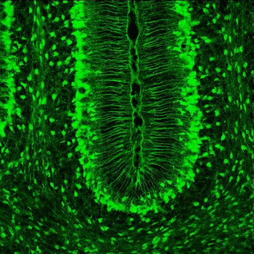
Radial glial cells, or radial glial progenitor cells (RGPs), are bipolar-shaped progenitor cells that are responsible for producing all of the neurons in the cerebral cortex. RGPs also produce certain lineages of glia, including astrocytes and oligodendrocytes. Their cell bodies (somata) reside in the embryonic ventricular zone, which lies next to the developing ventricular system. During development, newborn neurons use radial glia as scaffolds, traveling along the radial glial fibers in order to reach their final destinations. Despite the various possible fates of the radial glial population, it has been demonstrated through clonal analysis that most radial glia have restricted, unipotent or multipotent, fates. Radial glia can be found during the neurogenic phase in all vertebrates (studied to date). The term "radial glia" refers to the morphological characteristics of these cells that were first observed: namely, their radial processes and their similarity to astrocytes, an ... [...More Info...] [...Related Items...] OR: [Wikipedia] [Google] [Baidu] |
Bergmann Glia
Radial glial cells, or radial glial progenitor cells (RGPs), are bipolar-shaped progenitor cells that are responsible for producing all of the neurons in the cerebral cortex. RGPs also produce certain lineages of glia, including astrocytes and oligodendrocytes. Their cell bodies (somata) reside in the embryonic ventricular zone, which lies next to the developing ventricular system. During development, newborn neurons use radial glia as scaffolds, traveling along the radial glial fibers in order to reach their final destinations. Despite the various possible fates of the radial glial population, it has been demonstrated through clonal analysis that most radial glia have restricted, unipotent or multipotent, fates. Radial glia can be found during the neurogenic phase in all vertebrates (studied to date). The term "radial glia" refers to the morphological characteristics of these cells that were first observed: namely, their radial processes and their similarity to astrocytes, an ... [...More Info...] [...Related Items...] OR: [Wikipedia] [Google] [Baidu] |
Basket Cell
Basket cells are inhibitory GABAergic interneurons of the brain, found throughout different regions of the cortex and cerebellum. Anatomy and physiology Basket cells are multipolar GABAergic interneurons that function to make inhibitory synapses and control the overall potentials of target cells. In general, dendrites of basket cells are free branching, contain smooth spines, and extend from 3 to 9 mm. Axons are highly branched, ranging in total from 20 to 50mm in total length. The branched axonal arborizations give rise to the name as they appear as baskets surrounding the soma of the target cell. Basket cells form axo-somatic synapses, meaning their synapses target somas of other cells. By controlling the somas of other neurons, basket cells can directly control the action potential discharge rate of target cells. Basket cells can be found throughout the brain, in among other the cortex, hippocampus, amygdala, basal ganglia, and the cerebellum. Cortex In the cortex, basket cell ... [...More Info...] [...Related Items...] OR: [Wikipedia] [Google] [Baidu] |
Primary Visual Cortex
The visual cortex of the brain is the area of the cerebral cortex that processes visual information. It is located in the occipital lobe. Sensory input originating from the eyes travels through the lateral geniculate nucleus in the thalamus and then reaches the visual cortex. The area of the visual cortex that receives the sensory input from the lateral geniculate nucleus is the primary visual cortex, also known as visual area 1 ( V1), Brodmann area 17, or the striate cortex. The extrastriate areas consist of visual areas 2, 3, 4, and 5 (also known as V2, V3, V4, and V5, or Brodmann area 18 and all Brodmann area 19). Both hemispheres of the brain include a visual cortex; the visual cortex in the left hemisphere receives signals from the right visual field, and the visual cortex in the right hemisphere receives signals from the left visual field. Introduction The primary visual cortex (V1) is located in and around the calcarine fissure in the occipital lobe. Each hemisphere's V1 ... [...More Info...] [...Related Items...] OR: [Wikipedia] [Google] [Baidu] |
Dendritic Tree
Dendrites (from Greek δένδρον ''déndron'', "tree"), also dendrons, are branched protoplasmic extensions of a nerve cell that propagate the electrochemical stimulation received from other neural cells to the cell body, or soma, of the neuron from which the dendrites project. Electrical stimulation is transmitted onto dendrites by upstream neurons (usually via their axons) via synapses which are located at various points throughout the dendritic tree. Dendrites play a critical role in integrating these synaptic inputs and in determining the extent to which action potentials are produced by the neuron. Dendritic arborization, also known as dendritic branching, is a multi-step biological process by which neurons form new dendritic trees and branches to create new synapses. The morphology of dendrites such as branch density and grouping patterns are highly correlated to the function of the neuron. Malformation of dendrites is also tightly correlated to impaired nervous syste ... [...More Info...] [...Related Items...] OR: [Wikipedia] [Google] [Baidu] |
Interneurons
Interneurons (also called internuncial neurons, relay neurons, association neurons, connector neurons, intermediate neurons or local circuit neurons) are neurons that connect two brain regions, i.e. not direct motor neurons or sensory neurons. Interneurons are the central nodes of neural circuits, enabling communication between sensory or motor neurons and the central nervous system (CNS). They play vital roles in reflexes, neuronal oscillations, and neurogenesis in the adult mammalian brain. Interneurons can be further broken down into two groups: local interneurons and relay interneurons. Local interneurons have short axons and form circuits with nearby neurons to analyze small pieces of information. Relay interneurons have long axons and connect circuits of neurons in one region of the brain with those in other regions. However, interneurons are generally considered to operate mainly within local brain areas. The interaction between interneurons allow the brain to perform compl ... [...More Info...] [...Related Items...] OR: [Wikipedia] [Google] [Baidu] |


