|
Stargardt Disease
Stargardt disease is the most common inherited single-gene retinal disease. In terms of the first description of the disease, it follows an autosomal recessive inheritance pattern, which has been later linked to bi-allelic ABCA4 gene variants (STGD1). However, there are Stargardt-like diseases with mimicking phenotypes that are referred to as STGD3 and STGD4, and have a autosomal dominant inheritance due to defects with ELOVL4 or PROM1 genes, respectively. It is characterized by macular degeneration that begins in childhood, adolescence or adulthood, resulting in progressive loss of vision. Signs and symptoms The presentation usually occurs in childhood or adolescence, though there is no upper age limit for presentation and late-onset is possible. The main symptom is loss of visual acuity, uncorrectable with glasses. This manifests as the lack of the ability to see fine details when reading or viewing distant objects. Symptoms typically develop before age 20 (median age of ons ... [...More Info...] [...Related Items...] OR: [Wikipedia] [Google] [Baidu] |
Optical Coherence Tomography
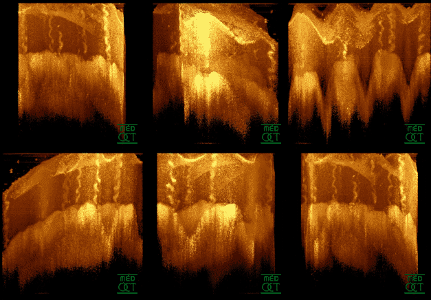
Optical coherence tomography (OCT) is an imaging technique that uses low-coherence light to capture micrometer-resolution, two- and three-dimensional images from within optical scattering media (e.g., biological tissue). It is used for medical imaging and industrial nondestructive testing (NDT). Optical coherence tomography is based on low-coherence interferometry, typically employing near-infrared light. The use of relatively long wavelength light allows it to penetrate into the scattering medium. Confocal microscopy, another optical technique, typically penetrates less deeply into the sample but with higher resolution. Depending on the properties of the light source ( superluminescent diodes, ultrashort pulsed lasers, and supercontinuum lasers have been employed), optical coherence tomography has achieved sub-micrometer resolution (with very wide-spectrum sources emitting over a ~100 nm wavelength range). Optical coherence tomography is one of a class of optical tom ... [...More Info...] [...Related Items...] OR: [Wikipedia] [Google] [Baidu] |
Genotype
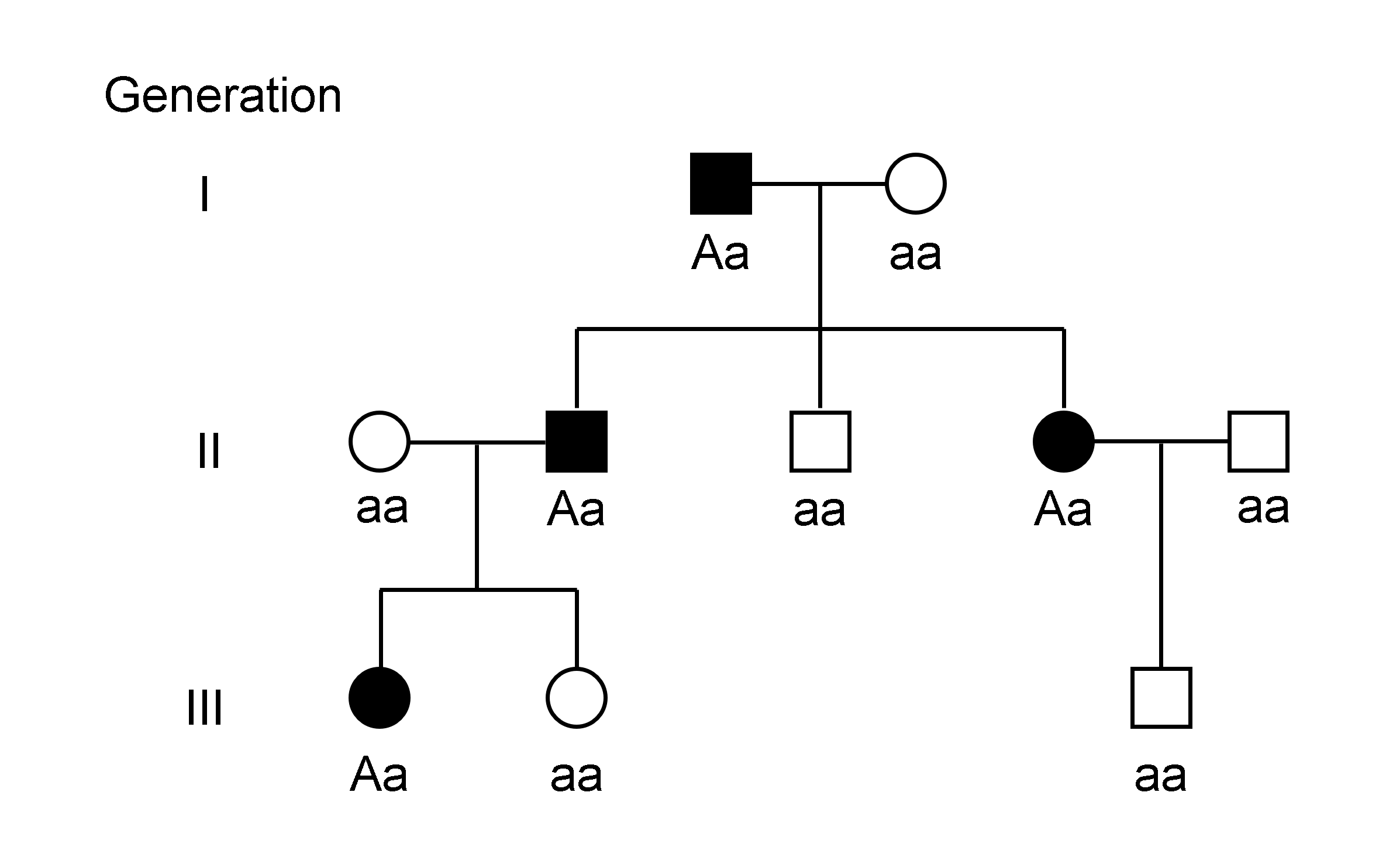
The genotype of an organism is its complete set of genetic material. Genotype can also be used to refer to the alleles or variants an individual carries in a particular gene or genetic location. The number of alleles an individual can have in a specific gene depends on the number of copies of each chromosome found in that species, also referred to as ploidy. In diploid species like humans, two full sets of chromosomes are present, meaning each individual has two alleles for any given gene. If both alleles are the same, the genotype is referred to as homozygous. If the alleles are different, the genotype is referred to as heterozygous. Genotype contributes to phenotype, the observable traits and characteristics in an individual or organism. The degree to which genotype affects phenotype depends on the trait. For example, the petal color in a pea plant is exclusively determined by genotype. The petals can be purple or white depending on the alleles present in the pea plant. Howev ... [...More Info...] [...Related Items...] OR: [Wikipedia] [Google] [Baidu] |
Autofluorescence
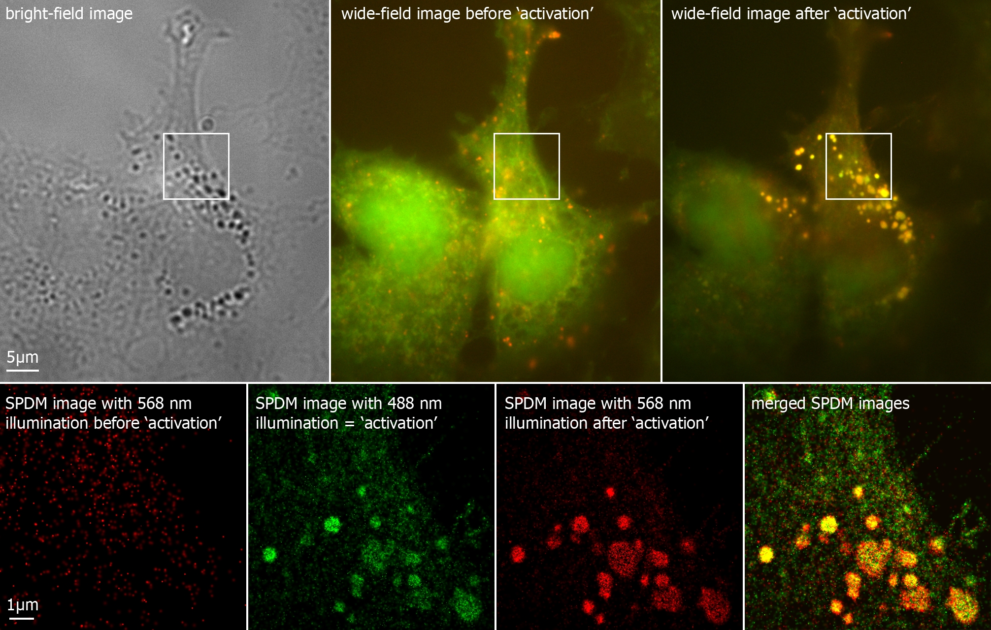
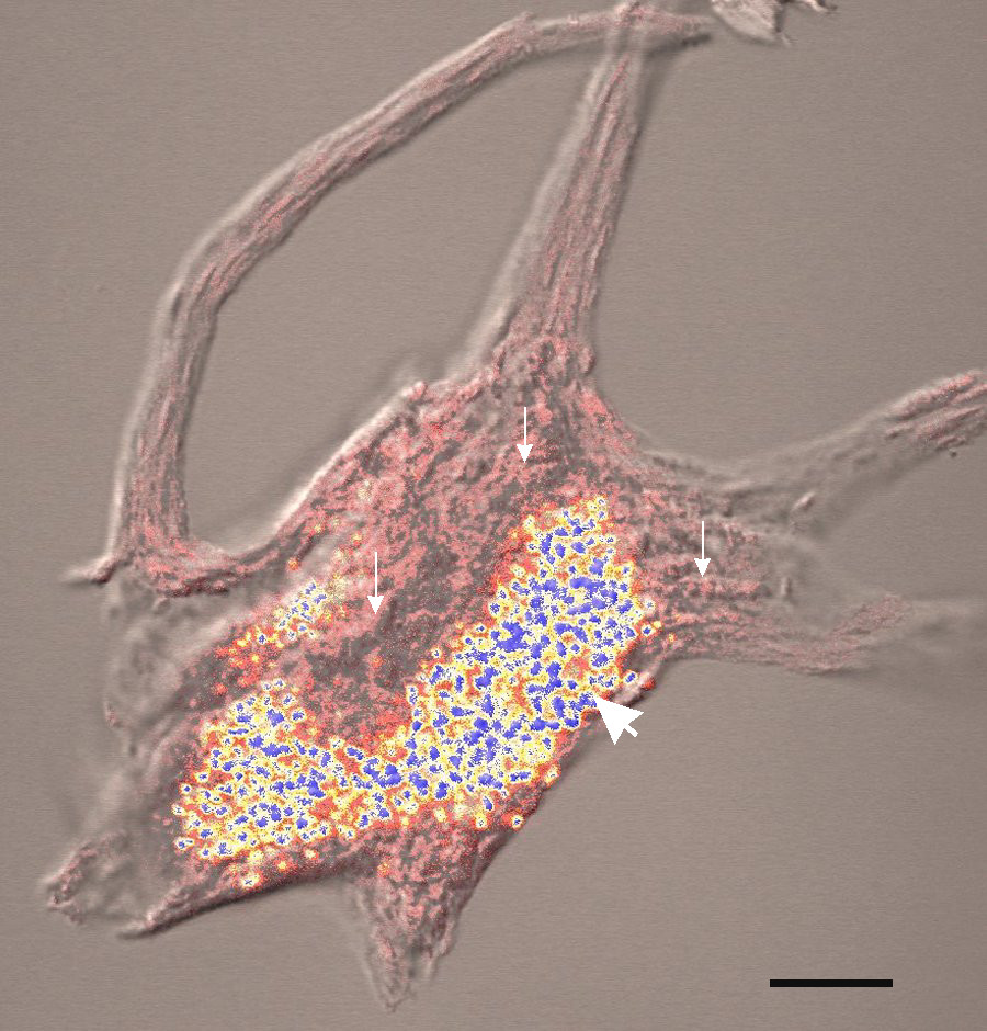
Autofluorescence is the natural emission of light by biological structures such as mitochondria and lysosomes when they have absorbed light, and is used to distinguish the light originating from artificially added fluorescent markers (fluorophores). The most commonly observed autofluorescencing molecules are NADPH and flavins; the extracellular matrix can also contribute to autofluorescence because of the intrinsic properties of collagen and elastin. Generally, proteins containing an increased amount of the amino acids tryptophan, tyrosine, and phenylalanine show some degree of autofluorescence. Autofluorescence also occurs in non-biological materials found in many papers and textiles. Autofluorescence from U.S. paper money has been demonstrated as a means for discerning counterfeit currency from authentic currency. Microscopy Autofluorescence can be problematic in fluorescence microscopy. Light-emitting stains (such as fluorescently labelled antibodies) are applied to sampl ... [...More Info...] [...Related Items...] OR: [Wikipedia] [Google] [Baidu] |
Scanning Laser Ophthalmoscopy
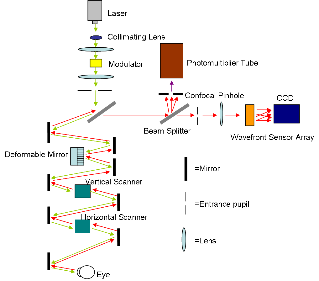
Scanning laser ophthalmoscopy (SLO) is a method of examination of the eye. It uses the technique of confocal laser scanning microscopy for diagnostic imaging of the retina or cornea of the human eye. As a method used to image the retina with a high degree of spatial sensitivity, it is helpful in the diagnosis of glaucoma, macular degeneration, and other retinal disorders. It has further been combined with adaptive optics technology to provide sharper images of the retina."Roorda Lab" — (last accessed: 9 December 2006) Published on October 25, 2006—(last accessed: 9 December 2006) Scanning laser ophthalmoscopy SLO utilizes horizontal and vertical scanning mirrors to scan a specific region of ...[...More Info...] [...Related Items...] OR: [Wikipedia] [Google] [Baidu] |
Slit Lamp
A slit lamp is an instrument consisting of a high-intensity light source that can be focused to shine a thin sheet of light into the eye. It is used in conjunction with a biomicroscope. The lamp facilitates an examination of the anterior segment and posterior segment of the human eye, which includes the eyelid, sclera, conjunctiva, iris, natural crystalline lens, and cornea. The binocular slit-lamp examination provides a stereoscopic magnified view of the eye structures in detail, enabling anatomical diagnoses to be made for a variety of eye conditions. A second, hand-held lens is used to examine the retina. History Two conflicting trends emerged in the development of the slit lamp. One trend originated from clinical research and aimed to apply the increasingly complex and advanced technology of the time. [...More Info...] [...Related Items...] OR: [Wikipedia] [Google] [Baidu] |
Very Long Chain Fatty Acid
A very-long-chain fatty acid (VLCFA) is a fatty acid with 22 or more carbons. Their biosynthesis occurs in the endoplasmic reticulum. VLCFA's can represent up to a few percent of the total fatty acid content of a cell. Unlike most fatty acids, VLCFAs are too long to be metabolized in the mitochondria, in the endoplasmic reticulum (ER) in plants and must be metabolized in peroxisomes. Certain peroxisomal disorders, such as adrenoleukodystrophy and Zellweger syndrome, can be associated with an accumulation of VLCFAs. Enzymes that produce VLCFAs are the targets of herbicides including pyroxasulfone. Major VLCFAs Some of the more common saturated VLCFAs: lignoceric acid (C24), cerotic acid (C26), montanic acid (C28), melissic acid (C30), lacceroic acid (C32), ghedoic acid (C34), and the odd-chain fatty acid ceroplastic acid (C35). Several monounsaturated VLCFAs are also known: nervonic acid (Δ15-24:1), ximenic acid (Δ17-26:1), and lumequeic acid (Δ21-30:1). See also * AC ... [...More Info...] [...Related Items...] OR: [Wikipedia] [Google] [Baidu] |
Dystrophy
Dystrophy is the degeneration of tissue, due to disease or malnutrition, most likely due to heredity. Types * Muscular dystrophy ** Duchenne muscular dystrophy ** Becker's muscular dystrophy ** Myotonic dystrophy * Reflex neurovascular dystrophy * Retinal dystrophy * Cone dystrophy * Corneal dystrophy * Lipodystrophy * Nail dystrophy See also * Muscle weakness * Muscle atrophy * Myotonia * List of biological development disorders References Bibliography * Reece JB, Urry LA, Cain ML, Wasserman SA, Minorsky PV, Jackson RB. Campbell Biology (10th ed.). Addison Wesley Longman; 2014. {{DEFAULTSORT:Biological development disorders Lists of diseases Disability-related l ... {{Authority control Medical terminology ... [...More Info...] [...Related Items...] OR: [Wikipedia] [Google] [Baidu] |
Retinal Pigmented Epithelium
The pigmented layer of retina or retinal pigment epithelium (RPE) is the pigmented cell layer just outside the neurosensory retina that nourishes retinal visual cells, and is firmly attached to the underlying choroid and overlying retinal visual cells. History The RPE was known in the 18th and 19th centuries as the pigmentum nigrum, referring to the observation that the RPE is dark (black in many animals, brown in humans); and as the tapetum nigrum, referring to the observation that in animals with a tapetum lucidum, in the region of the tapetum lucidum the RPE is not pigmented. Anatomy The RPE is composed of a single layer of hexagonal cells that are densely packed with pigment granules. When viewed from the outer surface, these cells are smooth and hexagonal in shape. When seen in section, each cell consists of an outer non-pigmented part containing a large oval nucleus and an inner pigmented portion which extends as a series of straight thread-like processes between the rods, ... [...More Info...] [...Related Items...] OR: [Wikipedia] [Google] [Baidu] |
Lipofuscin
Lipofuscin is the name given to fine yellow-brown pigment granules composed of lipid-containing residues of lysosomal digestion. It is considered to be one of the aging or "wear-and-tear" pigments, found in the liver, kidney, heart muscle, retina, adrenals, nerve cells, and ganglion cells. Formation and turnover Lipofuscin appears to be the product of the oxidation of unsaturated fatty acids and may be symptomatic of membrane damage, or damage to mitochondria and lysosomes. Aside from a large lipid content, lipofuscin is known to contain sugars and metals, including mercury, aluminium, iron, copper and zinc.Chris Gaugler,Lipofuscin", ''Stanislaus Journal of Biochemical Reviews'' May 1997 Lipofuscin is also accepted as consisting of oxidized proteins (30–70%) as well as lipids (20–50%). It is a type of lipochrome and is specifically arranged around the nucleus. The accumulation of lipofuscin-like material may be the result of an imbalance between formation and disposal mech ... [...More Info...] [...Related Items...] OR: [Wikipedia] [Google] [Baidu] |
Visual Phototransduction
Visual phototransduction is the sensory transduction process of the visual system by which light is detected to yield nerve impulses in the rod cells and cone cells in the retina of the eye in humans and other vertebrates. It relies on the visual cycle, a sequence of biochemical reactions in which a molecule of retinal bound to opsin undergoes photoisomerization, initiates a cascade that signals detection of the photon, and is indirectly restored to its photosensitive isomer for reuse. Phototransduction in some invertebrates such as fruit flies relies on similar processes. Photoreceptors The photoreceptor cells involved in vertebrate vision are the rods, the cones, and the photosensitive ganglion cells (ipRGCs). These cells contain a chromophore ( 11-''cis''-retinal, the aldehyde of vitamin A1 and light-absorbing portion) that is bound to a cell membrane protein, opsin. Rods deal with low light level and do not mediate color vision. Cones, on the other hand, can code the color o ... [...More Info...] [...Related Items...] OR: [Wikipedia] [Google] [Baidu] |
ATP-binding Cassette Transporter
The ATP-binding cassette transporters (ABC transporters) are a transport system superfamily that is one of the largest and possibly one of the oldest gene families. It is represented in all extant phyla, from prokaryotes to humans. ABC transporters belong to translocases. ABC transporters often consist of multiple subunits, one or two of which are transmembrane proteins and one or two of which are membrane-associated AAA ATPases. The ATPase subunits utilize the energy of adenosine triphosphate (ATP) binding and hydrolysis to provide the energy needed for the translocation of substrates across membranes, either for uptake or for export of the substrate. Most of the uptake systems also have an extracytoplasmic receptor, a solute binding protein. Some homologous ATPases function in non-transport-related processes such as translation of RNA and DNA repair. ABC transporters are considered to be an ABC superfamily based on the similarities of the sequence and organization of their ... [...More Info...] [...Related Items...] OR: [Wikipedia] [Google] [Baidu] |
Missense
In genetics, a missense mutation is a point mutation in which a single nucleotide change results in a codon that codes for a different amino acid. It is a type of nonsynonymous substitution. Substitution of protein from DNA mutations Missense mutation refers to a change in one amino acid in a protein, arising from a point mutation in a single nucleotide. Missense mutation is a type of nonsynonymous substitution in a DNA sequence. Two other types of nonsynonymous substitution are the nonsense mutations, in which a codon is changed to a premature stop codon that results in truncation of the resulting protein, and the nonstop mutations, in which a stop codon erasement results in a longer, nonfunctional protein. Missense mutations can render the resulting protein nonfunctional, and such mutations are responsible for human diseases such as Epidermolysis bullosa, sickle-cell disease, SOD1 mediated ALS, and a substantial number of cancers. In the most common variant of sickle-cell dis ... [...More Info...] [...Related Items...] OR: [Wikipedia] [Google] [Baidu] |
