|
Skeletal System Of The Horse
The skeletal system of the horse has three major functions in the body. It protects vital organs, provides framework, and supports soft parts of the body. Horses typically have 205 bones. The pelvic limb typically contains 19 bones, while the thoracic limb contains 20 bones. Functions of bones Bones serve three major functions in the skeletal system; they act as levers, they store minerals, and they are the site of red blood cell formation. Bones can be classified into five categories # Long bones: aid in locomotion, store minerals, and act as levers. They are found mainly in the limbs. # Short bones: Absorb concussion. Found in joints such as the knee, hock, and fetlock. # Flat bones: Enclose body cavities containing organs. The ribs are examples of flat bones. # Irregular bones: Protect the central nervous system. The vertebral column consists of irregular bones. # Sesamoid bones: Bones embedded within a tendon. The horse's proximal digital sesamoids are simply called the "se ... [...More Info...] [...Related Items...] OR: [Wikipedia] [Google] [Baidu] |
Horse Anatomy
Equine anatomy encompasses the gross and microscopic anatomy of horses, ponies and other equids, including donkeys, mules and zebras. While all anatomical features of equids are described in the same terms as for other animals by the International Committee on Veterinary Gross Anatomical Nomenclature in the book ''Nomina Anatomica Veterinaria'', there are many horse-specific colloquial terms used by equestrians. External anatomy * Back: the area where the saddle sits, beginning at the end of the withers, extending to the last thoracic vertebrae (colloquially includes the loin or "coupling," though technically incorrect usage) * Barrel: the body of the horse, enclosing the rib cage and the major internal organs * Buttock: the part of the hindquarters behind the thighs and below the root of the tail * Cannon or cannon bone: the area between the knee or hock and the fetlock joint, sometimes called the "shin" of the horse, though technically it is the third metacarpal * Chestnut: ... [...More Info...] [...Related Items...] OR: [Wikipedia] [Google] [Baidu] |
Interosseous Muscle
{{short description, Muscles between certain bones Interossei refer to muscles between certain bones. There are many interossei in a human body. Specific interossei include: On the hands * Dorsal interossei muscles of the hand * Palmar interossei muscles File:Gray428.png, Dorsal interossei muscles of the hand File:Gray429.png, Palmar interossei muscles On the feet * Dorsal interossei muscles of the foot * Plantar interossei muscles In human anatomy, plantar interossei muscles are three muscles located between the metatarsal bones in the foot. Structure The three plantar interosseous muscles are unipennate, as opposed to the bipennate structure of dorsal interosseous muscles ... File:Gray446.png, Dorsal interossei muscles of the foot File:Gray447.png, Plantar interossei muscles Muscular system ... [...More Info...] [...Related Items...] OR: [Wikipedia] [Google] [Baidu] |
Withers
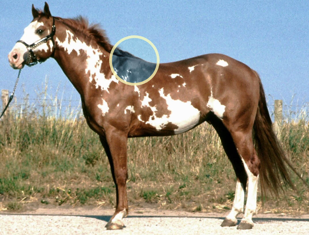
The withers is the ridge between the shoulder blades of an animal, typically a quadruped. In many species, it is the tallest point of the body. In horses and dogs, it is the standard place to measure the animal's height. In contrast, cattle are often measured to the top of the hips. The term (pronounced ) derives from Old English ''wither'' (“against”), because it is the part of a draft animal that pushes against a load. Horses The withers in horses are formed by the dorsal spinal processes of roughly the 3rd through 11th thoracic vertebrae, which are unusually long in this area. Most horses have 18 thoracic vertebrae. The processes at the withers can be more than long. Since they do not move relative to the ground as the horse's head does, the withers are used as the measuring point for the height of a horse. Horses are sometimes measured in hands – one hand is . Horse heights are extremely variable, from small pony breeds to large draft breeds. The height at the ... [...More Info...] [...Related Items...] OR: [Wikipedia] [Google] [Baidu] |
Arabian (horse)
The Arabian or Arab horse ( ar, الحصان العربي , DIN 31635, DMG ''ḥiṣān ʿarabī'') is a horse breed, breed of horse that originated on the Arabian Peninsula. With a distinctive head shape and high tail carriage, the Arabian is one of the most easily recognizable horse breeds in the world. It is also one of the oldest breeds, with archaeological evidence of horses in the Middle East that resemble modern Arabians dating back 4,500 years. Throughout history, Arabian horses have spread around the world by both war and trade, used to improve other breeds by adding speed, refinement, endurance, and strong bone. Today, Arabian bloodlines are found in almost every modern breed of riding horse. The Arabian developed in a desert climate and was prized by the nomadic Bedouin people, often being brought inside the family tent for shelter and protection from theft. Selective breeding for traits, including an ability to form a cooperative relationship with humans, create ... [...More Info...] [...Related Items...] OR: [Wikipedia] [Google] [Baidu] |
Atlas (anatomy)
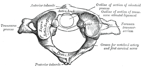
In anatomy, the atlas (C1) is the most superior (first) cervical vertebra of the spine and is located in the neck. It is named for Atlas of Greek mythology because, just as Atlas supported the globe, it supports the entire head. The atlas is the topmost vertebra and, with the axis (the vertebra below it), forms the joint connecting the skull and spine. The atlas and axis are specialized to allow a greater range of motion than normal vertebrae. They are responsible for the nodding and rotation movements of the head. The atlanto-occipital joint allows the head to nod up and down on the vertebral column. The dens acts as a pivot that allows the atlas and attached head to rotate on the axis, side to side. The atlas's chief peculiarity is that it has no body. It is ring-like and consists of an anterior and a posterior arch and two lateral masses. The atlas and axis are important neurologically because the brainstem extends down to the axis. Structure Anterior arch The anterio ... [...More Info...] [...Related Items...] OR: [Wikipedia] [Google] [Baidu] |
Sternebrae
The sternum or breastbone is a long flat bone located in the central part of the chest. It connects to the ribs via cartilage and forms the front of the rib cage, thus helping to protect the heart, lungs, and major blood vessels from injury. Shaped roughly like a necktie, it is one of the largest and longest flat bones of the body. Its three regions are the manubrium, the body, and the xiphoid process. The word "sternum" originates from the Ancient Greek στέρνον (stérnon), meaning "chest". Structure The sternum is a narrow, flat bone, forming the middle portion of the front of the chest. The top of the sternum supports the clavicles (collarbones) and its edges join with the costal cartilages of the first two pairs of ribs. The inner surface of the sternum is also the attachment of the sternopericardial ligaments. Its top is also connected to the sternocleidomastoid muscle. The sternum consists of three main parts, listed from the top: * Manubrium * Body (gladiolus) * Xip ... [...More Info...] [...Related Items...] OR: [Wikipedia] [Google] [Baidu] |
Ribs
The rib cage, as an enclosure that comprises the ribs, vertebral column and sternum in the thorax of most vertebrates, protects vital organs such as the heart, lungs and great vessels. The sternum, together known as the thoracic cage, is a semi-rigid bony and cartilaginous structure which surrounds the thoracic cavity and supports the shoulder girdle to form the core part of the human skeleton. A typical human thoracic cage consists of 12 pairs of ribs and the adjoining costal cartilages, the sternum (along with the manubrium and xiphoid process), and the 12 thoracic vertebrae articulating with the ribs. Together with the skin and associated fascia and muscles, the thoracic cage makes up the thoracic wall and provides attachments for extrinsic skeletal muscles of the neck, upper limbs, upper abdomen and back. The rib cage intrinsically holds the muscles of respiration ( diaphragm, intercostal muscles, etc.) that are crucial for active inhalation and forced exhalation, and t ... [...More Info...] [...Related Items...] OR: [Wikipedia] [Google] [Baidu] |
Sternum
The sternum or breastbone is a long flat bone located in the central part of the chest. It connects to the ribs via cartilage and forms the front of the rib cage, thus helping to protect the heart, lungs, and major blood vessels from injury. Shaped roughly like a necktie, it is one of the largest and longest flat bones of the body. Its three regions are the manubrium, the body, and the xiphoid process. The word "sternum" originates from the Ancient Greek στέρνον (stérnon), meaning "chest". Structure The sternum is a narrow, flat bone, forming the middle portion of the front of the chest. The top of the sternum supports the clavicles (collarbones) and its edges join with the costal cartilages of the first two pairs of ribs. The inner surface of the sternum is also the attachment of the sternopericardial ligaments. Its top is also connected to the sternocleidomastoid muscle. The sternum consists of three main parts, listed from the top: * Manubrium * Body (gladiolus) * ... [...More Info...] [...Related Items...] OR: [Wikipedia] [Google] [Baidu] |
Vertebral Column
The vertebral column, also known as the backbone or spine, is part of the axial skeleton. The vertebral column is the defining characteristic of a vertebrate in which the notochord (a flexible rod of uniform composition) found in all chordata, chordates has been replaced by a segmented series of bone: vertebrae separated by intervertebral discs. Individual vertebrae are named according to their region and position, and can be used as anatomical landmarks in order to guide procedures such as Lumbar puncture, lumbar punctures. The vertebral column houses the spinal canal, a cavity that encloses and protects the spinal cord. There are about 50,000 species of animals that have a vertebral column. The human vertebral column is one of the most-studied examples. Many different diseases in humans can affect the spine, with spina bifida and scoliosis being recognisable examples. The general structure of human vertebrae is fairly typical of that found in mammals, reptiles, and birds. Th ... [...More Info...] [...Related Items...] OR: [Wikipedia] [Google] [Baidu] |
Skull
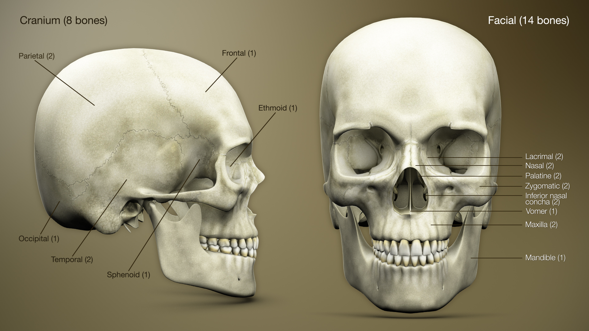
The skull is a bone protective cavity for the brain. The skull is composed of four types of bone i.e., cranial bones, facial bones, ear ossicles and hyoid bone. However two parts are more prominent: the cranium and the mandible. In humans, these two parts are the neurocranium and the viscerocranium ( facial skeleton) that includes the mandible as its largest bone. The skull forms the anterior-most portion of the skeleton and is a product of cephalisation—housing the brain, and several sensory structures such as the eyes, ears, nose, and mouth. In humans these sensory structures are part of the facial skeleton. Functions of the skull include protection of the brain, fixing the distance between the eyes to allow stereoscopic vision, and fixing the position of the ears to enable sound localisation of the direction and distance of sounds. In some animals, such as horned ungulates (mammals with hooves), the skull also has a defensive function by providing the mount (on the front ... [...More Info...] [...Related Items...] OR: [Wikipedia] [Google] [Baidu] |
Axial Skeleton
The axial skeleton is the part of the skeleton that consists of the bones of the head and trunk (anatomy), trunk of a vertebrate. In the human skeleton, it consists of 80 bones and is composed of six parts; the human skull, skull (22 bones), also the ossicles of the middle ear, the hyoid bone, the rib cage, sternum and the vertebral column. The axial skeleton together with the appendicular skeleton form the complete skeleton. Another definition of axial skeleton is the bones including the vertebrae, sacrum, coccyx, skull, ribs, and sternum. Structure Flat bones house the brain and other vital organs. This article mainly deals with the axial skeletons of humans; however, it is important to understand the evolutionary lineage of the axial skeleton. The human axial skeleton consists of 81 different bones. It is the medial core of the body and connects the pelvis to the body, where the appendix skeleton attaches. As the skeleton grows older the bones get weaker with the exception ... [...More Info...] [...Related Items...] OR: [Wikipedia] [Google] [Baidu] |
Horse Skull 02
The horse (''Equus ferus caballus'') is a domesticated, one-toed, hoofed mammal. It belongs to the taxonomic family Equidae and is one of two extant subspecies of ''Equus ferus''. The horse has evolved over the past 45 to 55 million years from a small multi-toed creature, ''Eohippus'', into the large, single-toed animal of today. Humans began domesticating horses around 4000 BCE, and their domestication is believed to have been widespread by 3000 BCE. Horses in the subspecies ''caballus'' are domesticated, although some domesticated populations live in the wild as feral horses. These feral populations are not true wild horses, as this term is used to describe horses that have never been domesticated. There is an extensive, specialized vocabulary used to describe equine-related concepts, covering everything from anatomy to life stages, size, colors, markings, breeds, locomotion, and behavior. Horses are adapted to run, allowing them to quickly escape predators, and poss ... [...More Info...] [...Related Items...] OR: [Wikipedia] [Google] [Baidu] |


