|
Siamese Twins
Conjoined twins – sometimes popularly referred to as Siamese twins – are twins joined ''in utero''. A very rare phenomenon, the occurrence is estimated to range from 1 in 49,000 births to 1 in 189,000 births, with a somewhat higher incidence in Southwest Asia and Africa. Approximately half are stillborn, and an additional one-third die within 24 hours. Most live births are female, with a ratio of 3:1. Two theories exist to explain the origins of conjoined twins. The more generally accepted theory is ''fission'', in which the fertilized egg splits partially. The other theory, no longer believed to be the basis of conjoined twinning, is ''fusion'', in which a fertilized egg completely separates, but stem cells (which search for similar cells) find similar stem cells on the other twin and fuse the twins together. Conjoined twins share a single common chorion, placenta, and amniotic sac, although these characteristics are not exclusive to conjoined twins, as there are some monozyg ... [...More Info...] [...Related Items...] OR: [Wikipedia] [Google] [Baidu] |
Uterus
The uterus (from Latin ''uterus'', plural ''uteri'') or womb () is the organ in the reproductive system of most female mammals, including humans that accommodates the embryonic and fetal development of one or more embryos until birth. The uterus is a hormone-responsive sex organ that contains glands in its lining that secrete uterine milk for embryonic nourishment. In the human, the lower end of the uterus, is a narrow part known as the isthmus that connects to the cervix, leading to the vagina. The upper end, the body of the uterus, is connected to the fallopian tubes, at the uterine horns, and the rounded part above the openings to the fallopian tubes is the fundus. The connection of the uterine cavity with a fallopian tube is called the uterotubal junction. The fertilized egg is carried to the uterus along the fallopian tube. It will have divided on its journey to form a blastocyst that will implant itself into the lining of the uterus – the endometrium, where it will ... [...More Info...] [...Related Items...] OR: [Wikipedia] [Google] [Baidu] |
Diaphragm (anatomy)
The thoracic diaphragm, or simply the diaphragm ( grc, διάφραγμα, diáphragma, partition), is a sheet of internal skeletal muscle in humans and other mammals that extends across the bottom of the thoracic cavity. The diaphragm is the most important muscle of respiration, and separates the thoracic cavity, containing the heart and lungs, from the abdominal cavity: as the diaphragm contracts, the volume of the thoracic cavity increases, creating a negative pressure there, which draws air into the lungs. Its high oxygen consumption is noted by the many mitochondria and capillaries present; more than in any other skeletal muscle. The term ''diaphragm'' in anatomy, created by Gerard of Cremona, can refer to other flat structures such as the urogenital diaphragm or pelvic diaphragm, but "the diaphragm" generally refers to the thoracic diaphragm. In humans, the diaphragm is slightly asymmetric—its right half is higher up (superior) to the left half, since the large liver res ... [...More Info...] [...Related Items...] OR: [Wikipedia] [Google] [Baidu] |
Craniopagus Parasiticus
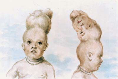
Craniopagus parasiticus is an extremely rare type of parasitic twinning occurring in about 2 to 3 of 5,000,000 births. In craniopagus parasiticus, a parasitic twin head with an undeveloped body is attached to the head of a developed twin. Fewer than a dozen cases of this type of conjoined twin have been documented in literature. Development The exact development of craniopagus parasiticus is not well known. However, it is known that the underdeveloped twin is a parasitic twin. Parasitic twins are known to occur ''in utero'' when monozygotic twins start to develop as an embryo, but the embryo fails to completely split. When this happens, one embryo will dominate development, while the other's development is severely altered. The key difference between a parasitic twin and conjoined twins is that in parasitic twins, one twin, the parasite, stops development during gestation, whereas the other twin, the autosite, develops completely. In normal monozygotic twin development, one egg ... [...More Info...] [...Related Items...] OR: [Wikipedia] [Google] [Baidu] |
Dicephalic Parapagus
Skeletal structure of dicephalic twins. B. C. Hirst & G. A. Piersol, Human monstrosities. Wellcome L0027955. (1893) Dicephalic parapagus () is a rare form of partial twinning with two heads side by side on one torso. Infants conjoined this way are sometimes called "two-headed babies" in popular media. The condition is also called parapagus dicephalus. If carried to term, most dicephalic twins are stillborn, or die soon after birth. A small number are known to have survived to adulthood. The extent to which limbs and organs are duplicated varies from case to case. One head may be only partially developed (anencephalic), or both may be complete. In some cases, two complete hearts are present as well, which improves their chances of survival. The total number of arms may be two, three or four. Their prospects are best if no attempt is made to separate them, except in cases in which one twin is clearly dying. Terminology Dicephalus means two-headed. Parapagus means joined side b ... [...More Info...] [...Related Items...] OR: [Wikipedia] [Google] [Baidu] |
Diprosopic Parapagus
Diprosopus ( el, διπρόσωπος, "two-faced", from , , "two" and , euter "face", "person"; with Latin ending), also known as craniofacial duplication (cranio- from Greek , "skull", the other parts Latin), is an extremely rare congenital disorder whereby parts (accessories) or all of the face are duplicated on the head.Definition of diprosopus at MedicineNet. Accessed 8 January 2006.'Miracle baby' is feted in India a BBC News Accessed 10 April 2008. Development Although classically considered conjoined twinning (whi ...[...More Info...] [...Related Items...] OR: [Wikipedia] [Google] [Baidu] |
Human Pelvis
The pelvis (plural pelves or pelvises) is the lower part of the Trunk (anatomy), trunk, between the human abdomen, abdomen and the thighs (sometimes also called pelvic region), together with its embedded skeleton (sometimes also called bony pelvis, or pelvic skeleton). The pelvic region of the trunk includes the bony pelvis, the pelvic cavity (the space enclosed by the bony pelvis), the pelvic floor, below the pelvic cavity, and the perineum, below the pelvic floor. The pelvic skeleton is formed in the area of the back, by the sacrum and the coccyx and anteriorly and to the left and right sides, by a pair of hip bones. The two hip bones connect the spine with the lower limbs. They are attached to the sacrum posteriorly, connected to each other anteriorly, and joined with the two femurs at the hip joints. The gap enclosed by the bony pelvis, called the pelvic cavity, is the section of the body underneath the abdomen and mainly consists of the reproductive organs (sex organs) and ... [...More Info...] [...Related Items...] OR: [Wikipedia] [Google] [Baidu] |
Anus
The anus (Latin, 'ring' or 'circle') is an opening at the opposite end of an animal's digestive tract from the mouth. Its function is to control the expulsion of feces, the residual semi-solid waste that remains after food digestion, which, depending on the type of animal, includes: matter which the animal cannot digest, such as bones; Summary at food material after the nutrients have been extracted, for example cellulose or lignin; ingested matter which would be toxic if it remained in the digestive tract; and dead or excess gut bacteria and other endosymbionts. Amphibians, reptiles, and birds use the same orifice (known as the cloaca) for excreting liquid and solid wastes, for copulation and egg-laying. Monotreme mammals also have a cloaca, which is thought to be a feature inherited from the earliest amniotes via the therapsids. Marsupials have a single orifice for excreting both solids and liquids and, in females, a separate vagina for reproduction. Female placental mamm ... [...More Info...] [...Related Items...] OR: [Wikipedia] [Google] [Baidu] |
Genitalia
A sex organ (or reproductive organ) is any part of an animal or plant that is involved in sexual reproduction. The reproductive organs together constitute the reproductive system. In animals, the testis in the male, and the ovary in the female, are called the ''primary sex organs''. All others are called ''secondary sex organs'', divided between the external sex organs—the genitals or external genitalia, visible at birth in both sexes—and the internal sex organs. Mosses, ferns, and some similar plants have gametangia for reproductive organs, which are part of the gametophyte. The flowers of flowering plants produce pollen and egg cells, but the sex organs themselves are inside the gametophytes within the pollen and the ovule. Coniferous plants likewise produce their sexually reproductive structures within the gametophytes contained within the cones and pollen. The cones and pollen are not themselves sexual organs. Terminology The ''primary sex organs'' are the gonads, a p ... [...More Info...] [...Related Items...] OR: [Wikipedia] [Google] [Baidu] |
Vertebral Column
The vertebral column, also known as the backbone or spine, is part of the axial skeleton. The vertebral column is the defining characteristic of a vertebrate in which the notochord (a flexible rod of uniform composition) found in all chordata, chordates has been replaced by a segmented series of bone: vertebrae separated by intervertebral discs. Individual vertebrae are named according to their region and position, and can be used as anatomical landmarks in order to guide procedures such as Lumbar puncture, lumbar punctures. The vertebral column houses the spinal canal, a cavity that encloses and protects the spinal cord. There are about 50,000 species of animals that have a vertebral column. The human vertebral column is one of the most-studied examples. Many different diseases in humans can affect the spine, with spina bifida and scoliosis being recognisable examples. The general structure of human vertebrae is fairly typical of that found in mammals, reptiles, and birds. Th ... [...More Info...] [...Related Items...] OR: [Wikipedia] [Google] [Baidu] |
Ischiopagi
Ischiopagi comes from the Greek word ''ischio-'' meaning hip (ilium) and ''-pagus'' meaning fixed or united. It is the medical term used for conjoined twins (Class V) who are united at the pelvis. The twins are classically joined with the vertebral axis at 180°. However, the most frequent cases usually structures the ischiopagus twins with two separate spines forming a lateral angle smaller than 90°. The conjoined twins usually have four arms; two, three or four legs; and typically one external genitalia and anus. It is mostly confused with pygopagus where the twins are joined dorsally at the buttocks facing away from each other, whereas ischiopagus twins are joined ventrally and caudally at the sacrum and coccyx. Parapagus is also similar to ischiopagus; however, parapagus twins are joined side-by-side whereas ischiopagus twins typically have spines connected at a 180° angle, facing away from one another. Classification Ischiopagus Dipus: This is the rarest variety with the t ... [...More Info...] [...Related Items...] OR: [Wikipedia] [Google] [Baidu] |
Xiphoid Process
The xiphoid process , or xiphisternum or metasternum, is a small cartilaginous process (extension) of the inferior (lower) part of the sternum, which is usually ossified in the adult human. It may also be referred to as the ensiform process. Both the Greek-derived ''xiphoid'' and its Latin equivalent ''ensiform'' mean 'swordlike' or 'sword-shaped' Structure The xiphoid process is considered to be at the level of the 9th thoracic vertebra and the T7 dermatome. Development In newborns and young (especially small) infants, the tip of the xiphoid process may be both seen and felt as a lump just below the sternal notch. At 15 to 29 years old, the xiphoid usually fuses to the body of the sternum with a fibrous joint. Unlike the synovial articulation of major joints, this is non-movable. Ossification of the xiphoid process occurs around age 40. Variation The xiphoid process can be naturally bifurcated or sometimes perforated (xiphoidal foramen). These variances in morphology are inher ... [...More Info...] [...Related Items...] OR: [Wikipedia] [Google] [Baidu] |
Janus (mythology)
In ancient Roman religion and myth, Janus ( ; la, Ianvs ) is the god of beginnings, gates, transitions, time, duality, doorways, passages, frames, and endings. He is usually depicted as having two faces. The month of January is named for Janus (''Ianuarius''). According to ancient Roman farmers' almanacs, Juno was mistaken as the tutelary deity of the month of January; but, Juno is the tutelary deity of the month of June. Janus presided over the beginning and ending of conflict, and hence war and peace. The gates of a building in Rome named after him (not a temple, as it is often called, but an open enclosure with gates at each end) were opened in time of war, and closed to mark the arrival of peace. As a god of transitions, he had functions pertaining to birth and to journeys and exchange, and in his association with Portunus, a similar harbor and gateway god, he was concerned with travelling, trading and shipping. Janus had no flamen or specialised priest ''( sacerdos)'' a ... [...More Info...] [...Related Items...] OR: [Wikipedia] [Google] [Baidu] |


