|
Scapular Anastomosis
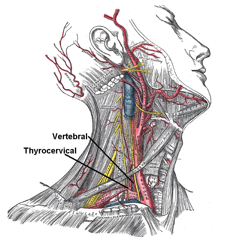
The scapular anastomosis is a system connecting certain subclavian artery and their corresponding axillary artery, forming a circulatory anastomosis around the scapula. It allows blood to flow past the joint in case of occlusion, damage, or pinching of the following scapular arteries: * Transverse cervical artery * Dorsal scapular artery (the anastomosing branch of the transverse cervical) * Suprascapular artery * Branches of subscapular artery * Branches of thoracic aorta The transverse cervical artery gives off a branch, ''the dorsal scapular artery'', which accompanies the dorsal scapular nerve and runs down the vertebral border of the scapula to its medial edge and inferior angle. The dorsal scapular artery anastomoses with the subscapular artery, providing an alternate route to the 3rd part of the axillary artery in the event of a slowly forming occlusion. The suprascapular artery branches off from the thyrocervical trunk, which in turn arises from the first part of the s ... [...More Info...] [...Related Items...] OR: [Wikipedia] [Google] [Baidu] |
Subclavian Artery
In human anatomy, the subclavian arteries are paired major arteries of the upper thorax, below the clavicle. They receive blood from the aortic arch. The left subclavian artery supplies blood to the left arm and the right subclavian artery supplies blood to the right arm, with some branches supplying the head and thorax. On the left side of the body, the subclavian comes directly off the aortic arch, while on the right side it arises from the relatively short brachiocephalic artery when it bifurcates into the subclavian and the right common carotid artery. The usual branches of the subclavian on both sides of the body are the vertebral artery, the internal thoracic artery, the thyrocervical trunk, the costocervical trunk and the dorsal scapular artery, which may branch off the transverse cervical artery, which is a branch of the thyrocervical trunk. The subclavian becomes the axillary artery at the lateral border of the first rib. Structure From its origin, the subclavian artery t ... [...More Info...] [...Related Items...] OR: [Wikipedia] [Google] [Baidu] |
Axillary Artery
In human anatomy, the axillary artery is a large blood vessel that conveys oxygenated blood to the lateral aspect of the thorax, the axilla (armpit) and the upper limb. Its origin is at the lateral margin of the first rib, before which it is called the subclavian artery. After passing the lower margin of teres major it becomes the brachial artery. Structure The axillary artery is often referred to as having three parts, with these divisions based on its location relative to the Pectoralis minor muscle, which is superficial to the artery. * First part – the part of the artery superior to the pectoralis minor * Second part – the part of the artery posterior to the pectoralis minor * Third part – the part of the artery inferior to the pectoralis minor. Relations The axillary artery is accompanied by the axillary vein, which lies medial to the artery, along its length. In the axilla, the axillary artery is surrounded by the brachial plexus. The second part of the axi ... [...More Info...] [...Related Items...] OR: [Wikipedia] [Google] [Baidu] |
Circulatory Anastomosis
A circulatory anastomosis is a connection (an anastomosis) between two blood vessels, such as between arteries (arterio-arterial anastomosis), between veins (veno-venous anastomosis) or between an artery and a vein (arterio-venous anastomosis). Anastomoses between arteries and between veins result in a multitude of arteries and veins, respectively, serving the same volume of tissue. Such anastomoses occur normally in the body in the circulatory system, serving as backup routes for blood to flow if one link is blocked or otherwise compromised, but may also occur pathologically. Physiologic Arterio-arterial anastomoses include actual (e.g., palmar and plantar arches) and potential varieties (e.g., coronary arteries and cortical branch of cerebral arteries). There are many examples of normal arterio-arterial anastomoses in the body. Clinically important examples include: *Circle of Willis (in the brain) *Coronary: anterior interventricular artery and posterior interventricular art ... [...More Info...] [...Related Items...] OR: [Wikipedia] [Google] [Baidu] |
Scapula
The scapula (plural scapulae or scapulas), also known as the shoulder blade, is the bone that connects the humerus (upper arm bone) with the clavicle (collar bone). Like their connected bones, the scapulae are paired, with each scapula on either side of the body being roughly a mirror image of the other. The name derives from the Classical Latin word for trowel or small shovel, which it was thought to resemble. In compound terms, the prefix omo- is used for the shoulder blade in medical terminology. This prefix is derived from ὦμος (ōmos), the Ancient Greek word for shoulder, and is cognate with the Latin , which in Latin signifies either the shoulder or the upper arm bone. The scapula forms the back of the shoulder girdle. In humans, it is a flat bone, roughly triangular in shape, placed on a posterolateral aspect of the thoracic cage. Structure The scapula is a thick, flat bone lying on the thoracic wall that provides an attachment for three groups of muscles: intrin ... [...More Info...] [...Related Items...] OR: [Wikipedia] [Google] [Baidu] |
Transverse Cervical Artery
The transverse cervical artery (transverse artery of neck or transversa colli artery) is an artery in the neck and a branch of the thyrocervical trunk, running at a higher level than the suprascapular artery. Structure It passes transversely below the inferior belly of the omohyoid muscle to the anterior margin of the trapezius, beneath which it divides into a superficial and a deep branch. It crosses in front of the phrenic nerve and the scalene muscles, and in front of or between the divisions of the brachial plexus, and is covered by the platysma and sternocleidomastoid muscles, and crossed by the omohyoid and trapezius. The transverse cervical artery originates from the thyrocervical trunk, it passes through the posterior triangle of the neck to the anterior border of the levator scapulae muscle, where it divides into deep and superficial branches. * Superficial branch ** Ascending branch ** Descending branch (also known as superficial cervical artery, which supplies th ... [...More Info...] [...Related Items...] OR: [Wikipedia] [Google] [Baidu] |
Dorsal Scapular Artery
The transverse cervical artery (transverse artery of neck or transversa colli artery) is an artery in the neck and a branch of the thyrocervical trunk, running at a higher level than the suprascapular artery. Structure It passes transversely below the inferior belly of the omohyoid muscle to the anterior margin of the trapezius, beneath which it divides into a superficial and a deep branch. It crosses in front of the phrenic nerve and the scalene muscles, and in front of or between the divisions of the brachial plexus, and is covered by the platysma and sternocleidomastoid muscles, and crossed by the omohyoid and trapezius. The transverse cervical artery originates from the thyrocervical trunk, it passes through the posterior triangle of the neck to the anterior border of the levator scapulae muscle, where it divides into deep and superficial branches. * Superficial branch ** Ascending branch ** Descending branch (also known as superficial cervical artery, which supplies t ... [...More Info...] [...Related Items...] OR: [Wikipedia] [Google] [Baidu] |
Suprascapular Artery
The suprascapular artery is a branch of the thyrocervical trunk on the neck. Structure At first, it passes downward and laterally across the scalenus anterior and phrenic nerve, being covered by the sternocleidomastoid muscle; it then crosses the subclavian artery and the brachial plexus, running behind and parallel with the clavicle and subclavius muscle and beneath the inferior belly of the omohyoid to the superior border of the scapula. It passes over the superior transverse scapular ligament in most of the cases while below it through the suprascapular notch in some cases. The artery then enters the supraspinous fossa of the scapula. It travels close to the bone, running through the suprascapular canal underneath the supraspinatus muscle, to which it supplies branches. It then descends behind the neck of the scapula, through the great scapular notch and under cover of the inferior transverse ligament, to reach the infraspinatous fossa, where it supplies infraspinatus a ... [...More Info...] [...Related Items...] OR: [Wikipedia] [Google] [Baidu] |
Subscapular Artery
The subscapular artery, the largest branch of the axillary artery, arises from the third part of the axillary artery at the lower border of the subscapularis muscle, which it follows to the inferior angle of the scapula, where it anastomoses with the lateral thoracic and intercostal arteries, and with the descending branch of the dorsal scapular artery (a.k.a. deep branch of the transverse cervical artery if it arises from the cervical trunk), and ends in the neighboring muscles. About 4 cm from its origin it gives off two branches, first the scapular circumflex artery and then the thoracodorsal artery. From the thoracodorsal artery it supplies latissimus dorsi, while the scapular circumflex artery participates in the scapular anastamosis. It terminates in an anastomosis with the dorsal scapular artery The transverse cervical artery (transverse artery of neck or transversa colli artery) is an artery in the neck and a branch of the thyrocervical trunk, running at a h ... [...More Info...] [...Related Items...] OR: [Wikipedia] [Google] [Baidu] |
Thoracic Aorta
The descending thoracic aorta is a part of the aorta located in the thorax. It is a continuation of the aortic arch. It is located within the posterior mediastinal cavity, but frequently bulges into the left pleural cavity. The descending thoracic aorta begins at the lower border of the fourth thoracic vertebra and ends in front of the lower border of the twelfth thoracic vertebra, at the aortic hiatus in the diaphragm where it becomes the abdominal aorta. At its commencement, it is situated on the left of the vertebral column; it approaches the median line as it descends; and, at its termination, lies directly in front of the column. The descending thoracic aorta has a curved shape that faces forward, and has small branches. It has a radius of approximately 1.16 cm. Structure The descending thoracic aorta is part of the aorta, which has different parts named according to their structure or location. The descending thoracic aorta is a continuation of the descending aorta ... [...More Info...] [...Related Items...] OR: [Wikipedia] [Google] [Baidu] |
Dorsal Scapular Artery
The transverse cervical artery (transverse artery of neck or transversa colli artery) is an artery in the neck and a branch of the thyrocervical trunk, running at a higher level than the suprascapular artery. Structure It passes transversely below the inferior belly of the omohyoid muscle to the anterior margin of the trapezius, beneath which it divides into a superficial and a deep branch. It crosses in front of the phrenic nerve and the scalene muscles, and in front of or between the divisions of the brachial plexus, and is covered by the platysma and sternocleidomastoid muscles, and crossed by the omohyoid and trapezius. The transverse cervical artery originates from the thyrocervical trunk, it passes through the posterior triangle of the neck to the anterior border of the levator scapulae muscle, where it divides into deep and superficial branches. * Superficial branch ** Ascending branch ** Descending branch (also known as superficial cervical artery, which supplies t ... [...More Info...] [...Related Items...] OR: [Wikipedia] [Google] [Baidu] |
Circulatory Anastomosis
A circulatory anastomosis is a connection (an anastomosis) between two blood vessels, such as between arteries (arterio-arterial anastomosis), between veins (veno-venous anastomosis) or between an artery and a vein (arterio-venous anastomosis). Anastomoses between arteries and between veins result in a multitude of arteries and veins, respectively, serving the same volume of tissue. Such anastomoses occur normally in the body in the circulatory system, serving as backup routes for blood to flow if one link is blocked or otherwise compromised, but may also occur pathologically. Physiologic Arterio-arterial anastomoses include actual (e.g., palmar and plantar arches) and potential varieties (e.g., coronary arteries and cortical branch of cerebral arteries). There are many examples of normal arterio-arterial anastomoses in the body. Clinically important examples include: *Circle of Willis (in the brain) *Coronary: anterior interventricular artery and posterior interventricular art ... [...More Info...] [...Related Items...] OR: [Wikipedia] [Google] [Baidu] |


_Victoria_blue-HE.jpg)