|
SNAP-25
Synaptosomal-Associated Protein, 25kDa (SNAP-25) is a Target Soluble NSF (''N''-ethylmaleimide-sensitive factor) Attachment Protein Receptor (t-SNARE) protein encoded by the ''SNAP25'' gene found on chromosome 20p12.2 in humans. SNAP-25 is a component of the ''trans''-SNARE complex, which accounts for membrane fusion specificity and directly executes fusion by forming a tight complex that brings the synaptic vesicle and plasma membranes together. Structure and function SNAP-25, a Q-SNARE protein, is anchored to the cytosolic face of membranes via palmitoyl side chains covalently bound to cysteine amino acid residues in the central linker domain of the molecule. This means that SNAP-25 does not contain a trans-membrane domain. SNAP-25 has been identified to contribute two α-helices to the SNARE complex, a four-α-helix domain complex. The SNARE complex participates in vesicle fusion, which involves the docking, priming and merging of a vesicle with the cell membrane to in ... [...More Info...] [...Related Items...] OR: [Wikipedia] [Google] [Baidu] |
SNARE (protein)
SNARE proteins – " SNAP REceptor" – are a large protein family consisting of at least 24 members in yeasts, more than 60 members in mammalian cells, and some numbers in plants. The primary role of SNARE proteins is to mediate vesicle fusion – the fusion of vesicles with the target membrane; this notably mediates exocytosis, but can also mediate the fusion of vesicles with membrane-bound compartments (such as a lysosome). The best studied SNAREs are those that mediate the neurotransmitter release of synaptic vesicles in neurons. These neuronal SNAREs are the targets of the neurotoxins responsible for botulism and tetanus produced by certain bacteria. Types SNAREs can be divided into two categories: ''vesicle'' or ''v-SNAREs'', which are incorporated into the membranes of transport vesicles during budding, and ''target'' or ''t-SNAREs'', which are associated with nerve terminal membranes. Evidence suggests that t-SNAREs form stable subcomplexes which serve as guides f ... [...More Info...] [...Related Items...] OR: [Wikipedia] [Google] [Baidu] |
Q-SNARE
SNARE proteins – " SNAP REceptor" – are a large protein family consisting of at least 24 members in yeasts, more than 60 members in mammalian cells, and some numbers in plants. The primary role of SNARE proteins is to mediate vesicle fusion – the fusion of vesicles with the target membrane; this notably mediates exocytosis, but can also mediate the fusion of vesicles with membrane-bound compartments (such as a lysosome). The best studied SNAREs are those that mediate the neurotransmitter release of synaptic vesicles in neurons. These neuronal SNAREs are the targets of the neurotoxins responsible for botulism and tetanus produced by certain bacteria. Types SNAREs can be divided into two categories: ''vesicle'' or ''v-SNAREs'', which are incorporated into the membranes of transport vesicles during budding, and ''target'' or ''t-SNAREs'', which are associated with nerve terminal membranes. Evidence suggests that t-SNAREs form stable subcomplexes which serve as guides f ... [...More Info...] [...Related Items...] OR: [Wikipedia] [Google] [Baidu] |
Synaptotagmin
Synaptotagmins (SYTs) constitute a family of membrane-trafficking proteins that are characterized by an N-terminal transmembrane region (TMR), a variable linker, and two C-terminal C2 domains - C2A and C2B. There are 17 isoforms in the mammalian synaptotagmin family. There are several C2-domain containing protein families that are related to synaptotagmins, including transmembrane (Ferlins, Extended-Synaptotagmin (E-Syt) membrane proteins, and MCTPs) and soluble (RIMS1 and RIMS2, UNC13D, synaptotagmin-related proteins and B/K) proteins. The family includes synaptotagmin 1, a Ca2+ sensor in the membrane of the pre-synaptic axon terminal, coded by gene SYT1. Functions Based on their brain/endocrine distribution and biochemical properties, in particular C2 domains of certain synaptotagmins bound to calcium, synaptotagmins were proposed to function as calcium sensors in the regulation of neurotransmitter release and hormone secretion. Although synaptotagmins share a similar domain st ... [...More Info...] [...Related Items...] OR: [Wikipedia] [Google] [Baidu] |
Synaptic Vesicle
In a neuron, synaptic vesicles (or neurotransmitter vesicles) store various neurotransmitters that are released at the synapse. The release is regulated by a voltage-dependent calcium channel. Vesicles are essential for propagating nerve impulses between neurons and are constantly recreated by the cell. The area in the axon that holds groups of vesicles is an axon terminal or "terminal bouton". Up to 130 vesicles can be released per bouton over a ten-minute period of stimulation at 0.2 Hz. In the visual cortex of the human brain, synaptic vesicles have an average diameter of 39.5 nanometers (nm) with a standard deviation of 5.1 nm. Structure Synaptic vesicles are relatively simple because only a limited number of proteins fit into a sphere of 40 nm diameter. Purified vesicles have a protein:phospholipid ratio of 1:3 with a lipid composition of 40% phosphatidylcholine, 32% phosphatidylethanolamine, 12% phosphatidylserine, 5% phosphatidylinositol, and 10% chole ... [...More Info...] [...Related Items...] OR: [Wikipedia] [Google] [Baidu] |
Syntaxin
Syntaxins are a family of membrane integrated Q-SNARE proteins participating in exocytosis. Domains Syntaxins possess a single C-terminal transmembrane domain, a SNARE domain (known as H3), and an N-terminal regulatory domain (Habc). Syntaxin 17 may have two transmembrane domains. * The SNARE (H3) domain binds to both synaptobrevin and SNAP-25 forming the core SNARE complex. Formation of this stable SNARE core complex is believed to generate the free energy required to initiate fusion between the vesicle membrane and plasma membrane. * The N-terminal Habc domain is formed by 3 α-helices and when collapsed onto its own H3 helix forms an inactive "closed" syntaxin conformation. This closed conformation of syntaxin is believed to be stabilized by binding of Munc-18 (nSec1), although more recent data suggests that nSec1 may bind to other conformations of syntaxin, as well. The "open" syntaxin conformation is the conformation that is competent to form into SNARE core complexes. ... [...More Info...] [...Related Items...] OR: [Wikipedia] [Google] [Baidu] |
Vesicle Fusion
Vesicle fusion is the merging of a vesicle with other vesicles or a part of a cell membrane. In the latter case, it is the end stage of secretion from secretory vesicles, where their contents are expelled from the cell through exocytosis. Vesicles can also fuse with other target cell compartments, such as a lysosome. Exocytosis occurs when secretory vesicles transiently dock and fuse at the base of cup-shaped structures at the cell plasma membrane called porosome, the universal secretory machinery in cells. Vesicle fusion may depend on SNARE proteins in the presence of increased intracellular calcium (Ca2+) concentration. Triggers Stimuli that trigger vesicle fusion act by increasing intracellular Ca2+. * Synaptic vesicles commit vesicle fusion by a nerve impulse reaching the synapse, activating voltage-dependent calcium channels that cause influx of Ca2+ into the cell. * In the endocrine system, many hormones are released by their releasing hormones binding to G protein coupled rec ... [...More Info...] [...Related Items...] OR: [Wikipedia] [Google] [Baidu] |
Synaptobrevin
Synaptobrevins (''synaptobrevin isotypes 1-2'') are small integral membrane proteins of secretory vesicles with molecular weight of 18 kilodalton (kDa) that are part of the vesicle-associated membrane protein (VAMP) family. Synaptobrevin is one of the SNARE proteins involved in formation of the SNARE complexes. Structure Out of four α-helices of the core SNARE complex one is contributed by synaptobrevin, one by syntaxin, and two by SNAP-25 (in neurons). Function SNARE proteins are the key components of the molecular machinery that drives fusion of membranes in exocytosis. Their function however is subject to fine-tuning by various regulatory proteins collectively referred to as ''SNARE masters''. Classification In the Q/R nomenclature for organizing SNARE proteins, VAMP/synaptobrevin family members are classified as R-SNAREs, so named for the presence of an arginine at a specific location within the primary sequence of the protein (as opposed to the SNAREs of the ta ... [...More Info...] [...Related Items...] OR: [Wikipedia] [Google] [Baidu] |
Exocytosis
Exocytosis () is a form of active transport and bulk transport in which a cell transports molecules (e.g., neurotransmitters and proteins) out of the cell ('' exo-'' + ''cytosis''). As an active transport mechanism, exocytosis requires the use of energy to transport material. Exocytosis and its counterpart, endocytosis, are used by all cells because most chemical substances important to them are large polar molecules that cannot pass through the hydrophobic portion of the cell membrane by passive means. Exocytosis is the process by which a large amount of molecules are released; thus it is a form of bulk transport. Exocytosis occurs via secretory portals at the cell plasma membrane called porosomes. Porosomes are permanent cup-shaped lipoprotein structure at the cell plasma membrane, where secretory vesicles transiently dock and fuse to release intra-vesicular contents from the cell. In exocytosis, membrane-bound secretory vesicles are carried to the cell membrane, where they ... [...More Info...] [...Related Items...] OR: [Wikipedia] [Google] [Baidu] |
Vesicle-associated Membrane Protein
Vesicle associated membrane proteins (VAMP) are a family of SNARE proteins with similar structure, and are mostly involved in vesicle fusion. * VAMP1 and VAMP2 proteins known as synaptobrevins are expressed in brain and are constituents of the synaptic vesicles, where they participate in neurotransmitter release. * VAMP3 (known as cellubrevin) is ubiquitously expressed and participates in regulated and constitutive exocytosis as a constituent of secretory granules and secretory vesicles. * VAMP5 and VAMP7 ( SYBL1) participate in constitutive exocytosis. ** VAMP5 is a constituent of secretory vesicles, myotubes and tubulovesicular structures. ** VAMP7 is found both in secretory granules and endosomes. * VAMP8 (known as endobrevin) participates in endocytosis and is found in early endosomes. VAMP8 also participates the regulated exocytosis in pancreatic acinar cells. *VAMP4 Vesicle-associated membrane protein 4 is a protein that in humans is encoded by the ''VAMP4'' gene. ... [...More Info...] [...Related Items...] OR: [Wikipedia] [Google] [Baidu] |
Palmitoyl Chain
Palmitoylation is the covalent attachment of fatty acids, such as palmitic acid, to cysteine (''S''-palmitoylation) and less frequently to serine and threonine (''O''-palmitoylation) residues of proteins, which are typically membrane proteins. The precise function of palmitoylation depends on the particular protein being considered. Palmitoylation enhances the hydrophobicity of proteins and contributes to their membrane association. Palmitoylation also appears to play a significant role in subcellular trafficking of proteins between membrane compartments, as well as in modulating protein–protein interactions. In contrast to prenylation and myristoylation, palmitoylation is usually reversible (because the bond between palmitic acid and protein is often a thioester bond). The reverse reaction in mammalian cells is catalyzed by acyl-protein thioesterases (APTs) in the cytosol and palmitoyl protein thioesterases in lysosomes. Because palmitoylation is a dynamic, post-translatio ... [...More Info...] [...Related Items...] OR: [Wikipedia] [Google] [Baidu] |
Q-type Calcium Channel
The Q-type calcium channel is a type of voltage-dependent calcium channel. Like the others of this class, the α1 subunit is the one that determines most of the channel's properties. They are poorly understood, but like R-type calcium channels, they appear to be present in cerebellar The cerebellum (Latin for "little brain") is a major feature of the hindbrain of all vertebrates. Although usually smaller than the cerebrum, in some animals such as the mormyrid fishes it may be as large as or even larger. In humans, the cerebel ... granule cells. They have a high threshold of activation and relatively slow kinetics. External links * {{Ion channels, g1 Ion channels Electrophysiology Membrane biology Integral membrane proteins Calcium channels ... [...More Info...] [...Related Items...] OR: [Wikipedia] [Google] [Baidu] |
P-type Calcium Channel
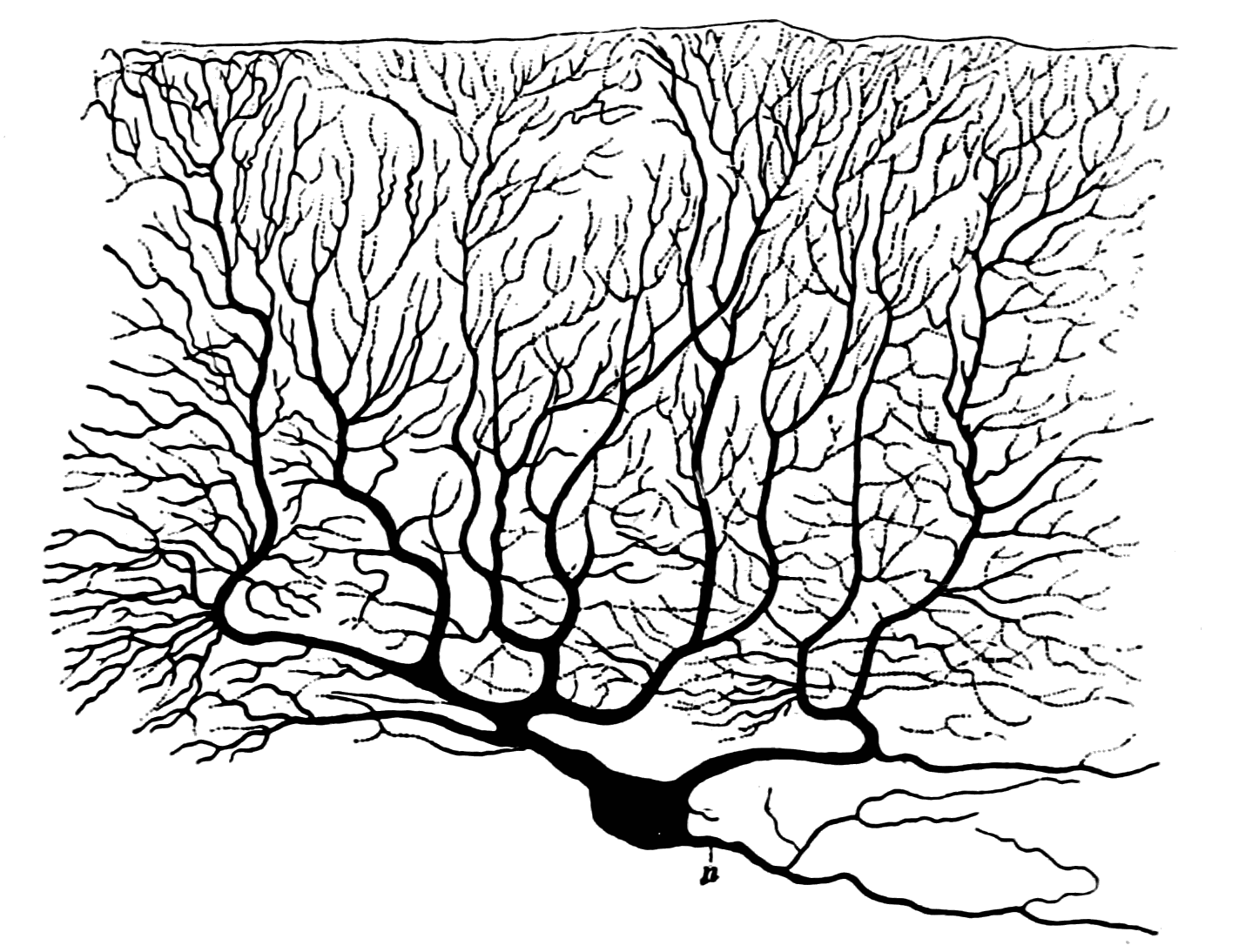
The P-type calcium channel is a type of voltage-dependent calcium channel. Similar to many other high-voltage-gated calcium channels, the α1 subunit determines most of the channel's properties. The 'P' signifies cerebellar Purkinje cells, referring to the channel's initial site of discovery. P-type calcium channels play a similar role to the N-type calcium channel in neurotransmitter release at the presynaptic terminal and in neuronal integration in many neuronal types. History The calcium channel experiments that led to the discovery of P-type calcium channels were initially completed by Rodolfo Llinas, Llinás and Sugimori in 1980. P type calcium channels were named in 1989 because they were discovered within mammalian Purkinje neurons. They were able to use an ''in vitro'' preparation to examine the ionic currents that account for Purkinje cells' electrophysiology, electrophysiological properties. They found that there are calcium dependent action potentials which rise slowly a ... [...More Info...] [...Related Items...] OR: [Wikipedia] [Google] [Baidu] |


