|
SA Nodal Artery
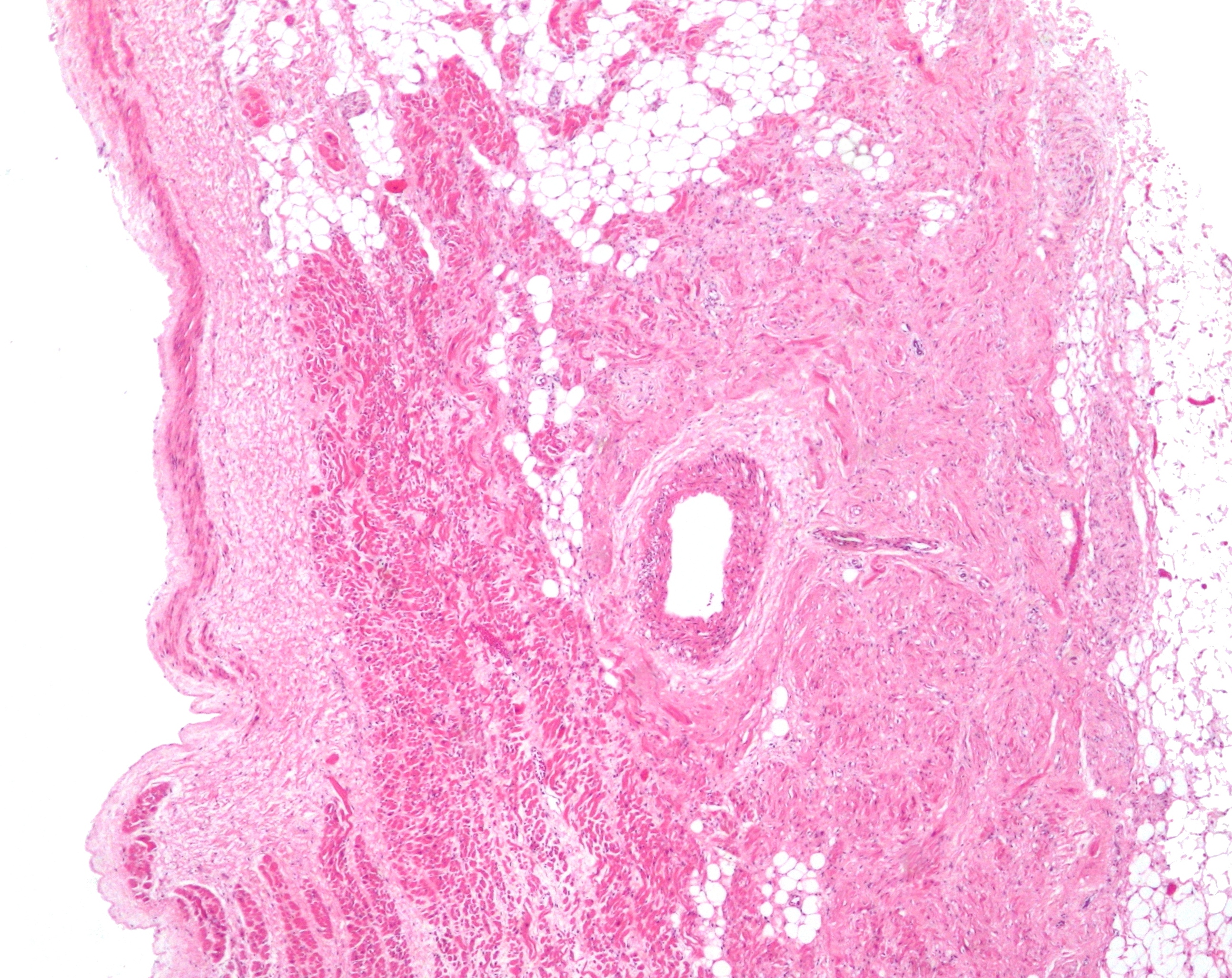
The sinoatrial nodal artery (or sinuatrial nodal artery or sinoatrial artery) is an artery of the heart which supplies the sinoatrial node, the natural pacemaker center of the heart, and arises from the right coronary artery in around 60% of people. In about 40% of cases, the sinoatrial artery is a branch of the left circumflex coronary artery. In less than 1% of humans, the artery has an anomalous origin directly from the coronary sinus, descending aorta, or distal right coronary artery. In more than 50% of human hearts, the artery actually courses close to the superior posterior aspect of the interatrial septum. The origin of the sinoatrial node artery is not related to coronary artery dominance, which means the side (right or left) that provides the circulation to the back of the heart. In contrast, the atrioventricular nodal branch, that is the artery that brings blood to the atrioventricular node, depends on coronary artery dominance. The sinoatrial node surrounds the sinoatri ... [...More Info...] [...Related Items...] OR: [Wikipedia] [Google] [Baidu] |
Micrograph
A micrograph or photomicrograph is a photograph or digital image taken through a microscope or similar device to show a magnified image of an object. This is opposed to a macrograph or photomacrograph, an image which is also taken on a microscope but is only slightly magnified, usually less than 10 times. Micrography is the practice or art of using microscopes to make photographs. A micrograph contains extensive details of microstructure. A wealth of information can be obtained from a simple micrograph like behavior of the material under different conditions, the phases found in the system, failure analysis, grain size estimation, elemental analysis and so on. Micrographs are widely used in all fields of microscopy. Types Photomicrograph A light micrograph or photomicrograph is a micrograph prepared using an optical microscope, a process referred to as ''photomicroscopy''. At a basic level, photomicroscopy may be performed simply by connecting a camera to a microscope, th ... [...More Info...] [...Related Items...] OR: [Wikipedia] [Google] [Baidu] |
Middle Cardiac Vein
The middle cardiac vein commences at the apex of the heart; ascends in the posterior longitudinal sulcus, and ends in the coronary sinus In anatomy, the coronary sinus () is a collection of veins joined together to form a large vessel that collects blood from the heart muscle (myocardium). It delivers deoxygenated blood to the right atrium, as do the superior and inferior vena ... near its right extremity. Structure Variation The middle cardiac vein has a constant location on the surface of the ventricles. Clinical significance The middle cardiac vein is useful for epicardial access to the inferior side of the ventricles. References External links * - "Posterior view of the heart." Veins of the torso {{circulatory-stub ... [...More Info...] [...Related Items...] OR: [Wikipedia] [Google] [Baidu] |
Ablation Of Atrial Fibrillation
Catheter ablation is a procedure used to remove or terminate a faulty electrical pathway from sections of the heart of those who are prone to developing cardiac arrhythmias such as atrial fibrillation, atrial flutter and Wolff-Parkinson-White syndrome. If not controlled, such arrhythmias increase the risk of ventricular fibrillation and sudden cardiac arrest. The ablation procedure can be classified by energy source: radiofrequency ablation and cryoablation. Medical uses Catheter ablation may be recommended for a recurrent or persistent arrhythmia resulting in symptoms or other dysfunction. Typically, catheter ablation is used only when pharmacologic treatment has been ineffective. Effectiveness Catheter ablation of most arrhythmias has a high success rate. Success rates for WPW syndrome have been as high as 95% For SVT, single procedure success is 91% to 96% (95% Confidence Interval) and multiple procedure success is 92% to 97% (95% Confidence Interval). For atrial flutter, sin ... [...More Info...] [...Related Items...] OR: [Wikipedia] [Google] [Baidu] |
Left Atrial Appendage
The atrium ( la, ātrium, , entry hall) is one of two upper chambers in the heart that receives blood from the circulatory system. The blood in the atria is pumped into the heart ventricles through the atrioventricular valves. There are two atria in the human heart – the left atrium receives blood from the pulmonary circulation, and the right atrium receives blood from the venae cavae of the systemic circulation. During the cardiac cycle the atria receive blood while relaxed in diastole, then contract in systole to move blood to the ventricles. Each atrium is roughly cube-shaped except for an ear-shaped projection called an atrial appendage, sometimes known as an auricle. All animals with a closed circulatory system have at least one atrium. The atrium was formerly called the 'auricle'. That term is still used to describe this chamber in some other animals, such as the '' Mollusca''. They have thicker muscular walls than the atria do. Structure Humans have a four-chamber ... [...More Info...] [...Related Items...] OR: [Wikipedia] [Google] [Baidu] |
Sinus Node
The sinoatrial node (also known as the sinuatrial node, SA node or sinus node) is an oval shaped region of special cardiac muscle in the upper back wall of the right atrium made up of cells known as pacemaker cells. The sinus node is approximately fifteen mm long, three mm wide, and one mm thick, located directly below and to the side of the superior vena cava. These cells can produce an electrical impulse an action potential known as a cardiac action potential that travels through the electrical conduction system of the heart, causing it to contract. In a healthy heart, the SA node continuously produces action potentials, setting the rhythm of the heart (sinus rhythm), and so is known as the heart's natural pacemaker. The rate of action potentials produced (and therefore the heart rate) is influenced by the nerves that supply it. Structure The sinoatrial node is a oval-shaped structure that is approximately fifteen mm long, three mm wide, and one mm thick, located directly ... [...More Info...] [...Related Items...] OR: [Wikipedia] [Google] [Baidu] |
Left Atrium
The atrium ( la, ātrium, , entry hall) is one of two upper chambers in the heart that receives blood from the circulatory system. The blood in the atria is pumped into the heart ventricles through the atrioventricular valves. There are two atria in the human heart – the left atrium receives blood from the pulmonary circulation, and the right atrium receives blood from the venae cavae of the systemic circulation. During the cardiac cycle the atria receive blood while relaxed in diastole, then contract in systole to move blood to the ventricles. Each atrium is roughly cube-shaped except for an ear-shaped projection called an atrial appendage, sometimes known as an auricle. All animals with a closed circulatory system have at least one atrium. The atrium was formerly called the 'auricle'. That term is still used to describe this chamber in some other animals, such as the ''Mollusca''. They have thicker muscular walls than the atria do. Structure Humans have a four-chambered ... [...More Info...] [...Related Items...] OR: [Wikipedia] [Google] [Baidu] |
Atrioventricular Node
The atrioventricular node or AV node electrically connects the heart's atria and ventricles to coordinate beating in the top of the heart; it is part of the electrical conduction system of the heart. The AV node lies at the lower back section of the interatrial septum near the opening of the coronary sinus, and conducts the normal electrical impulse from the atria to the ventricles. The AV node is quite compact (~1 x 3 x 5 mm).Full Size Picture triangle of-Koch.jpg Retrieved on 2008-12-22 Structure Location The AV node lies at the lower back section of the |
Interatrial Septum
The interatrial septum is the wall of tissue that separates the right and left atria of the heart. Structure The interatrial septum is a that lies between the left atrium and right atrium of the human heart. The interatrial septum lies at angle of 65 degrees from right posterior to left anterior because right atrium is located at the right side of the body while left atrium is located at the left side of the body. Development The interatrial septum forms during the first and second months of fetal development. Formation of the septum occurs in several stages. The first is the development of the septum primum, a crescent-shaped piece of tissue forming the initial divider between the right and left atria. Because of its crescent shape, the septum primum does not fully occlude the space between the left and right atria; the opening that remains is called the ostium primum. During fetal development, this opening allows blood to be shunted from the right atrium to the left. As the ... [...More Info...] [...Related Items...] OR: [Wikipedia] [Google] [Baidu] |
Descending Aorta
In human anatomy, the descending aorta is part of the aorta, the largest artery in the body. The descending aorta begins at the aortic arch and runs down through the chest and abdomen. The descending aorta anatomically consists of two portions or segments, the thoracic and the abdominal aorta, in correspondence with the two great cavities of the trunk in which it is situated. Within the abdomen, the descending aorta branches into the two common iliac arteries which serve the pelvis and eventually legs. The ductus arteriosus connects to the junction between the pulmonary artery and the descending aorta in foetal life. This artery later regresses as the ligamentum arteriosum. See also *Abbott artery Abbott's artery describes an anomalous artery that arises from the posteromedial aspect of the proximal part of the descending aorta. Normally a minor congenital abnormality, its presence is important during surgical repair of coarctation of the ... References External links ... [...More Info...] [...Related Items...] OR: [Wikipedia] [Google] [Baidu] |
Left Circumflex Coronary Artery
The circumflex branch of left coronary artery, or left circumflex artery or circumflex artery, is a branch of the left coronary artery. Description The left circumflex artery follows the left part of the coronary sulcus, running first to the left and then to the right, reaching nearly as far as the posterior longitudinal sulcus. There have been multiple anomalies described, for example the left circumflex having an aberrant course from the right coronary artery. Branches The circumflex artery curves to the left around the heart within the coronary sulcus, giving rise to one or more left marginal arteries (also called obtuse marginal branches) as it curves toward the posterior surface of the heart. It helps form the posterior left ''ventricular branch'' or posterolateral artery. The circumflex artery ends at the point where it joins to form to the posterior interventricular artery in 15% of all cases, which lies in the posterior interventricular sulcus. In the other 85% of all ... [...More Info...] [...Related Items...] OR: [Wikipedia] [Google] [Baidu] |
Heart
The heart is a muscular organ in most animals. This organ pumps blood through the blood vessels of the circulatory system. The pumped blood carries oxygen and nutrients to the body, while carrying metabolic waste such as carbon dioxide to the lungs. In humans, the heart is approximately the size of a closed fist and is located between the lungs, in the middle compartment of the chest. In humans, other mammals, and birds, the heart is divided into four chambers: upper left and right atria and lower left and right ventricles. Commonly the right atrium and ventricle are referred together as the right heart and their left counterparts as the left heart. Fish, in contrast, have two chambers, an atrium and a ventricle, while most reptiles have three chambers. In a healthy heart blood flows one way through the heart due to heart valves, which prevent backflow. The heart is enclosed in a protective sac, the pericardium, which also contains a small amount of fluid. The wall ... [...More Info...] [...Related Items...] OR: [Wikipedia] [Google] [Baidu] |
Artery
An artery (plural arteries) () is a blood vessel in humans and most animals that takes blood away from the heart to one or more parts of the body (tissues, lungs, brain etc.). Most arteries carry oxygenated blood; the two exceptions are the pulmonary and the umbilical arteries, which carry deoxygenated blood to the organs that oxygenate it (lungs and placenta, respectively). The effective arterial blood volume is that extracellular fluid which fills the arterial system. The arteries are part of the circulatory system, that is responsible for the delivery of oxygen and nutrients to all cells, as well as the removal of carbon dioxide and waste products, the maintenance of optimum blood pH, and the circulation of proteins and cells of the immune system. Arteries contrast with veins, which carry blood back towards the heart. Structure The anatomy of arteries can be separated into gross anatomy, at the macroscopic level, and microanatomy, which must be studied with a microscop ... [...More Info...] [...Related Items...] OR: [Wikipedia] [Google] [Baidu] |


