|
Supreme Intercostal Vein
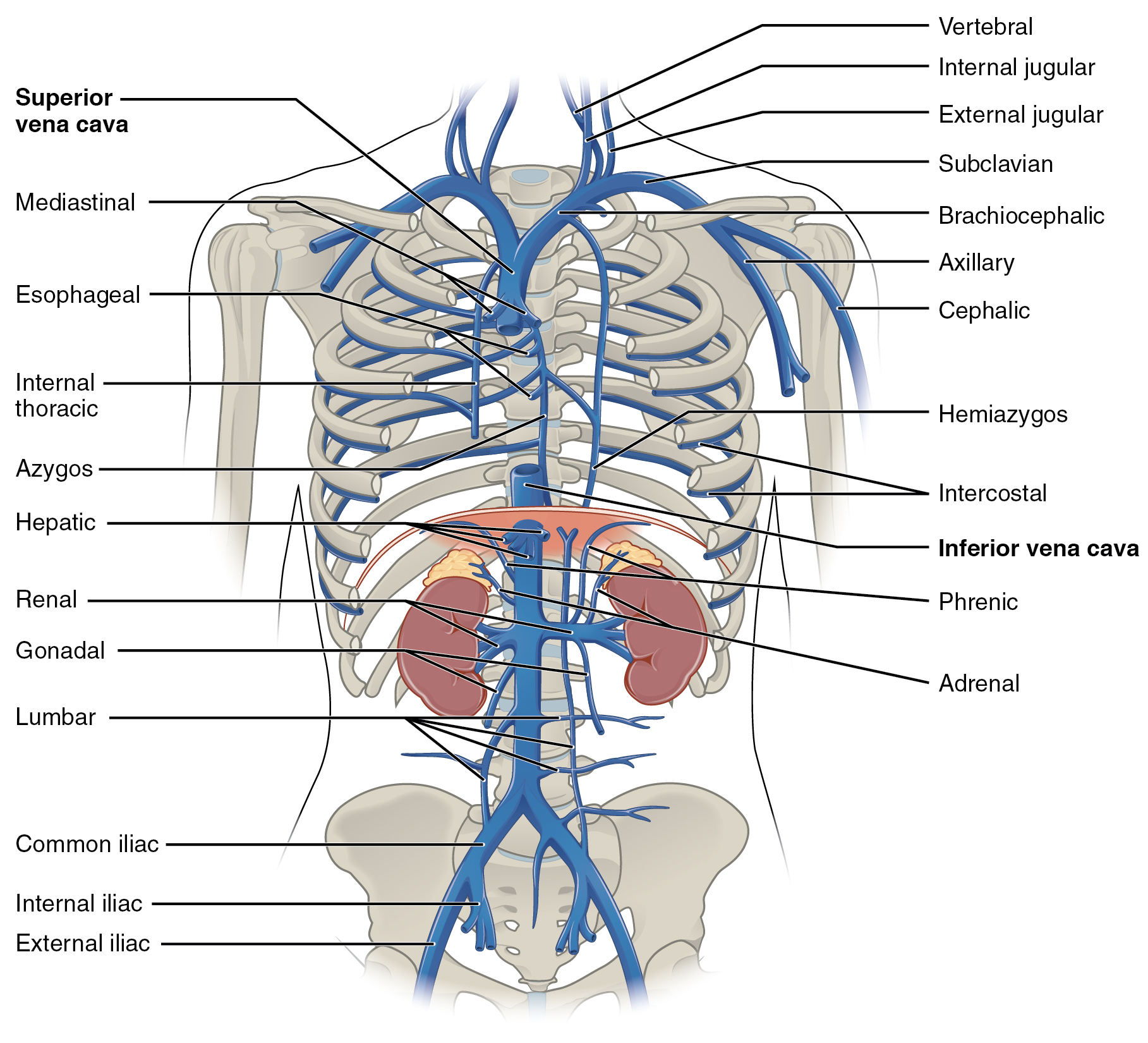
The supreme intercostal vein (highest intercostal vein) is a paired vein that drains the first intercostal space on its corresponding side. It usually drains into the brachiocephalic vein. Alternatively, it drains into the superior intercostal vein, or the vertebral vein of its corresponding side. Clinical significance This vein does not have valves, this is an important point when it comes to spread of cancerous secondaries. Additional images File:Gray480.png, Diagram showing completion of development of the parietal veins. See also * superior intercostal vein * posterior intercostal vein * azygos vein The azygos vein is a vein running up the right side of the thoracic vertebral column draining itself towards the superior vena cava. It connects the systems of superior vena cava and inferior vena cava and can provide an alternative path for blood ... References Veins of the torso {{circulatory-stub ... [...More Info...] [...Related Items...] OR: [Wikipedia] [Google] [Baidu] |
Venæ Cavæ
In anatomy, the venae cavae (; singular: vena cava ; ) are two large veins (great vessels) that return deoxygenated blood from the body into the heart. In humans they are the superior vena cava and the inferior vena cava, and both empty into the right atrium. They are located slightly off-center, toward the right side of the body. The right atrium receives deoxygenated blood through coronary sinus and two large veins called venae cavae. The inferior vena cava (or caudal vena cava in some animals) travels up alongside the abdominal aorta with blood from the lower part of the body. It is the largest vein in the human body. MadSci Network: Anatomy. Retrieved 19 September 2013. The superior vena cava (or cranial vena cava in animals) is above the heart, and ... [...More Info...] [...Related Items...] OR: [Wikipedia] [Google] [Baidu] |
Azygos Veins
The azygos vein is a vein running up the right side of the thoracic vertebral column draining itself towards the superior vena cava. It connects the systems of superior vena cava and inferior vena cava and can provide an alternative path for blood to the right atrium when either of the venae cavae is blocked. Structure The azygos vein transports deoxygenated blood from the posterior walls of the thorax and abdomen into the superior vena cava. It is formed by the union of the ascending lumbar veins with the right subcostal veins at the level of the 12th thoracic vertebra, ascending to the right of the descending aorta and thoracic duct, passing behind the right crus of diaphragm, anterior to the vertebral bodies of T12 to T5 and right posterior intercostal arteries. At the level of T4 vertebrae, it arches over the root of the right lung from behind to the front to join the superior vena cava. The trachea and oesophagus is located medially to the arch of the azygous vein. The ... [...More Info...] [...Related Items...] OR: [Wikipedia] [Google] [Baidu] |
Brachiocephalic Vein
The left and right brachiocephalic veins (previously called innominate veins) are major veins in the upper chest, formed by the union of each corresponding internal jugular vein and subclavian vein. This is at the level of the sternoclavicular joint. The left brachiocephalic vein is nearly always longer than the right. These veins merge to form the superior vena cava, a great vessel, posterior to the junction of the first costal cartilage with the manubrium of the sternum. The brachiocephalic veins are the major veins returning blood to the superior vena cava. Tributaries The brachiocephalic vein is formed by the confluence of the subclavian and internal jugular veins. In addition it receives drainage from: * Left and right internal thoracic vein (Also called internal mammary veins): drain into the inferior border of their corresponding vein * Left and right inferior thyroid veins: drain into the superior aspect of their corresponding veins near the confluence * Left and righ ... [...More Info...] [...Related Items...] OR: [Wikipedia] [Google] [Baidu] |
Intercostal Arteries
The intercostal arteries are a group of arteries that supply the area between the ribs ("costae"), called the intercostal space. The highest intercostal artery (supreme intercostal artery or superior intercostal artery) is an artery in the human body that usually gives rise to the first and second posterior intercostal arteries, which supply blood to their corresponding intercostal space. It usually arises from the costocervical trunk, which is a branch of the subclavian artery. Some anatomists may contend that there is no supreme intercostal artery, only a supreme intercostal vein. The anterior intercostal branches of internal thoracic artery supply the upper five or six intercostal spaces. The internal thoracic artery (previously called as internal mammary artery) then divides into the superior epigastric artery and musculophrenic artery. The latter gives out the remaining anterior intercostal branches. Two in number in each space, these small vessels pass lateralward, one l ... [...More Info...] [...Related Items...] OR: [Wikipedia] [Google] [Baidu] |
Intercostal Space
The intercostal space (ICS) is the anatomic space between two ribs (Lat. costa). Since there are 12 ribs on each side, there are 11 intercostal spaces, each numbered for the rib superior to it. Structures in intercostal space * several kinds of intercostal muscle * intercostal arteries and intercostal veins * intercostal lymph nodes * intercostal nerves Order of components Muscles There are 3 muscular layers in each intercostal space, consisting of the external intercostal muscle, the internal intercostal muscle, and the thinner innermost intercostal muscle. These muscles help to move the ribs during breathing. Neurovascular bundles Neurovascular bundles are located between the internal intercostal muscle and the innermost intercostal muscle. The neurovascular bundle has a strict order of vein-artery- nerve (VAN), from top to bottom. This neurovascular bundle runs high in the intercostal space, and the smaller collateral neurovascular bundle runs just superior ... [...More Info...] [...Related Items...] OR: [Wikipedia] [Google] [Baidu] |
Superior Intercostal Vein
The superior intercostal veins are two veins that drain the 2nd, 3rd, and 4th intercostal spaces, one vein for each side of the body. Right superior intercostal vein The right superior intercostal vein drains the 2nd, 3rd, and 4th posterior intercostal veins on the right side of the body. It flows into the azygos vein. Left superior intercostal vein The left superior intercostal vein drains the 2nd and 3rd posterior intercostal veins on the left side of the body. It usually drains into the left brachiocephalic vein. It may also communicate with the accessory hemiazygos vein. As it passes posteriorly above the aortic arch, it crosses deep to the phrenic nerve and the pericardiacophrenic vessels and then superficial to the vagus nerve. See also * Supreme intercostal vein * Posterior intercostal veins The posterior intercostal veins are veins that drain the intercostal spaces posteriorly. They run with their corresponding posterior intercostal artery on the underside of the rib, ... [...More Info...] [...Related Items...] OR: [Wikipedia] [Google] [Baidu] |
Vertebral Vein
The vertebral vein is formed in the suboccipital triangle, from numerous small tributaries which spring from the internal vertebral venous plexuses and issue from the vertebral canal above the posterior arch of the Atlas (anatomy), atlas. They unite with small veins from the deep muscles at the upper part of the back of the neck, and form a vessel which enters the foramen in the transverse process of the atlas, and descends, forming a dense plexus around the vertebral artery, in the canal formed by the transverse foramina of the upper six cervical vertebrae. This plexus ends in a single trunk, which emerges from the transverse foramina of the sixth cervical vertebra, and opens at the root of the neck into the back part of the innominate vein near its origin, its mouth being guarded by a pair of valves. On the right side, it crosses the first part of the subclavian artery. Additional images File:Gray384.png, Section of the neck at about the level of the sixth cervical vertebra. ... [...More Info...] [...Related Items...] OR: [Wikipedia] [Google] [Baidu] |
Cancerous
Cancer is a group of diseases involving Cell growth#Disorders, abnormal cell growth with the potential to Invasion (cancer), invade or Metastasis, spread to other parts of the body. These contrast with benign tumors, which do not spread. Possible Cancer signs and symptoms, signs and symptoms include a lump, abnormal bleeding, prolonged cough, unexplained weight loss, and a change in defecation, bowel movements. While these symptoms may indicate cancer, they can also have other causes. Over List of cancer types, 100 types of cancers affect humans. Tobacco use is the cause of about 22% of cancer deaths. Another 10% are due to obesity, poor Diet (nutrition), diet, sedentary lifestyle, lack of physical activity or Alcohol abuse, excessive drinking of alcohol. Other factors include certain infections, exposure to ionizing radiation, and environmental pollutants. In the Developing country, developing world, 15% of cancers are due to infections such as ''Helicobacter pylori'', hepat ... [...More Info...] [...Related Items...] OR: [Wikipedia] [Google] [Baidu] |
Posterior Intercostal Vein
The posterior intercostal veins are veins that drain the intercostal spaces posteriorly. They run with their corresponding posterior intercostal artery on the underside of the rib, the vein superior to the artery. Each vein also gives off a dorsal branch that drains blood from the muscles of the back. There are eleven posterior intercostal veins on each side. Their patterns are variable, but they are commonly arranged as: * The 1st posterior intercostal vein, supreme intercostal vein, drains into the brachiocephalic vein or the vertebral vein. * The 2nd and 3rd (and often 4th) posterior intercostal veins drain into the superior intercostal vein. * The remaining posterior intercostal veins drain into the azygos vein on the right, or the hemiazygos and accessory hemiazygos vein The accessory hemiazygos vein, also called the superior hemiazygous vein, is a vein on the left side of the vertebral column that generally drains the fourth through eighth intercostal spaces on the left ... [...More Info...] [...Related Items...] OR: [Wikipedia] [Google] [Baidu] |
Azygos Vein
The azygos vein is a vein running up the right side of the thoracic vertebral column draining itself towards the superior vena cava. It connects the systems of superior vena cava and inferior vena cava and can provide an alternative path for blood to the right atrium when either of the venae cavae is blocked. Structure The azygos vein transports deoxygenated blood from the posterior walls of the thorax and abdomen into the superior vena cava. It is formed by the union of the ascending lumbar veins with the right subcostal veins at the level of the 12th thoracic vertebra, ascending to the right of the descending aorta and thoracic duct, passing behind the right crus of diaphragm, anterior to the vertebral bodies of T12 to T5 and right posterior intercostal arteries. At the level of T4 vertebrae, it arches over the root of the right lung from behind to the front to join the superior vena cava. The trachea and oesophagus is located medially to the arch of the azygous vein. The ... [...More Info...] [...Related Items...] OR: [Wikipedia] [Google] [Baidu] |

