|
Subgaleal Hematoma
Subgaleal hemorrhage, also known as subgaleal hematoma, is bleeding in the potential space between the skull periosteum and the scalp galea aponeurosis (dense fibrous tissue surrounding the skull). Symptoms The diagnosis is generally clinical, with a fluctuant boggy mass developing over the scalp (especially over the occiput) with superficial skin bruising. The swelling develops gradually 12–72 hours after delivery, although it may be noted immediately after delivery in severe cases. Subgaleal hematoma growth is insidious, as it spreads across the whole calvaria and may not be recognized for hours to days. If enough blood accumulates, a visible fluid wave may be seen. Patients may develop periorbital ecchymosis (" raccoon eyes"). Patients with subgaleal hematoma may present with hemorrhagic shock given the volume of blood that can be lost into the potential space between the skull periosteum and the scalp galea aponeurosis, which has been found to be as high as 20-40% of t ... [...More Info...] [...Related Items...] OR: [Wikipedia] [Google] [Baidu] |
Bleeding
Bleeding, hemorrhage, haemorrhage or blood loss, is blood escaping from the circulatory system from damaged blood vessels. Bleeding can occur internally, or externally either through a natural opening such as the mouth, nose, ear, urethra, vagina, or anus, or through a puncture in the skin. Hypovolemia is a massive decrease in blood volume, and death by excessive loss of blood is referred to as exsanguination. Typically, a healthy person can endure a loss of 10–15% of the total blood volume without serious medical difficulties (by comparison, blood donation typically takes 8–10% of the donor's blood volume). The stopping or controlling of bleeding is called hemostasis and is an important part of both first aid and surgery. Types * Upper head ** Intracranial hemorrhage — bleeding in the skull. ** Cerebral hemorrhage — a type of intracranial hemorrhage, bleeding within the brain tissue itself. ** Intracerebral hemorrhage — bleeding in the brain caused by ... [...More Info...] [...Related Items...] OR: [Wikipedia] [Google] [Baidu] |
Cephalohematoma
A cephalohematoma (American English), also spelled cephalohaematoma (British English), is a hemorrhage of blood between the skull and the periosteum at any age, including a newborn baby secondary to rupture of blood vessels crossing the periosteum. Because the swelling is subperiosteal, its boundaries are limited by the individual bones, in contrast to a caput succedaneum. Symptoms and signs Swelling appears 2-3 days after birth. If severe the child may develop jaundice, anemia or hypotension. In some cases it may be an indication of a linear skull fracture or be at risk of an infection leading to osteomyelitis or meningitis. The swelling of a cephalohematoma takes weeks to resolve as the blood clot is slowly absorbed from the periphery towards the centre. In time the swelling hardens (calcification) leaving a relatively softer centre so that it appears as a 'depressed fracture'. Cephalohematoma should be distinguished from another scalp bleeding called subgaleal hemorrhage ... [...More Info...] [...Related Items...] OR: [Wikipedia] [Google] [Baidu] |
Racoon Eyes
Raccoon eyes, also known as panda eyes or periorbital ecchymosis, is a sign of basal skull fracture or subgaleal hematoma, a craniotomy that ruptured the meninges, or (rarely) certain cancers. Bilateral hemorrhage occurs when damage at the time of a facial fracture tears the meninges and causes the venous sinuses to bleed into the arachnoid villi and the cranial sinuses. In lay terms, blood from skull fracture seeps into the soft tissue around the eyes. Raccoon eyes may be accompanied by Battle's sign, an ecchymosis behind the ear. These signs may be the only sign of a skull fracture, as it may not show on an X-ray. They normally appear between 48 and 72 hours (2-3 days) after the injury. It is recommended that the patient not blow their nose, cough vigorously, or strain, to prevent further tearing of the meninges. Raccoon eyes may be bilateral or unilateral. If unilateral, it is highly suggestive of basilar skull fracture, with a positive predictive value of 85%. They are ... [...More Info...] [...Related Items...] OR: [Wikipedia] [Google] [Baidu] |
Hematoma
A hematoma, also spelled haematoma, or blood suffusion is a localized bleeding outside of blood vessels, due to either disease or trauma including injury or surgery and may involve blood continuing to seep from broken capillaries. A hematoma is benign and is initially in liquid form spread among the tissues including in sacs between tissues where it may coagulate and solidify before blood is reabsorbed into blood vessels. An ecchymosis is a hematoma of the skin larger than 10 mm. They may occur among and or within many areas such as skin and other organs, connective tissues, bone, joints and muscle. A collection of blood (or even a hemorrhage) may be aggravated by anticoagulant medication (blood thinner). Blood seepage and collection of blood may occur if heparin is given via an intramuscular route; to avoid this, heparin must be given intravenously or subcutaneously. Signs and symptoms Some hematomas are visible under the surface of the skin (commonly called bruise ... [...More Info...] [...Related Items...] OR: [Wikipedia] [Google] [Baidu] |
Cephalohematoma
A cephalohematoma (American English), also spelled cephalohaematoma (British English), is a hemorrhage of blood between the skull and the periosteum at any age, including a newborn baby secondary to rupture of blood vessels crossing the periosteum. Because the swelling is subperiosteal, its boundaries are limited by the individual bones, in contrast to a caput succedaneum. Symptoms and signs Swelling appears 2-3 days after birth. If severe the child may develop jaundice, anemia or hypotension. In some cases it may be an indication of a linear skull fracture or be at risk of an infection leading to osteomyelitis or meningitis. The swelling of a cephalohematoma takes weeks to resolve as the blood clot is slowly absorbed from the periphery towards the centre. In time the swelling hardens (calcification) leaving a relatively softer centre so that it appears as a 'depressed fracture'. Cephalohematoma should be distinguished from another scalp bleeding called subgaleal hemorrhage ... [...More Info...] [...Related Items...] OR: [Wikipedia] [Google] [Baidu] |
Cephalic
A head is the part of an organism which usually includes the ears, brain, forehead, cheeks, chin, eyes, nose, and mouth, each of which aid in various sensory functions such as sight, hearing, smell, and taste. Some very simple animals may not have a head, but many bilaterally symmetric forms do, regardless of size. Heads develop in animals by an evolutionary trend known as cephalization. In bilaterally symmetrical animals, nervous tissue concentrate at the anterior region, forming structures responsible for information processing. Through biological evolution, sense organs and feeding structures also concentrate into the anterior region; these collectively form the head. Human head The human head is an anatomical unit that consists of the skull, hyoid bone and cervical vertebrae. The skull consists of the brain case which encloses the cranial cavity, and the facial skeleton, which includes the mandible. There are eight bones in the brain case and fourteen in the faci ... [...More Info...] [...Related Items...] OR: [Wikipedia] [Google] [Baidu] |
Caput Succedaneum
Caput succedaneum is a benign neonatal condition involving a serosanguinous (containing blood and serum), subcutaneous, extra- periosteal fluid collection with poorly defined margins caused by the pressure on the presenting part of the fetal scalp by the vaginal walls and uterus as the infant passes through a narrowed cervix during delivery. It involves bleeding below the scalp and above the periosteum. Etiology Risk factors for development of caput succedaneum include vertex presentation, vacuum-assisted delivery, forceps-assisted delivery, prolonged labor, maternal nulliparity, oligohydramnios, premature rupture of membranes, and macrosomia. The cause of the fluid collection is due to pressure on the fetal scalp by the vaginal walls and uterus during vaginal delivery. As one side of the infant's head is compressed during delivery, blood and lymph flow to the compressed side is obstructed. As a result, blood and lymph flow to the scalp opposite the side being compres ... [...More Info...] [...Related Items...] OR: [Wikipedia] [Google] [Baidu] |
Coagulopathy
Coagulopathy (also called a bleeding disorder) is a condition in which the blood's ability to coagulate (form clots) is impaired. This condition can cause a tendency toward prolonged or excessive bleeding ( bleeding diathesis), which may occur spontaneously or following an injury or medical and dental procedures. Coagulopathies are sometimes erroneously referred to as "clotting disorders", but a clotting disorder is the opposite, defined as a predisposition to excessive clot formation (thrombus), also known as a hypercoagulable state or thrombophilia. Signs and symptoms Coagulopathy may cause uncontrolled internal or external bleeding. Left untreated, uncontrolled bleeding may cause damage to joints, muscles, or internal organs and may be life-threatening. People should seek immediate medical care for serious symptoms, including heavy external bleeding, blood in the urine or stool, double vision, severe head or neck pain, repeated vomiting, difficulty walking, convulsions, or ... [...More Info...] [...Related Items...] OR: [Wikipedia] [Google] [Baidu] |
Jaundice
Jaundice, also known as icterus, is a yellowish or, less frequently, greenish pigmentation of the skin and sclera due to high bilirubin levels. Jaundice in adults is typically a sign indicating the presence of underlying diseases involving abnormal heme metabolism, liver dysfunction, or biliary-tract obstruction. The prevalence of jaundice in adults is rare, while jaundice in babies is common, with an estimated 80% affected during their first week of life. The most commonly associated symptoms of jaundice are itchiness, pale feces, and dark urine. Normal levels of bilirubin in blood are below 1.0 mg/ dl (17 μmol/ L), while levels over 2–3 mg/dl (34–51 μmol/L) typically result in jaundice. High blood bilirubin is divided into two types: unconjugated and conjugated bilirubin. Causes of jaundice vary from relatively benign to potentially fatal. High unconjugated bilirubin may be due to excess red blood cell breakdown, large bruises, gen ... [...More Info...] [...Related Items...] OR: [Wikipedia] [Google] [Baidu] |
Emissary Vein
The emissary veins connect the extracranial venous system with the intracranial venous sinuses. They connect the veins outside the cranium to the venous sinuses inside the cranium. They drain from the scalp, through the skull, into the larger meningeal veins and dural venous sinuses. They may also connect to diploic veins within the skull. Emissary veins have an important role in selective cooling of the head. They also serve as routes where infections are carried into the cranial cavity from the extracranial veins to the intracranial veins. There are several types of emissary veins including the posterior condyloid, mastoid, occipital and parietal emissary veins. Structure There are also emissary veins passing through the foramen ovale, jugular foramen, foramen lacerum, and hypoglossal canal. Function Because the emissary veins are valveless, they are an important part in selective brain cooling through bidirectional flow of cooler blood from the evaporating surface of ... [...More Info...] [...Related Items...] OR: [Wikipedia] [Google] [Baidu] |
Ventouse
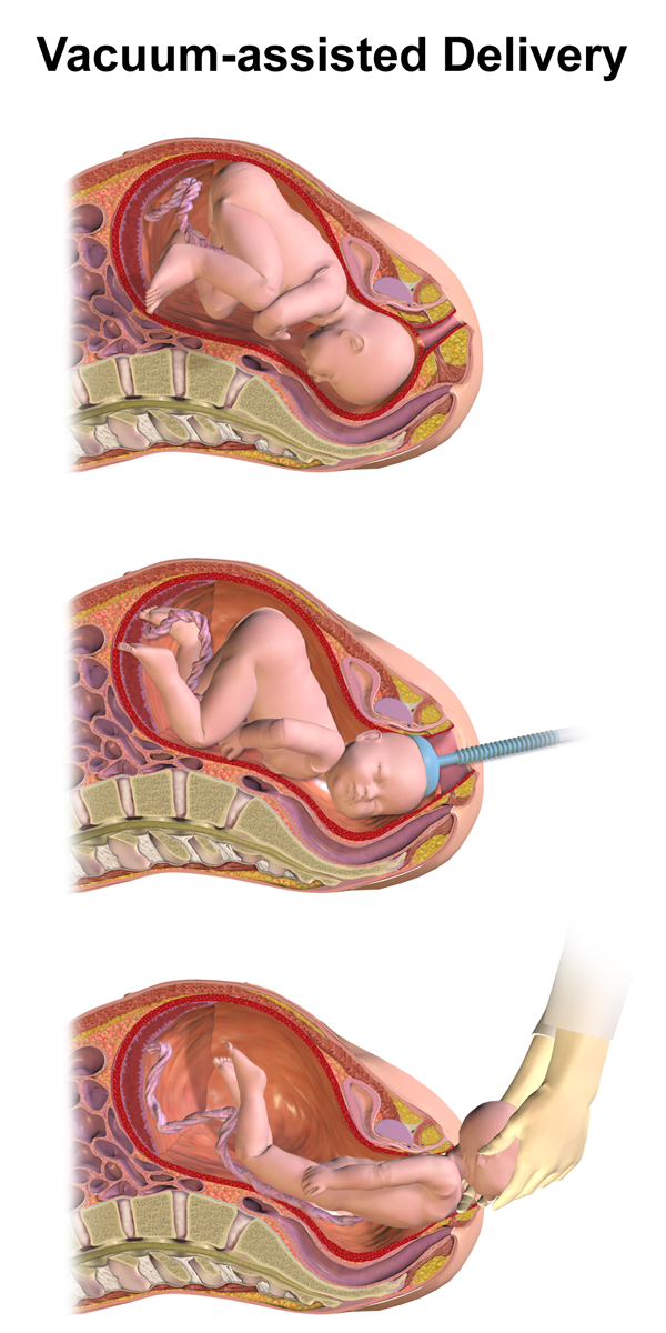
Vacuum extraction (VE), also known as ventouse, is a method to assist delivery of a baby using a vacuum device. It is used in the second stage of labor if it has not progressed adequately. It may be an alternative to a forceps delivery and caesarean section. It cannot be used when the baby is in the breech position or for premature births. The use of VE is generally safe, but it can occasionally have negative effects on either the mother or the child. The term ''ventouse'' comes from the French word for "suction cup". Medical uses There are several indications to use a vacuum extraction to aid delivery: * Maternal exhaustion * Prolonged second stage of labor * Foetal distress in the second stage of labor, generally indicated by changes in the foetal heart-rate (usually measured on a CTG) * Maternal illness where prolonged "bearing down" or pushing efforts would be risky (e.g. cardiac conditions, blood pressure, aneurysm, glaucoma). If these conditions are known about before ... [...More Info...] [...Related Items...] OR: [Wikipedia] [Google] [Baidu] |
Hyperbilirubinemia
Bilirubin (BR) (adopted from German, originally bili—bile—plus ruber—red—from Latin) is a red-orange compound that occurs in the normcomponent of the straw-yellow color in urine. Another breakdown product, stercobilin, causes the brown color of feces. Although bilirubin is usually found in animals rather than plants, at least one plant species, ''Strelitzia nicolai'', is known to contain the pigment. Structure Bilirubin consists of an open-chain tetrapyrrole. It is formed by oxidative cleavage of a porphyrin in heme, which affords biliverdin. Biliverdin is reduced to bilirubin. After conjugation with glucuronic acid, bilirubin is water-soluble and can be excreted. Bilirubin is structurally similar to the pigment phycobilin used by certain algae to capture light energy, and to the pigment phytochrome used by plants to sense light. All of these contain an open chain of four pyrrolic rings. Like these other pigments, some of the double-bonds in bilirubin isomerize whe ... [...More Info...] [...Related Items...] OR: [Wikipedia] [Google] [Baidu] |


