|
Stygiella
''Stygiella'' /ˌstɪ.d͡ʒiˈɛ.lə/ is a genus of free-living marine flagellates belonging to the family Stygiellidae in the jakobids (excavata). The genus currently includes four species, all of which are secondary obligate anaerobes. The species are all unicellular and crescent-shaped.Bernard, C, Simpson, A. G. B. & Patterson, D. J. (2000) Some free-living flagellates (protista) from anoxic habitats, Ophelia, 52:2, 113-142, DOI: 10.1080/00785236.1999.10409422. All members possess hydrogenosomes, a type of acristate mitochondrion-derived organelle (MRO) that produces hydrogen gas as a metabolic product.Leger, M. M., Eme, L., Hug, L. A., & Roger, A. J. (2016). ovel hydrogenosomes in the microaerophilic jakobid ''Stygiella incarcerata'' Molecular Biology and Evolution, 33(9), 2318-2336. Retrieved April 28, 2020. doi: doi.org/10.1093/2Fmolbev/2Fmsw103. Stygiella is a deep-branching lineage within excavates, and hydrogenosome genes sometimes show eubacterium-like mechanisms th ... [...More Info...] [...Related Items...] OR: [Wikipedia] [Google] [Baidu] |
Jakoba
''Jakoba'' is a genus in the taxon Excavata, and currently has a single described species, ''Jakoba libera'' described by Patterson in 1990, and named in honour of Dutch botanist (Algology, Myology and Lichenology) Jakoba Ruinen. (Previously described ''Jakoba incarcerata'' has been renamed ''Andalucia incarcerata'', and ''Jakoba bahamensis'' /''Jakoba bahamiensis'' is not formally described.) Appearance and characteristics ''Jakoba'' are small bacterivorous zooflagellates (jakobids) found in marine and hypersaline environments. They are free swimming trophic cells with two flagella and range between five and ten micrometers in length. Cells rotate along their longitudinal axis to allow for swimming in straight lines unless deformation and “squirming” occurs due to compression in debris. During feeding, bacteria are removed from the water column by a current created by the posterior flagellum. This current causes the bacteria to collect in the groove on the right ventra ... [...More Info...] [...Related Items...] OR: [Wikipedia] [Google] [Baidu] |
Stygioides
''Stygioides'' is a genus of moths in the family Cossidae. Species * ''Stygioides aethiops'' (Staudinger, 1887) * ''Stygioides colchica'' (Herrich-Schäffer, 1851) * ''Stygioides ivinskisi'' Saldaitis et Yakovlev in Saldaitis, Yakovlev et Ivinskis, 2007 * ''Stygioides nupponenorum'' Yakovlev et Saldaitis, 2011 * ''Stygioides persephone'' (Reisser, 1962) * ''Stygioides psyche'' (Grum-Grshimailo, 1893) Former species * ''Stygioides tricolor'' References Natural History Museum Lepidoptera generic names catalog Cossinae Cossidae genera {{Cossinae-stub ... [...More Info...] [...Related Items...] OR: [Wikipedia] [Google] [Baidu] |
Axoneme
An axoneme, also called an axial filament is the microtubule-based cytoskeletal structure that forms the core of a cilium or flagellum. Cilia and flagella are found on many cells, organisms, and microorganisms, to provide motility. The axoneme serves as the "skeleton" of these organelles, both giving support to the structure and, in some cases, the ability to bend. Though distinctions of function and length may be made between cilia and flagella, the internal structure of the axoneme is common to both. Structure Inside a cilium and a flagellum is a microtubule-based cytoskeleton called the axoneme. The axoneme of a primary cilium typically has a ring of nine outer microtubule doublets (called a 9+0 axoneme), and the axoneme of a motile cilium has two central microtubules in addition to the nine outer doublets (called a 9+2 axoneme). The axonemal cytoskeleton acts as a scaffolding for various protein complexes and provides binding sites for molecular motor proteins such as ... [...More Info...] [...Related Items...] OR: [Wikipedia] [Google] [Baidu] |
Oxidative Stress
Oxidative stress reflects an imbalance between the systemic manifestation of reactive oxygen species and a biological system's ability to readily Detoxification, detoxify the reactive intermediates or to repair the resulting damage. Disturbances in the normal redox state of cells can cause toxic effects through the production of peroxides and free radicals that damage all components of the cell, including proteins, lipids, and DNA. Oxidative stress from Cellular respiration, oxidative metabolism causes base damage, as well as DNA damage (naturally occurring), strand breaks in DNA. Base damage is mostly indirect and caused by the reactive oxygen species generated, e.g., O2− (superoxide radical), OH (hydroxyl radical) and H2O2 (hydrogen peroxide). Further, some reactive oxidative species act as cellular messengers in redox signaling. Thus, oxidative stress can cause disruptions in normal mechanisms of cellular signaling. In humans, oxidative stress is thought to be involved in the ... [...More Info...] [...Related Items...] OR: [Wikipedia] [Google] [Baidu] |
Trichomonas Vaginalis
''Trichomonas vaginalis'' is an anaerobic, flagellated protozoan parasite and the causative agent of a sexually transmitted disease called trichomoniasis. It is the most common pathogenic protozoan that infects humans in industrialized countries. Infection rates in men and women are similar but women are usually symptomatic, while infections in men are usually asymptomatic. Transmission usually occurs via direct, skin-to-skin contact with an infected individual, most often through vaginal intercourse. The WHO has estimated that 160 million cases of infection are acquired annually worldwide. The estimates for North America alone are between 5 and 8 million new infections each year, with an estimated rate of asymptomatic cases as high as 50%. Usually treatment consists of metronidazole and tinidazole. Clinical History Alfred Francois Donné (1801–1878) was the first to describe a procedure to diagnose trichomoniasis through "the microscopic observation of motile protozoa i ... [...More Info...] [...Related Items...] OR: [Wikipedia] [Google] [Baidu] |
Hydrogenase
A hydrogenase is an enzyme that catalyses the reversible oxidation of molecular hydrogen (H2), as shown below: Hydrogen uptake () is coupled to the reduction of electron acceptors such as oxygen, nitrate, sulfate, carbon dioxide (), and fumarate. On the other hand, proton reduction () is coupled to the oxidation of electron donors such as ferredoxin (FNR), and serves to dispose excess electrons in cells (essential in pyruvate fermentation). Both low-molecular weight compounds and proteins such as FNRs, cytochrome ''c''3, and cytochrome ''c''6 can act as physiological electron donors or acceptors for hydrogenases. Structural classification It has been estimated that 99% of all organisms utilize hydrogen, H2. Most of these species are microbes and their ability to use H2 as a metabolite arises from the expression of metalloenzymes known as hydrogenases. Hydrogenases are sub-classified into three different types based on the active site metal content: iron-iron hydrogenase, ni ... [...More Info...] [...Related Items...] OR: [Wikipedia] [Google] [Baidu] |
Ferredoxin Oxidoreductase
Ferredoxins (from Latin ''ferrum'': iron + redox, often abbreviated "fd") are iron–sulfur proteins that mediate electron transfer in a range of metabolic reactions. The term "ferredoxin" was coined by D.C. Wharton of the DuPont Co. and applied to the "iron protein" first purified in 1962 by Mortenson, Valentine, and Carnahan from the anaerobic bacterium '' Clostridium pasteurianum''. Another redox protein, isolated from spinach chloroplasts, was termed "chloroplast ferredoxin". The chloroplast ferredoxin is involved in both cyclic and non-cyclic photophosphorylation reactions of photosynthesis. In non-cyclic photophosphorylation, ferredoxin is the last electron acceptor thus reducing the enzyme NADP+ reductase. It accepts electrons produced from sunlight- excited chlorophyll and transfers them to the enzyme ferredoxin: NADP+ oxidoreductase . Ferredoxins are small proteins containing iron and sulfur atoms organized as iron–sulfur clusters. These biological " capacitors" can acc ... [...More Info...] [...Related Items...] OR: [Wikipedia] [Google] [Baidu] |
Electron Transport Chain
An electron transport chain (ETC) is a series of protein complexes and other molecules that transfer electrons from electron donors to electron acceptors via redox reactions (both reduction and oxidation occurring simultaneously) and couples this electron transfer with the transfer of protons (H+ ions) across a membrane. The electrons that transferred from NADH and FADH2 to the ETC involves 4 multi-subunit large enzymes complexes and 2 mobile electron carriers. Many of the enzymes in the electron transport chain are membrane-bound. The flow of electrons through the electron transport chain is an exergonic process. The energy from the redox reactions creates an electrochemical proton gradient that drives the synthesis of adenosine triphosphate (ATP). In aerobic respiration, the flow of electrons terminates with molecular oxygen as the final electron acceptor. In anaerobic respiration, other electron acceptors are used, such as sulfate. In an electron transport chain, the redo ... [...More Info...] [...Related Items...] OR: [Wikipedia] [Google] [Baidu] |
Cytosol
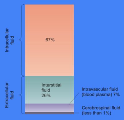
The cytosol, also known as cytoplasmic matrix or groundplasm, is one of the liquids found inside cells (intracellular fluid (ICF)). It is separated into compartments by membranes. For example, the mitochondrial matrix separates the mitochondrion into many compartments. In the eukaryotic cell, the cytosol is surrounded by the cell membrane and is part of the cytoplasm, which also comprises the mitochondria, plastids, and other organelles (but not their internal fluids and structures); the cell nucleus is separate. The cytosol is thus a liquid matrix around the organelles. In prokaryotes, most of the chemical reactions of metabolism take place in the cytosol, while a few take place in membranes or in the periplasmic space. In eukaryotes, while many metabolic pathways still occur in the cytosol, others take place within organelles. The cytosol is a complex mixture of substances dissolved in water. Although water forms the large majority of the cytosol, its structure and prope ... [...More Info...] [...Related Items...] OR: [Wikipedia] [Google] [Baidu] |
Ribosome
Ribosomes ( ) are macromolecular machines, found within all cells, that perform biological protein synthesis (mRNA translation). Ribosomes link amino acids together in the order specified by the codons of messenger RNA (mRNA) molecules to form polypeptide chains. Ribosomes consist of two major components: the small and large ribosomal subunits. Each subunit consists of one or more ribosomal RNA (rRNA) molecules and many ribosomal proteins (RPs or r-proteins). The ribosomes and associated molecules are also known as the ''translational apparatus''. Overview The sequence of DNA that encodes the sequence of the amino acids in a protein is transcribed into a messenger RNA chain. Ribosomes bind to messenger RNAs and use their sequences for determining the correct sequence of amino acids to generate a given protein. Amino acids are selected and carried to the ribosome by transfer RNA (tRNA) molecules, which enter the ribosome and bind to the messenger RNA chain via an anti-c ... [...More Info...] [...Related Items...] OR: [Wikipedia] [Google] [Baidu] |
Cisterna
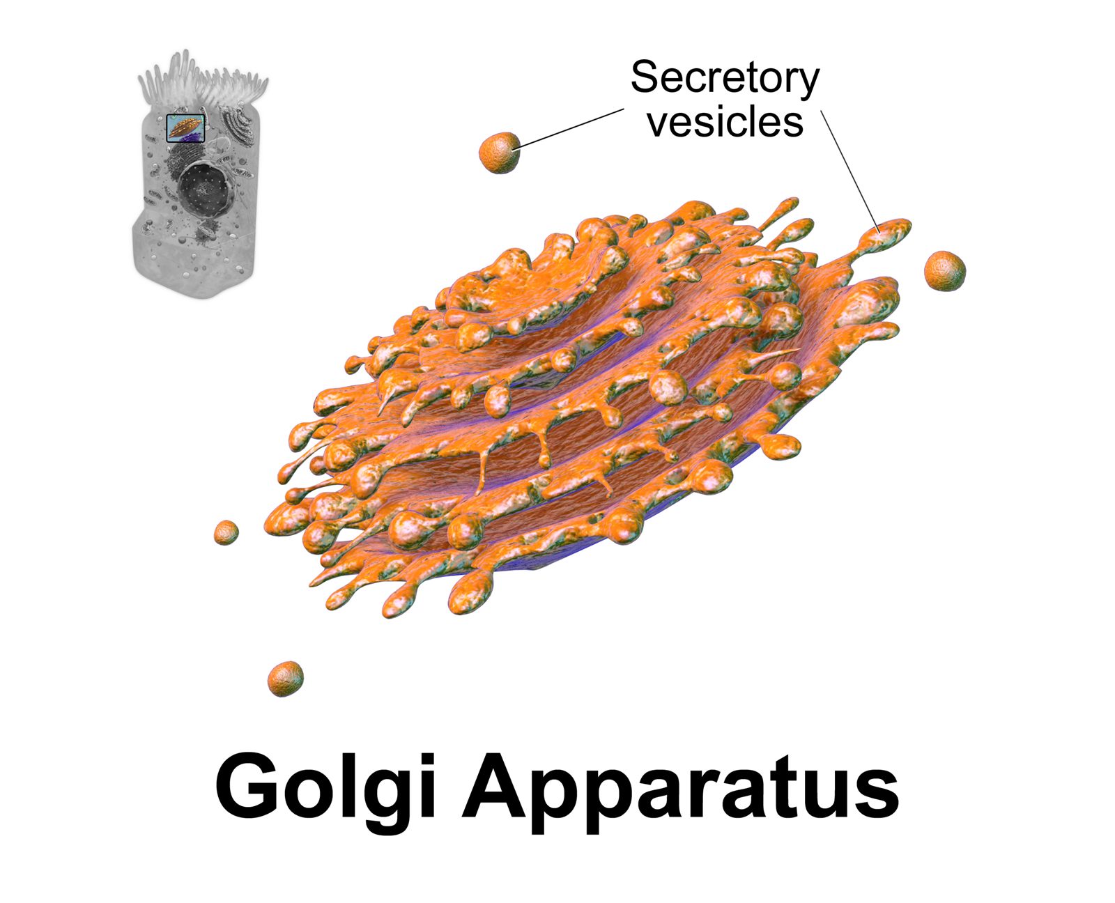
A cisterna (plural cisternae) is a flattened membrane vesicle found in the endoplasmic reticulum and Golgi apparatus. Cisternae are an integral part of the packaging and modification processes of proteins occurring in the Golgi. Function Proteins begin on the cis side of the Golgi (the side facing the ER) and exit on the trans side (the side facing the plasma membrane). Throughout their journey in the cisterna, the proteins are packaged and are modified for transport throughout the cell. The number of cisterna in the Golgi stack is dependent on the organism and cell type. The structure, composition, and function of each of the cisternae may be different inside the Golgi stack. These different variations of Golgi cisternae are categorized into 3 groups; cis Golgi network, medial, and trans Golgi network. The cis Golgi network is the first step in the cisternal structure of a protein being packaged, while the trans Golgi network is the last step in the cisternal structure when the ... [...More Info...] [...Related Items...] OR: [Wikipedia] [Google] [Baidu] |
Dictyosome
The Golgi apparatus (), also known as the Golgi complex, Golgi body, or simply the Golgi, is an organelle found in most eukaryotic cells. Part of the endomembrane system in the cytoplasm, it packages proteins into membrane-bound vesicles inside the cell before the vesicles are sent to their destination. It resides at the intersection of the secretory, lysosomal, and endocytic pathways. It is of particular importance in processing proteins for secretion, containing a set of glycosylation enzymes that attach various sugar monomers to proteins as the proteins move through the apparatus. It was identified in 1897 by the Italian scientist Camillo Golgi and was named after him in 1898. Discovery Owing to its large size and distinctive structure, the Golgi apparatus was one of the first organelles to be discovered and observed in detail. It was discovered in 1898 by Italian physician Camillo Golgi during an investigation of the nervous system. After first observing it under his micr ... [...More Info...] [...Related Items...] OR: [Wikipedia] [Google] [Baidu] |

.png)


