|
Stannius Ligature
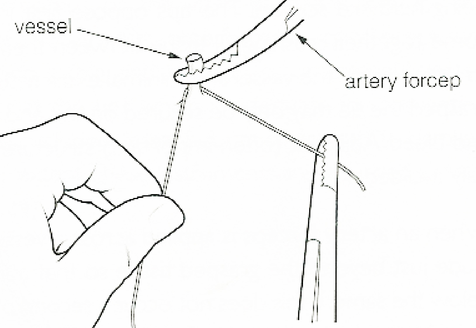
The Stannius Ligature was an experimental procedure that robustly illustrated impulse conduction in the frog heart. This procedure decisively demonstrated that the Sinoatrial Node is the intrinsic origin pacemaker of the heart. A ligature placed either around the junction between the sinus venosus and atrium of the frog or turtle heart (first stannius ligature) or around the atrioventricular junction (second stannius ligature); demonstrates that the cardiac impulse is conducted from sinus venosus to atria to ventricle, but that successive chambers possess automaticity since each may continue to beat, but the atria now have a slower rate than the sinus venosus, and the ventricle either does not contract or beats at a slower rate than the atria. History In 1852 H.F. Stannius experimented on the heart. By tying a ligature as a constriction between the sinus venosus and the atrium in the frog and also one around the atrioventricular groove The coronary sulcus (also called coronary g ... [...More Info...] [...Related Items...] OR: [Wikipedia] [Google] [Baidu] |
Frog
A frog is any member of a diverse and largely Carnivore, carnivorous group of short-bodied, tailless amphibians composing the order (biology), order Anura (ανοὐρά, literally ''without tail'' in Ancient Greek). The oldest fossil "proto-frog" ''Triadobatrachus'' is known from the Early Triassic of Madagascar, but molecular clock, molecular clock dating suggests their split from other amphibians may extend further back to the Permian, 265 Myr, million years ago. Frogs are widely distributed, ranging from the tropics to subarctic regions, but the greatest concentration of species diversity is in tropical rainforest. Frogs account for around 88% of extant amphibian species. They are also one of the five most diverse vertebrate orders. Warty frog species tend to be called toads, but the distinction between frogs and toads is informal, not from Taxonomy (biology), taxonomy or evolutionary history. An adult frog has a stout body, protruding eyes, anteriorly-attached tongue, limb ... [...More Info...] [...Related Items...] OR: [Wikipedia] [Google] [Baidu] |
Sinus Venosus
The sinus venosus is a large quadrangular cavity which precedes the atrium on the venous side of the chordate heart. In mammals, it exists distinctly only in the embryonic heart, where it is found between the two venae cavae. However, the sinus venosus persists in the adult. In the adult, it is incorporated into the wall of the right atrium to form a smooth part called the sinus venarum, which is separated from the rest of the atrium by a ridge of fibres called the crista terminalis. The sinus venosus also forms the sinoatrial node and the coronary sinus; in (most) mammals only. In the embryo, the thin walls of the sinus venosus are connected below with the right ventricle, and medially with the left atrium, but are free in the rest of their extent. It receives blood from the vitelline vein, umbilical vein and common cardinal vein. The sinus venosus originally starts as a paired structure but shifts towards associating only with the right atrium as the embryonic heart develops. T ... [...More Info...] [...Related Items...] OR: [Wikipedia] [Google] [Baidu] |
Turtle
Turtles are an order of reptiles known as Testudines, characterized by a special shell developed mainly from their ribs. Modern turtles are divided into two major groups, the Pleurodira (side necked turtles) and Cryptodira (hidden necked turtles), which differ in the way the head retracts. There are 360 living and recently extinct species of turtles, including land-dwelling tortoises and freshwater terrapins. They are found on most continents, some islands and, in the case of sea turtles, much of the ocean. Like other amniotes (reptiles, birds, and mammals) they breathe air and do not lay eggs underwater, although many species live in or around water. Turtle shells are made mostly of bone; the upper part is the domed carapace, while the underside is the flatter plastron or belly-plate. Its outer surface is covered in scales made of keratin, the material of hair, horns, and claws. The carapace bones develop from ribs that grow sideways and develop into broad flat plates th ... [...More Info...] [...Related Items...] OR: [Wikipedia] [Google] [Baidu] |
Atrium (heart)
The atrium ( la, ātrium, , entry hall) is one of two upper chambers in the heart that receives blood from the circulatory system. The blood in the atria is pumped into the heart ventricles through the atrioventricular valves. There are two atria in the human heart – the left atrium receives blood from the pulmonary circulation, and the right atrium receives blood from the venae cavae of the systemic circulation. During the cardiac cycle the atria receive blood while relaxed in diastole, then contract in systole to move blood to the ventricles. Each atrium is roughly cube-shaped except for an ear-shaped projection called an atrial appendage, sometimes known as an auricle. All animals with a closed circulatory system have at least one atrium. The atrium was formerly called the 'auricle'. That term is still used to describe this chamber in some other animals, such as the ''Mollusca''. They have thicker muscular walls than the atria do. Structure Humans have a four-chambered ... [...More Info...] [...Related Items...] OR: [Wikipedia] [Google] [Baidu] |
Ventricle (heart)
A ventricle is one of two large chambers toward the bottom of the heart that collect and expel blood towards the peripheral beds within the body and lungs. The blood pumped by a ventricle is supplied by an atrium, an adjacent chamber in the upper heart that is smaller than a ventricle. Interventricular means between the ventricles (for example the interventricular septum), while intraventricular means within one ventricle (for example an intraventricular block). In a four-chambered heart, such as that in humans, there are two ventricles that operate in a double circulatory system: the right ventricle pumps blood into the pulmonary circulation to the lungs, and the left ventricle pumps blood into the systemic circulation through the aorta. Structure Ventricles have thicker walls than atria and generate higher blood pressures. The physiological load on the ventricles requiring pumping of blood throughout the body and lungs is much greater than the pressure generated by the atria ... [...More Info...] [...Related Items...] OR: [Wikipedia] [Google] [Baidu] |
Hermann Friedrich Stannius
Hermann Friedrich Stannius (15 March 1808, Hamburg – 15 January 1883, Sachsenberg near Schwerin) was a German anatomist, physiologist and entomologist. He specialised in the insect order Diptera especially the family Dolichopodidae. Works Entomology * ''De speciebus nonnullis Mycethophila vel novis vel minus cognitis''.Bratislava, 1831. * Die europischen Arten der Zweyfluglergattung Dolichopus. ''Isis Oken'' 1831: 28–68, 122–144, 248–271, 1831. *''Beiträge zur Entomologie, besondere in Bezug auf Schlesien, gemeinschaftlich mit Schummel''. Breslau, 1832. *Über den Einfluss der Nerven auf den Blutumlauf. roriep's''Notizen aus dem Gebiete der Natur- und Heilkunde'', 1833, 36: 246–248. *Ueber einige Missbildungen an Insekten. üller's'' Archiv für Anatomie, Physiologie und wissenschaftliche Medizin'', Berlin, 1835: 295–310. Medical and Physiology *''Allgemeine Pathologie''. Berlin, I, 1837. *Ueber die Einwirkung des Strychnins auf das Nervensystem. ''Archiv ... [...More Info...] [...Related Items...] OR: [Wikipedia] [Google] [Baidu] |
Ligature (medicine)
In surgery or medical procedure, a ligature consists of a piece of thread ( suture) tied around an anatomical structure, usually a blood vessel or another hollow structure (e.g. urethra) to shut it off. History The principle of ligation is attributed to Hippocrates and Galen. In ancient Rome, ligatures were used to treat hemorrhoids. The concept of a ligature was reintroduced some 1,500 years later by Ambroise Paré, and finally it found its modern use in 1870–80, made popular by Jules-Émile Péan. Procedure With a blood vessel the surgeon will clamp the vessel perpendicular to the axis of the artery or vein with a hemostat, then secure it by ligating it; i.e. using a piece of suture around it before dividing the structure and releasing the hemostat. It is different from a tourniquet in that the tourniquet will not be secured by knots and it can therefore be released/tightened at will. Ligature is one of the remedies to treat skin tag, or acrochorda. It is done by tying str ... [...More Info...] [...Related Items...] OR: [Wikipedia] [Google] [Baidu] |
Coronary Sulcus
The coronary sulcus (also called coronary groove, auriculoventricular groove, atrioventricular groove, AV groove) is a groove on the surface of the heart at the base of right auricle that separates the atria from the ventricles. The structure contains the trunks of the nutrient vessels of the heart, and is deficient in front, where it is crossed by the root of the pulmonary trunk. On the posterior surface of the heart, the coronary sulcus contains the coronary sinus. Structure In relation to the rib cage, the coronary sulcus spans from the medial side of the 3rd left costal cartilage, to the middle of the right 6th costal cartilage. Epicardial fat tends to be concentrated along the coronary sulcus. There are two coronary sulci in the heart, including left and right coronary sulci. Left coronary sulcus The left coronary sulcus originates posterior to the pulmonary trunk, and travels inferiorly separating the left atrium and left ventricle. The location of the left coronary ... [...More Info...] [...Related Items...] OR: [Wikipedia] [Google] [Baidu] |
_Ranomafana.jpg)


