|
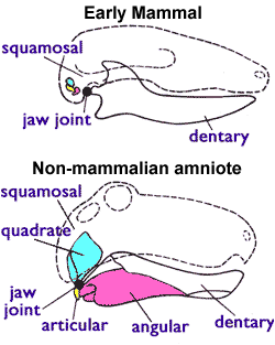
Squamosal Bone
The squamosal is a skull bone found in most reptiles, amphibians, and birds. In fishes, it is also called the pterotic bone. In most tetrapods, the squamosal and quadratojugal bones form the cheek series of the skull. The bone forms an ancestral component of the dermal roof and is typically thin compared to other skull bones. The squamosal bone lies ventral to the temporal series and otic notch, and is bordered anteriorly by the postorbital. Posteriorly, the squamosal articulates with the quadrate and pterygoid bones. The squamosal is bordered anteroventrally by the jugal and ventrally by the quadratojugal. Function in reptiles In reptiles, the quadrate and articular bones of the skull articulate to form the jaw joint. The squamosal bone lies anterior to the quadrate bone. Anatomy in synapsids Non-mammalian synapsids In non-mammalian synapsids, the jaw is composed of four bony elements and referred to as a quadro-articular jaw because the joint is between the art ... [...More Info...] [...Related Items...] OR: [Wikipedia] [Google] [Baidu] |
Skull Synapsida 1
The skull, or cranium, is typically a bony enclosure around the brain of a vertebrate. In some fish, and amphibians, the skull is of cartilage. The skull is at the head end of the vertebrate. In the human, the skull comprises two prominent parts: the neurocranium and the facial skeleton, which evolved from the first pharyngeal arch. The skull forms the frontmost portion of the axial skeleton and is a product of cephalization and vesicular enlargement of the brain, with several special senses structures such as the eyes, ears, nose, tongue and, in fish, specialized tactile organs such as barbels near the mouth. The skull is composed of three types of bone: cranial bones, facial bones and ossicles, which is made up of a number of fused flat and irregular bones. The cranial bones are joined at firm fibrous junctions called sutures and contains many foramina, fossae, processes, and sinuses. In zoology, the openings in the skull are called fenestrae, the most prominent ... [...More Info...] [...Related Items...] OR: [Wikipedia] [Google] [Baidu] |
Synapsid
Synapsida is a diverse group of tetrapod vertebrates that includes all mammals and their extinct relatives. It is one of the two major clades of the group Amniota, the other being the more diverse group Sauropsida (which includes all extant reptiles and therefore, birds). Unlike other amniotes, synapsids have a single temporal fenestra, an opening low in the skull roof behind each eye socket, leaving a zygomatic arch, bony arch beneath each; this accounts for the name "synapsid". The distinctive temporal fenestra developed about 318 million years ago during the Late Carboniferous period, when synapsids and sauropsids diverged, but was subsequently merged with the orbit in early mammals. The basal (phylogenetics), basal amniotes (reptiliomorphs) from which synapsids evolved were historically simply called "reptiles". Therefore, stem group synapsids were then described as mammal-like reptiles in classical systematics, and non-therapsid synapsids were also referred to as pelyco ... [...More Info...] [...Related Items...] OR: [Wikipedia] [Google] [Baidu] |
Malleus
The ''malleus'', or hammer, is a hammer-shaped small bone or ossicle of the middle ear. It connects with the incus, and is attached to the inner surface of the eardrum. The word is Latin for 'hammer' or 'mallet'. It transmits the sound vibrations from the eardrum to the ''incus'' (anvil). Structure The malleus is a bone situated in the middle ear. It is the first of the three ossicles, and attached to the eardrum (tympanic membrane). The head of the malleus is the large protruding section, which attaches to the incus. The head connects to the neck of malleus. The bone continues as the handle (or manubrium) of malleus, which connects to the tympanic membrane. Between the neck and handle of the malleus, lateral and anterior processes emerge from the bone. The bone is oriented so that the head is superior and the handle is inferior. Development Embryologically, the malleus is derived from the first pharyngeal arch along with the ''incus''. In humans it grows from Meckel's ... [...More Info...] [...Related Items...] OR: [Wikipedia] [Google] [Baidu] |
Ossicles
The ossicles (also called auditory ossicles) are three irregular bones in the middle ear of humans and other mammals, and are among the smallest bones in the human body. Although the term "ossicle" literally means "tiny bone" (from Latin ''ossiculum'') and may refer to any small bone throughout the body, it typically refers specifically to the malleus, incus and stapes ("hammer, anvil, and stirrup") of the middle ear. The auditory ossicles serve as a kinematic chain to transmit and amplify ( intensify) sound vibrations collected from the air by the ear drum to the fluid-filled labyrinth ( cochlea). The absence or pathology of the auditory ossicles would constitute a moderate-to-severe conductive hearing loss. Structure The ossicles are, in order from the eardrum to the inner ear (from superficial to deep): the malleus, incus, and stapes, terms that in Latin are translated as "the hammer, anvil, and stirrup". * The malleus () articulates with the incus through the ... [...More Info...] [...Related Items...] OR: [Wikipedia] [Google] [Baidu] |
Incus
The ''incus'' (: incudes) or anvil in the ear is one of three small bones (ossicles) in the middle ear. The incus receives vibrations from the malleus, to which it is connected laterally, and transmits these to the stapes medially. The incus is named for its resemblance to an anvil (). Structure The incus is the second of three ossicles, very small bones in the middle ear which act to transmit sound. It is shaped like an anvil, and has a long and short crus extending from the body, which articulates with the malleus. The short crus attaches to the posterior ligament of the incus. The long crus articulates with the stapes at the lenticular process. The superior ligament of the incus attaches at the body of the incus to the roof of the tympanic cavity. The incus is Homology (biology), homologous to the quadrate bone found in other tetrapods. Function Vibrations in the middle ear are received via the tympanic membrane. The malleus, resting on the membrane, conveys vibrations ... [...More Info...] [...Related Items...] OR: [Wikipedia] [Google] [Baidu] |
Auditory Bulla
The tympanic part of the temporal bone is a curved plate of bone lying below the squamous part of the temporal bone, in front of the mastoid process, and surrounding the external part of the ear canal. It originates as a separate bone (tympanic bone), which in some mammals stays separate through life. Evolutionarily, a portion of it is derived from the angular bone of the reptilian lower jaw. Surfaces Its postero-superior surface is concave, and forms the anterior wall, the floor, and part of the posterior wall of the bony ear canal. Medially, it presents a narrow furrow, the ''tympanic sulcus'', for the attachment of the tympanic membrane. Its antero-inferior surface is quadrilateral and slightly concave; it constitutes the posterior boundary of the mandibular fossa, and is in contact with the retromandibular part of the parotid gland. Borders Its lateral border is free and rough, and gives attachment to the cartilaginous part of the ear canal. Internally, the tympanic pa ... [...More Info...] [...Related Items...] OR: [Wikipedia] [Google] [Baidu] |
Periotic Bone
The periotic bone is the single bone that surrounds the inner ear of birds and mammals. It is formed from the fusion of the prootic, epiotic, and opisthotic bones, and in Cetacea forms a complex with the tympanic bone The tympanic part of the temporal bone is a curved plate of bone lying below the squamous part of the temporal bone, in front of the mastoid process, and surrounding the external part of the ear canal. It originates as a separate bone (tympanic .... References Skeletal system Mammal anatomy {{Vertebrate anatomy-stub ... [...More Info...] [...Related Items...] OR: [Wikipedia] [Google] [Baidu] |
Temporal Bone
The temporal bone is a paired bone situated at the sides and base of the skull, lateral to the temporal lobe of the cerebral cortex. The temporal bones are overlaid by the sides of the head known as the temples where four of the cranial bones fuse. Each temple is covered by a temporal muscle. The temporal bones house the structures of the ears. The lower seven cranial nerves and the major vessels to and from the brain traverse the temporal bone. Structure The temporal bone consists of four parts—the squamous, mastoid, petrous and tympanic parts. The squamous part is the largest and most superiorly positioned relative to the rest of the bone. The zygomatic process is a long, arched process projecting from the lower region of the squamous part and it articulates with the zygomatic bone. Posteroinferior to the squamous is the mastoid part. Fused with the squamous and mastoid parts and between the sphenoid and occipital bones lies the petrous part, which is shaped li ... [...More Info...] [...Related Items...] OR: [Wikipedia] [Google] [Baidu] |
Squama Temporalis
The squamous part of temporal bone, or temporal squama, forms the front and upper part of the temporal bone, and is scale-like, thin, and translucent. Surfaces Its outer surface is smooth and convex; it affords attachment to the temporal muscle, and forms part of the temporal fossa; on its hinder part is a vertical groove for the middle temporal artery. A curved line, the ''temporal line'', or ''supramastoid crest'', runs backward and upward across its posterior part; it serves for the attachment of the temporal fascia, and limits the origin of the temporalis muscle. The boundary between the squamous part and the mastoid portion of the bone, as indicated by traces of the original suture, lies about 1 cm. below this line. Projecting from the lower part of the squamous part is a long, arched process, the '' zygomatic process''. This process is at first directed lateralward, its two surfaces looking upward and downward; it then appears as if twisted inward upon itself, a ... [...More Info...] [...Related Items...] OR: [Wikipedia] [Google] [Baidu] |
Dentary
In jawed vertebrates, the mandible (from the Latin ''mandibula'', 'for chewing'), lower jaw, or jawbone is a bone that makes up the lowerand typically more mobilecomponent of the mouth (the upper jaw being known as the maxilla). The jawbone is the skull's only movable, posable bone, sharing joints with the cranium's temporal bones. The mandible hosts the lower teeth (their depth delineated by the alveolar process). Many muscles attach to the bone, which also hosts nerves (some connecting to the teeth) and blood vessels. Amongst other functions, the jawbone is essential for chewing food. Owing to the Neolithic advent of agriculture (), human jaws evolved to be smaller. Although it is the strongest bone of the facial skeleton, the mandible tends to deform in old age; it is also subject to fracturing. Surgery allows for the removal of jawbone fragments (or its entirety) as well as regenerative methods. Additionally, the bone is of great forensic significance. Structure ... [...More Info...] [...Related Items...] OR: [Wikipedia] [Google] [Baidu] |
Articular
The articular bone is part of the lower jaw of most vertebrates, including most jawed fish, amphibians, birds and various kinds of reptiles, as well as ancestral mammals. Anatomy In most vertebrates, the articular bone is connected to two other lower jaw bones, the suprangular and the angular. Developmentally, it originates from the embryonic mandibular cartilage. The most caudal portion of the mandibular cartilage ossifies to form the articular bone, while the remainder of the mandibular cartilage either remains cartilaginous or disappears. In snakes In snakes, the articular, surangular, and prearticular bones have fused to form the compound bone. The mandible is suspended from the quadrate bone and articulates at this compound bone. Function In amphibians and reptiles In most tetrapods, the articular bone forms the lower portion of the jaw joint. The upper jaw articulates at the quadrate bone. In mammals In mammals, the articular bone evolves to form the ... [...More Info...] [...Related Items...] OR: [Wikipedia] [Google] [Baidu] |


