|
Sphenoidal Conchae
The sphenoidal conchae (sphenoidal turbinated processes) are two thin, curved plates, situated at the anterior and lower part of the body of the sphenoid. An aperture of variable size exists in the anterior wall of each, and through this the sphenoidal sinus opens into the nasal cavity. ''General Anatomy and Osteology of Head and Neck'' (I. K. International Pvt Ltd, 2009; by Mahdi Hasan)- Retrieved 2018-08-29 Each is irregular in form, and tapers to a point behind, being broader and thinner in front. Its upper surface is concave, and looks toward the cavity of the sinus; its under surface is convex, and forms part of the roof of the corresponding nasal cavity. Each bone ... [...More Info...] [...Related Items...] OR: [Wikipedia] [Google] [Baidu] |
|
 |
Sphenoid Bone
The sphenoid bone is an unpaired bone of the neurocranium. It is situated in the middle of the skull towards the front, in front of the basilar part of the occipital bone. The sphenoid bone is one of the seven bones that articulate to form the orbit. Its shape somewhat resembles that of a butterfly or bat with its wings extended. Structure It is divided into the following parts: * a median portion, known as the body of sphenoid bone, containing the sella turcica, which houses the pituitary gland as well as the paired paranasal sinuses, the sphenoidal sinuses * two greater wings on the lateral side of the body and two lesser wings from the anterior side. * Pterygoid processes of the sphenoides, directed downwards from the junction of the body and the greater wings. Two sphenoidal conchae are situated at the anterior and inferior part of the body. Intrinsic ligaments of the sphenoid The more important of these are: * the pterygospinous, stretching between the spin ... [...More Info...] [...Related Items...] OR: [Wikipedia] [Google] [Baidu] |
|
Body Of The Sphenoid
The body of the sphenoid bone, more or less cubical in shape, is hollowed out in its interior to form two large cavities, the sphenoidal sinuses, which are separated from each other by a septum. Superior surface The superior surface of the body ig. 1presents in front a prominent spine, the ethmoidal spine, for articulation with the cribriform plate of the ethmoid bone; behind this is a smooth surface slightly raised in the middle line, and grooved on either side for the olfactory lobes of the brain. This surface is bounded behind by a ridge, which forms the anterior border of a narrow, transverse groove, the prechiasmatic groove, above and behind which lies the optic chiasma; the groove ends on either side in the optic foramen, which transmits the optic nerve and ophthalmic artery into the orbital cavity. Behind the chiasmatic groove is an elevation, the tuberculum sellae; and behind this is a deep depression, the saddle-shaped sella turcica (Turkish seat), the deepest part ... [...More Info...] [...Related Items...] OR: [Wikipedia] [Google] [Baidu] |
|
 |
Nasal Cavity
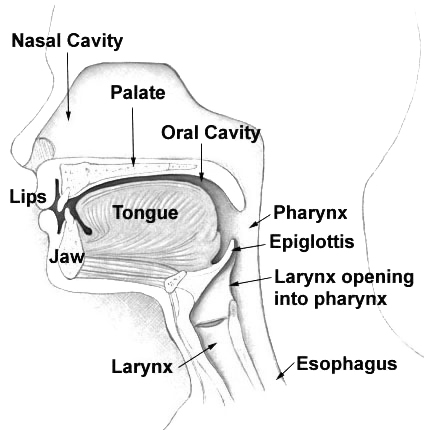
The nasal cavity is a large, air-filled space above and behind the nose in the middle of the face. The nasal septum divides the cavity into two cavities, also known as fossae. Each cavity is the continuation of one of the two nostrils. The nasal cavity is the uppermost part of the respiratory system and provides the nasal passage for inhaled air from the nostrils to the nasopharynx and rest of the respiratory tract. The paranasal sinuses surround and drain into the nasal cavity. Structure The term "nasal cavity" can refer to each of the two cavities of the nose, or to the two sides combined. The lateral wall of each nasal cavity mainly consists of the maxilla. However, there is a deficiency that is compensated for by the perpendicular plate of the palatine bone, the medial pterygoid plate, the labyrinth of ethmoid and the inferior concha. The paranasal sinuses are connected to the nasal cavity through small orifices called ostia. Most of these ostia communicate with ... [...More Info...] [...Related Items...] OR: [Wikipedia] [Google] [Baidu] |
|
Ethmoid
The ethmoid bone (; from grc, ἡθμός, hēthmós, sieve) is an unpaired bone in the skull that separates the nasal cavity from the brain. It is located at the roof of the nose, between the two orbits. The cubical bone is lightweight due to a spongy construction. The ethmoid bone is one of the bones that make up the orbit of the eye. Structure The ethmoid bone is an anterior cranial bone located between the eyes. It contributes to the medial wall of the orbit, the nasal cavity, and the nasal septum. The ethmoid has three parts: cribriform plate, ethmoidal labyrinth, and perpendicular plate. The cribriform plate forms the roof of the nasal cavity and also contributes to formation of the anterior cranial fossa, the ethmoidal labyrinth consists of a large mass on either side of the perpendicular plate, and the perpendicular plate forms the superior two-thirds of the nasal septum. Between the orbital plate and the nasal conchae are the ethmoidal sinuses or ethmoidal air cells ... [...More Info...] [...Related Items...] OR: [Wikipedia] [Google] [Baidu] |
|
|
Hard Palate
The hard palate is a thin horizontal bony plate made up of two bones of the facial skeleton, located in the roof of the mouth. The bones are the palatine process of the maxilla and the horizontal plate of palatine bone. The hard palate spans the alveolar arch formed by the alveolar process that holds the upper teeth (when these are developed). Structure The hard palate is formed by the palatine process of the maxilla and horizontal plate of palatine bone. It forms a partition between the nasal passages and the mouth. On the anterior portion of the hard palate are the plicae, irregular ridges in the mucous membrane that help facilitate the movement of food backward towards the larynx. This partition is continued deeper into the mouth by a fleshy extension called the soft palate. On the ventral surface of hard palate, some projections or transverse ridges are present which are called as palatine rugae. Function The hard palate is important for feeding and speech. Mammals with a ... [...More Info...] [...Related Items...] OR: [Wikipedia] [Google] [Baidu] |
|
|
Vomer
The vomer (; lat, vomer, lit=ploughshare) is one of the unpaired facial bones of the skull. It is located in the midsagittal line, and articulates with the sphenoid, the ethmoid, the left and right palatine bones, and the left and right maxillary bones. The vomer forms the inferior part of the nasal septum in humans, with the superior part formed by the perpendicular plate of the ethmoid bone. The name is derived from the Latin word for a ploughshare and the shape of the bone. In humans The vomer is situated in the median plane, but its anterior portion is frequently bent to one side. It is thin, somewhat quadrilateral in shape, and forms the hinder and lower part of the nasal septum; it has two surfaces and four borders. The surfaces are marked by small furrows for blood vessels, and on each is the nasopalatine groove, which runs obliquely downward and forward, and lodges the nasopalatine nerve and vessels. Borders The ''superior border'', the thickest, presents a d ... [...More Info...] [...Related Items...] OR: [Wikipedia] [Google] [Baidu] |
|
|
Rostrum Of The Sphenoid
Rostrum may refer to: * Any kind of a platform for a speaker: **dais **pulpit * Rostrum (anatomy), a beak, or anatomical structure resembling a beak, as in the mouthparts of many sucking insects * Rostrum (ship), a form of bow on naval ships * Rostrum Records, an American record label * ''The Rostrum'', the official monthly magazine of the National Forensic League *Australian Rostrum, public speaking clubs See also * Rastrum, a musical writing implement used to draw staff lines * Rostra The rostra ( it, Rostri, links=no) was a large platform built in the city of Rome that stood during the Roman Republic, republican and Roman Empire, imperial periods. Speakers would stand on the rostra and face the north side of the comitium tow ..., a large platform built in the ancient city of Rome * Rostral (other) * {{Disambiguation ... [...More Info...] [...Related Items...] OR: [Wikipedia] [Google] [Baidu] |
|
|
Lamina Papyracea
The orbital lamina of ethmoid bone, (or lamina papyracea or orbital lamina) is a smooth, oblong bone plate which forms the lateral surface of the labyrinth of the ethmoid bone in the skull. The plate covers in the middle and posterior ethmoidal cells and forms a large part of the medial wall of the orbit. It articulates above with the orbital plate of the frontal bone, below with the maxilla and the orbital process of palatine bone, in front with the lacrimal, and behind with the sphenoid. Its name lamina papyracea is an appropriate description, as this part of the ethmoid bone is paper-thin and fractures easily. A fracture here could cause entrapment of the medial rectus muscle. Additional Images File:Slide4fen.JPG, Orbital lamina of ethmoid bone References External links * - "Orbits and Eye: Bones" * Bones of the head and neck {{Portal bar, Anatomy ... [...More Info...] [...Related Items...] OR: [Wikipedia] [Google] [Baidu] |
|
|
Orbital Plate Of The Palatine
The orbital process of the palatine bone is placed on a higher level than the sphenoidal, and is directed upward and lateralward from the front of the vertical part, to which it is connected by a constricted neck. It presents five surfaces, which enclose an air cell. Of these surfaces, three are articular and two non-articular. The articular surfaces are: # the anterior or maxillary, directed forward, lateralward, and downward, of an oblong form, and rough for articulation with the maxilla # the posterior or sphenoidal, directed backward, upward, and medialward; it presents the opening of the air cell, which usually communicates with the sphenoidal sinus; the margins of the opening are serrated for articulation with the sphenoidal concha # the medial or ethmoidal, directed forward, articulates with the labyrinth of the ethmoid. In some cases the air cell opens on this surface of the bone and then communicates with the posterior ethmoidal cells. More rarely it opens on both surface ... [...More Info...] [...Related Items...] OR: [Wikipedia] [Google] [Baidu] |
|
.jpg) |
Frontal Bone
The frontal bone is a bone in the human skull. The bone consists of two portions.'' Gray's Anatomy'' (1918) These are the vertically oriented squamous part, and the horizontally oriented orbital part, making up the bony part of the forehead, part of the bony orbital cavity holding the eye, and part of the bony part of the nose respectively. The name comes from the Latin word ''frons'' (meaning " forehead"). Structure of the frontal bone The frontal bone is made up of two main parts. These are the squamous part, and the orbital part. The squamous part marks the vertical, flat, and also the biggest part, and the main region of the forehead. The orbital part is the horizontal and second biggest region of the frontal bone. It enters into the formation of the roofs of the orbital and nasal cavities. Sometimes a third part is included as the nasal part of the frontal bone, and sometimes this is included with the squamous part. The nasal part is between the brow ridges, and ends ... [...More Info...] [...Related Items...] OR: [Wikipedia] [Google] [Baidu] |