|
Smith's Fracture
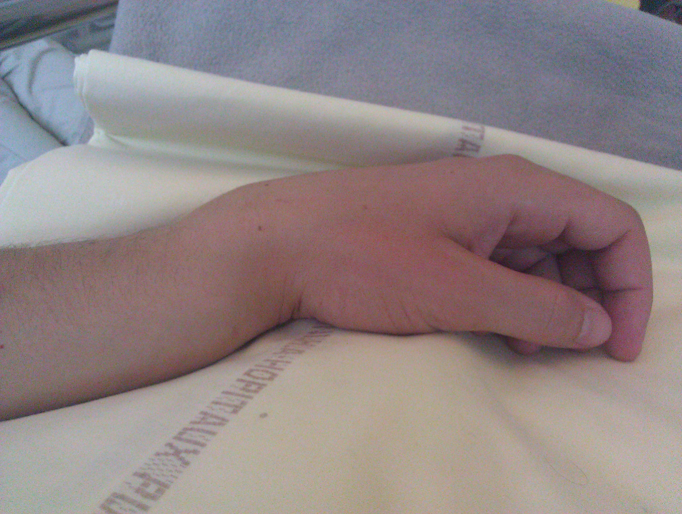
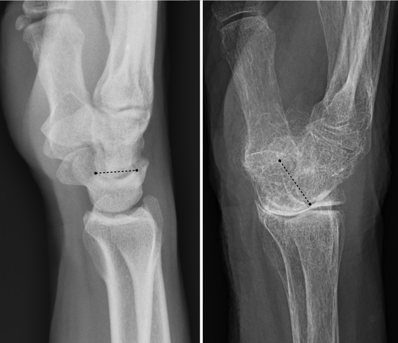
A Smith's fracture, is a fracture of the distal radius. Although it can also be caused by a direct blow to the dorsal forearm or by a fall with the wrist flexed, the most common mechanism of injury for Smith's fracture occurs in a palmar fall with the wrist joint slightly dorsiflexed. Smith's fractures are less common than Colles' fractures. The distal fracture fragment is displaced volarly ( ventrally), as opposed to a Colles' fracture which the fragment is displaced dorsally. Depending on the severity of the impact, there may be one or many fragments and it may or may not involve the articular surface of the wrist joint. Classification A commonly used classification of distal radial fractures is the Frykman classification: * Type I: Extra-articular * Type II: Type I, with fracture of distal ulna * Type III: Radiocarpal joint involvement * Type IV: Type III with fracture of distal ulna * Type V: Distal radioulnar joint involved. * Type VI: Type V with fracture of distal u ... [...More Info...] [...Related Items...] OR: [Wikipedia] [Google] [Baidu] |
Colles' Fracture
A Colles' fracture is a type of fracture of the distal forearm in which the broken end of the radius is bent backwards. Symptoms may include pain, swelling, deformity, and bruising. Complications may include damage to the median nerve. It typically occurs as a result of a fall on an outstretched hand. Risk factors include osteoporosis. The diagnosis may be confirmed via X-rays. The tip of the ulna may also be broken. Treatment may include casting or surgery. Surgical reduction and casting is possible in the majority of cases in people over the age of 50. Pain management can be achieved during the reduction with procedural sedation and analgesia or a hematoma block. A year or two may be required for healing to occur. About 15% of people have a Colles' fracture at some point in their life. They occur more commonly in young adults and older people than in children and middle-aged adults. Women are more frequently affected than men. The fracture is named after Abraham Colles ... [...More Info...] [...Related Items...] OR: [Wikipedia] [Google] [Baidu] |
Orthopedics
Orthopedic surgery or orthopedics (American and British English spelling differences, alternative spelling orthopaedics) is the branch of surgery concerned with conditions involving the musculoskeletal system. Orthopedic surgeons use both surgical and nonsurgical means to treat musculoskeletal Physical trauma, trauma, Spinal disease, spine diseases, Sports injury, sports injuries, degenerative diseases, infections, tumors and congenital disorders. Etymology Nicholas Andry coined the word in French as ', derived from the Ancient Greek words ("correct", "straight") and ("child"), and published ''Orthopedie'' (translated as ''Orthopædia: Or the Art of Correcting and Preventing Deformities in Children'') in 1741. The word was Assimilation (linguistics), assimilated into English as ''orthopædics''; the Typographic ligature, ligature ''æ'' was common in that era for ''ae'' in Greek- and Latin-based words. As the name implies, the discipline was initially developed with atte ... [...More Info...] [...Related Items...] OR: [Wikipedia] [Google] [Baidu] |
Anatomical Terms Of Location
Standard anatomical terms of location are used to describe unambiguously the anatomy of humans and other animals. The terms, typically derived from Latin or Greek roots, describe something in its standard anatomical position. This position provides a definition of what is at the front ("anterior"), behind ("posterior") and so on. As part of defining and describing terms, the body is described through the use of anatomical planes and axes. The meaning of terms that are used can change depending on whether a vertebrate is a biped or a quadruped, due to the difference in the neuraxis, or if an invertebrate is a non-bilaterian. A non-bilaterian has no anterior or posterior surface for example but can still have a descriptor used such as proximal or distal in relation to a body part that is nearest to, or furthest from its middle. International organisations have determined vocabularies that are often used as standards for subdisciplines of anatomy. For example, '' Termi ... [...More Info...] [...Related Items...] OR: [Wikipedia] [Google] [Baidu] |
Radius (bone)
The radius or radial bone (: radii or radiuses) is one of the two large bones of the forearm, the other being the ulna. It extends from the Anatomical terms of location, lateral side of the Elbow-joint, elbow to the thumb side of the wrist and runs parallel to the ulna. The ulna is longer than the radius, but the radius is thicker. The radius is a long bone, Prism (geometry), prism-shaped and slightly curved longitudinally. The radius is part of two joint (anatomy), joints: the elbow and the wrist. At the elbow, it joins with the capitulum of the humerus, and in a separate region, with the ulna at the radial notch. At the wrist, the radius forms a joint with the ulna bone. The corresponding bone in the human leg, lower leg is the tibia. Structure The long narrow medullary cavity is enclosed in a strong wall of compact bone. It is thickest along the interosseous border and thinnest at the extremities, same over the cup-shaped articular surface (fovea) of the head. The tra ... [...More Info...] [...Related Items...] OR: [Wikipedia] [Google] [Baidu] |
Wrist Joint
In human anatomy, the wrist is variously defined as (1) the carpus or carpal bones, the complex of eight bones forming the proximal skeletal segment of the hand; "The wrist contains eight bones, roughly aligned in two rows, known as the carpal bones." (2) the wrist joint or radiocarpal joint, the joint between the radius and the carpus and; (3) the anatomical region surrounding the carpus including the distal parts of the bones of the forearm and the proximal parts of the metacarpus or five metacarpal bones and the series of joints between these bones, thus referred to as ''wrist joints''. "With the large number of bones composing the wrist (ulna, radius, eight carpas, and five metacarpals), it makes sense that there are many, many joints that make up the structure known as the wrist." This region also includes the carpal tunnel, the anatomical snuff box, bracelet lines, the flexor retinaculum, and the extensor retinaculum. As a consequence of these various definitions, fra ... [...More Info...] [...Related Items...] OR: [Wikipedia] [Google] [Baidu] |
Frykman Classification
Frykman is a surname. Notable people with the surname include: * Anna-Lisa Frykman (1889–1960), Swedish composer, song lyricist, and teacher * John Frykman (1932–2017), American Lutheran minister and psychotherapist * Gösta Frykman (1909–1974), Swedish Army officer * Götrik Frykman (1891–1944), Swedish bandy player and footballer * Nils Frykman (1842–1911), Swedish teacher, evangelist, and hymnwriter * Per Frykman (born 1964), Swedish paralympic equestrian {{surname ... [...More Info...] [...Related Items...] OR: [Wikipedia] [Google] [Baidu] |
Ulna
The ulna or ulnar bone (: ulnae or ulnas) is a long bone in the forearm stretching from the elbow to the wrist. It is on the same side of the forearm as the little finger, running parallel to the Radius (bone), radius, the forearm's other long bone. Longer and thinner than the radius, the ulna is considered to be the smaller long bone of the lower arm. The corresponding bone in the Human leg#Structure, lower leg is the fibula. Structure The ulna is a long bone found in the forearm that stretches from the elbow to the wrist, and when in standard anatomical position, is found on the Medial (anatomy), medial side of the forearm. It is broader close to the elbow, and narrows as it approaches the wrist. Close to the elbow, the ulna has a bony Process (anatomy), process, the olecranon process, a hook-like structure that fits into the olecranon fossa of the humerus. This prevents hyperextension and forms a hinge joint with the trochlea of the humerus. There is also a radial notch for ... [...More Info...] [...Related Items...] OR: [Wikipedia] [Google] [Baidu] |
Radiocarpal Joint
In human anatomy, the wrist is variously defined as (1) the carpus or carpal bones, the complex of eight bones forming the proximal skeletal segment of the hand; "The wrist contains eight bones, roughly aligned in two rows, known as the carpal bones." (2) the wrist joint or radiocarpal joint, the joint between the radius and the carpus and; (3) the anatomical region surrounding the carpus including the distal parts of the bones of the forearm and the proximal parts of the metacarpus or five metacarpal bones and the series of joints between these bones, thus referred to as ''wrist joints''. "With the large number of bones composing the wrist (ulna, radius, eight carpas, and five metacarpals), it makes sense that there are many, many joints that make up the structure known as the wrist." This region also includes the carpal tunnel, the anatomical snuff box, bracelet lines, the flexor retinaculum, and the extensor retinaculum. As a consequence of these various definitions, fra ... [...More Info...] [...Related Items...] OR: [Wikipedia] [Google] [Baidu] |
Extensor Pollicis Longus Muscle
In human anatomy, the extensor pollicis longus muscle (EPL) is a skeletal muscle located dorsally on the forearm. It is much larger than the extensor pollicis brevis, the origin of which it partly covers and acts to stretch the thumb together with this muscle. Structure The extensor pollicis longus arises from the dorsal surface of the ulna and from the interosseous membrane, next to the origins of abductor pollicis longus and extensor pollicis brevis. Passing through the third tendon compartment, lying in a narrow, oblique groove on the back of the lower end of the radius,''Gray's Anatomy'' 1918, see infobox it crosses the wrist close to the dorsal midline before turning towards the thumb using Lister's tubercle on the distal end of the radius as a pulley. It obliquely crosses the tendons of the extensores carpi radialis longus and brevis, and is separated from the extensor pollicis brevis by a triangular interval, the anatomical snuff box in which the radial artery is foun ... [...More Info...] [...Related Items...] OR: [Wikipedia] [Google] [Baidu] |
Complex Regional Pain Syndrome
Complex regional pain syndrome (CRPS type 1 and type 2), sometimes referred to by the hyponyms reflex sympathetic dystrophy (RSD) or reflex neurovascular dystrophy (RND), is a rare and severe form of neuroinflammatory and dysautonomic disorder causing chronic pain, neurovascular, and neuropathic symptoms. Although it can vary widely, the classic presentation occurs when severe pain from a physical trauma or neurotropic viral infection outlasts the expected recovery time, and may subsequently spread to uninjured areas. The symptoms of types 1 and 2 are the same, except type 2 is associated with nerve injury. Usually starting in a single limb, CRPS often first manifests as pain, swelling, limited range of motion, or partial paralysis, and/or changes to the skin and bones. It may initially affect one limb and then spread throughout the body; 35% of affected individuals report symptoms throughout the body. Two types are thought to exist: CRPS type 1 (previously referred to as ' ... [...More Info...] [...Related Items...] OR: [Wikipedia] [Google] [Baidu] |
Reduction (orthopedic Surgery)
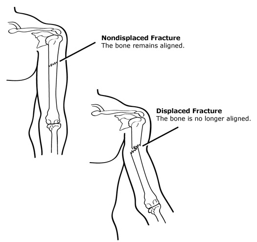
Reduction is a medical procedure to restore the correct anatomical alignment of a Fracture (bone), fracture or Dislocation (medicine), dislocation. When an injury results in a Bone fracture, fracture, or broken bone, the bone segments can sometimes become misaligned. This is referred to as a displaced fracture which requires the medical procedure called reduction. Some providers may refer to this as 'setting the bone'. When an injury results in a Joint dislocation, dislocation of a joint, or the misalignment of two connecting bones, a similar process of reduction must be performed to relocate the joint back into normal anatomical positioning. In the case of both displaced fractures and joint dislocation reduction is required for effective healing. Fracture Reduction There are two main categories of fracture reductions, closed reductions and open reductions. Both procedures require confirmatory imaging, such as x-ray, before the reduction to confirm the misalignment of bones and ... [...More Info...] [...Related Items...] OR: [Wikipedia] [Google] [Baidu] |
Open Reduction And Internal Fixation
Internal fixation is an operation in orthopedics that involves the surgical implementation of implants for the purpose of repairing a bone, a concept that dates to the mid-nineteenth century and was made applicable for routine treatment in the mid-twentieth century. An internal fixator may be made of stainless steel, titanium alloy, or cobalt-chrome alloy. Types of internal fixators include: * Plate and screws * Kirschner wires * Intramedullary nails Open reduction Open reduction internal fixation (ORIF) involves the implementation of implants to guide the healing process of a bone, as well as the open reduction, or setting, of the bone. ''Open reduction'' refers to open surgery to set bones, as is necessary for some fractures. ''Internal fixation'' refers to fixation of screws and/or plates, intramedullary rods and other devices to enable or facilitate healing. Rigid fixation prevents micro-motion across lines of fracture to enable healing and prevent infection, which h ... [...More Info...] [...Related Items...] OR: [Wikipedia] [Google] [Baidu] |


