|
Sinding-Larsen And Johansson Syndrome
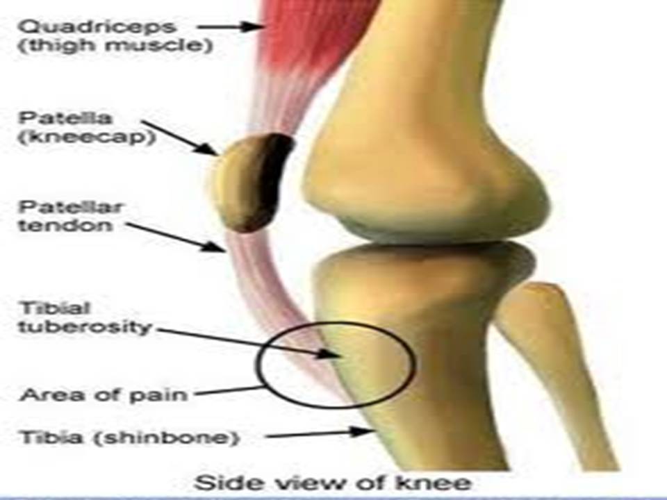
Sinding-Larsen and Johansson syndrome, named after Swedish surgeon Sven Christian Johansson (1880-1959), and Christian Magnus Falsen Sinding-Larsen (1866-1930), a Norwegian physician, is apophysitis of the inferior pole of the patella. It is analogous to Osgood–Schlatter disease which involves the upper margin of the tibia. This variant was discovered in 1908, during a winter indoor Olympic qualifier event in Scandinavia. Sever's disease is a similar condition affecting the heel. This condition called Sinding-Larsen and Johansson syndrome was described independently by Sinding-Larsen in 1921 and Johansson in 1922. Signs and symptoms The condition is usually seen in athletic individuals typically between 10 and 14 years of age. Following a strain or partial rupture of patellar ligament the patient develops a traction ‘tendinitis’ characterized by pain and point tenderness at the inferior (lower) pole of the patella associated with focal swelling. Children with cerebral palsy ... [...More Info...] [...Related Items...] OR: [Wikipedia] [Google] [Baidu] |
Apophysitis
In the skeleton of humans and other animals, a tubercle, tuberosity or apophysis is a Tubercle, protrusion or eminence that serves as an attachment for skeletal muscles. The muscles attach by tendons, where the enthesis is the connective tissue between the tendon and bone. A ''tuberosity'' is generally a larger tubercle. Main tubercles Humerus The humerus has two tubercles, the greater tubercle and the lesser tubercle. These are situated at the Anatomical terms of location#Proximal and distal, proximal end of the bone, that is the end that connects with the scapula. The greater/lesser tubercule is located from the top of the acromion laterally and inferiorly. The radius has two, the radial tuberosity and Lister's tubercle. Ribs On a rib cage, rib, tubercle is an eminence on the back surface, at the junction between the neck and the body of the rib. It consists of an articular and a non-articular area. The lower and more medial articular area is a small oval surface for articulati ... [...More Info...] [...Related Items...] OR: [Wikipedia] [Google] [Baidu] |
Patella
The patella, also known as the kneecap, is a flat, rounded triangular bone which articulates with the femur (thigh bone) and covers and protects the anterior articular surface of the knee joint. The patella is found in many tetrapods, such as mice, cats, birds and dogs, but not in whales, or most reptiles. In humans, the patella is the largest sesamoid bone (i.e., embedded within a tendon or a muscle) in the body. Babies are born with a patella of soft cartilage which begins to ossify into bone at about four years of age. Structure The patella is a sesamoid bone roughly triangular in shape, with the apex of the patella facing downwards. The apex is the most inferior (lowest) part of the patella. It is pointed in shape, and gives attachment to the patellar ligament. The front and back surfaces are joined by a thin margin and towards centre by a thicker margin. The tendon of the quadriceps femoris muscle attaches to the base of the patella., with the vastus intermedius muscle ... [...More Info...] [...Related Items...] OR: [Wikipedia] [Google] [Baidu] |
Osgood–Schlatter Disease
Osgood–Schlatter disease (OSD) is inflammation of the patellar ligament at the tibial tuberosity (apophysitis). It is characterized by a painful bump just below the knee that is worse with activity and better with rest. Episodes of pain typically last a few weeks to months. One or both knees may be affected and flares may recur. Risk factors include overuse, especially sports which involve frequent running or jumping. The underlying mechanism is repeated tension on the growth plate of the upper tibia. Diagnosis is typically based on the symptoms. A plain X-ray may be either normal or show fragmentation in the attachment area. Pain typically resolves with time. Applying cold to the affected area, rest, stretching, and strengthening exercises may help. NSAIDs such as ibuprofen may be used. Slightly less stressful activities such as swimming or walking may be recommended. Casting the leg for a period of time may help. After growth slows, typically age 16 in boys and 14 in girl ... [...More Info...] [...Related Items...] OR: [Wikipedia] [Google] [Baidu] |
Tibia
The tibia (; ), also known as the shinbone or shankbone, is the larger, stronger, and anterior (frontal) of the two bones in the leg below the knee in vertebrates (the other being the fibula, behind and to the outside of the tibia); it connects the knee with the ankle. The tibia is found on the medial side of the leg next to the fibula and closer to the median plane. The tibia is connected to the fibula by the interosseous membrane of leg, forming a type of fibrous joint called a syndesmosis with very little movement. The tibia is named for the flute ''tibia''. It is the second largest bone in the human body, after the femur. The leg bones are the strongest long bones as they support the rest of the body. Structure In human anatomy, the tibia is the second largest bone next to the femur. As in other vertebrates the tibia is one of two bones in the lower leg, the other being the fibula, and is a component of the knee and ankle joints. The ossification or formation of the bone ... [...More Info...] [...Related Items...] OR: [Wikipedia] [Google] [Baidu] |
Sever's Disease
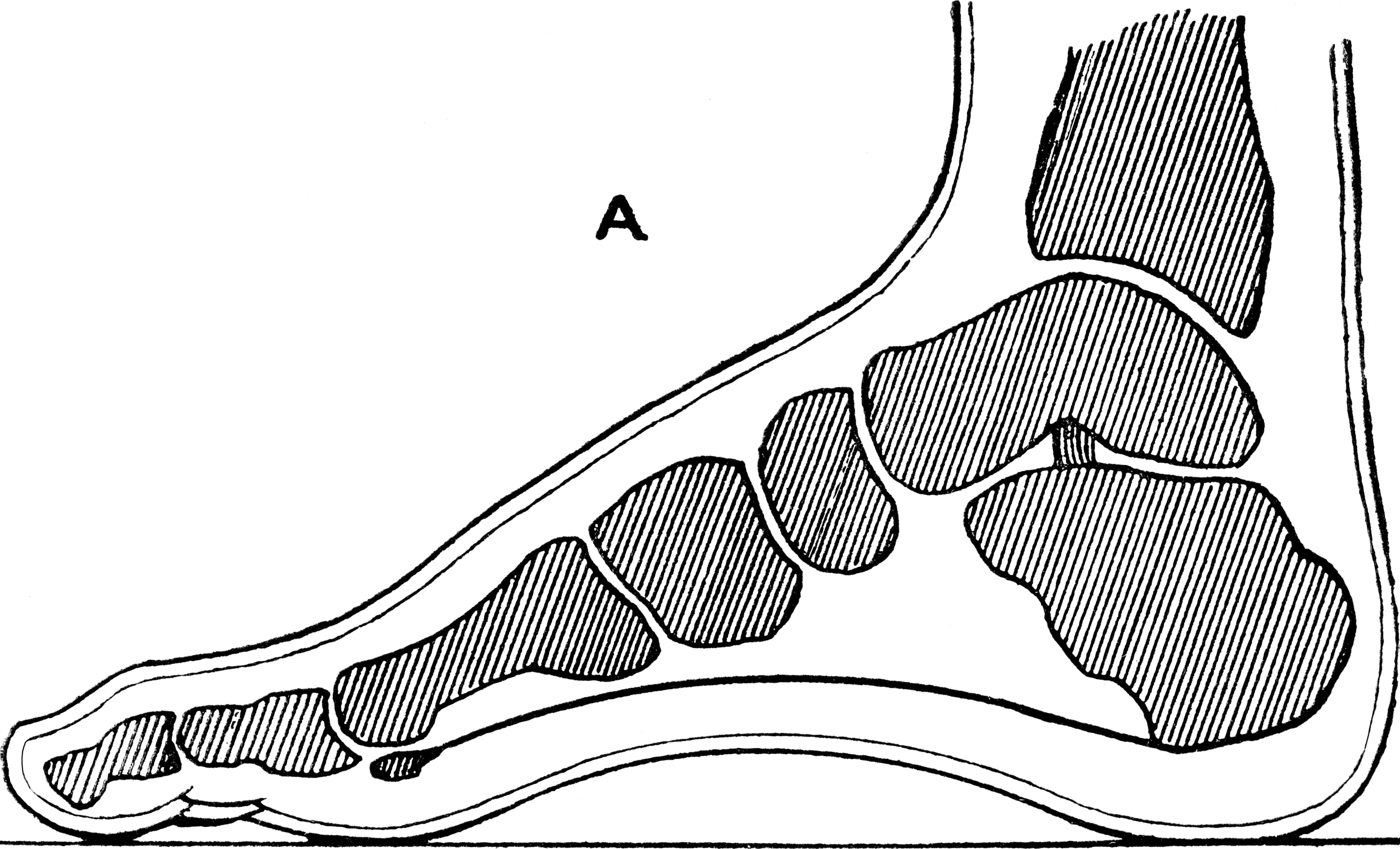
Sever's disease, also known as calcaneus apophysitis, is an inflammation at the back of the heel (or calcaneus) growth plate in growing children. The condition is thought to be caused by repetitive stress at the heel. This condition is benign and common and usually resolves when the growth plate has closed or during periods of less activity. It occurs in both males and females. There are a number of locations in the body that may get apophysitis pain. Another common location is at the front of the knee which is known as apophysitis of the tibial tuberosity or Osgood Schlatter's disease. Symptoms Children with calcaneal apophysitis commonly complain of pain at the back of the heel. This pain increases with jumping and some running sports. Sometimes, the pain makes children limp and may result in poor sports performance or them not wanting to participate in some sports. The back of the heel is never swollen or red, unless there has been shoe rubbing. When the back of the heel is squ ... [...More Info...] [...Related Items...] OR: [Wikipedia] [Google] [Baidu] |
Heel
The heel is the prominence at the posterior end of the foot. It is based on the projection of one bone, the calcaneus or heel bone, behind the articulation of the bones of the lower Human leg, leg. Structure To distribute the compressive forces exerted on the heel during gait, and especially the stance phase when the heel contacts the ground, the sole (foot), sole of the foot is covered by a layer of subcutaneous connective tissue up to 2 cm thick (under the heel). This tissue has a system of pressure chambers that both acts as a shock absorber and stabilises the sole. Each of these chambers contains fibrofatty tissue covered by a layer of tough connective tissue made of collagen fibers. These septum, septa ("walls") are firmly attached both to the plantar aponeurosis above and the sole's Dermis, skin below. The sole of the foot is one of the most highly vascularized regions of the body surface, and the dense system of blood vessels further stabilize the septa. The Ach ... [...More Info...] [...Related Items...] OR: [Wikipedia] [Google] [Baidu] |
Johansson
Johansson is a patronymic family name of Swedish origin meaning ''"son of Johan"'', or ''"Johan's son"''. It is the most common Swedish family name, followed by Andersson. (First 18 surnames ends -sson.) The Danish, Norwegian, German and Dutch variant is Johansen, while the most common spelling in the US is Johnson. There are still other spellings. Johansson is an uncommon given name. Geographical distribution As of 2014, 91.2% of all known bearers of the surname ''Johansson'' were residents of Sweden (frequency 1:39), 2.5% of Finland (1:802), 1.5% of Norway (1:1,274), 1.4% of the United States (1:93,010) and 1.0% of Denmark (1:2,022). In Sweden, the frequency of the surname was higher than national average (1:39) in the following counties: * 1. Kalmar (1:21) * 2. Kronoberg (1:21) * 3. Halland (1:21) * 4. Jönköping (1:25) * 5. Västra Götaland (1:26) * 6. Blekinge (1:29) * 7. Östergötland (1:30) * 8. Värmland (1:30) * 9. Västerbotten (1:36) * 10. Gotland (1:36) * 11. � ... [...More Info...] [...Related Items...] OR: [Wikipedia] [Google] [Baidu] |
Knee Anatomy
In humans and other primates, the knee joins the thigh with the leg and consists of two joints: one between the femur and tibia (tibiofemoral joint), and one between the femur and patella (patellofemoral joint). It is the largest joint in the human body. The knee is a modified hinge joint, which permits flexion and extension as well as slight internal and external rotation. The knee is vulnerable to injury and to the development of osteoarthritis. It is often termed a ''compound joint'' having tibiofemoral and patellofemoral components. (The fibular collateral ligament is often considered with tibiofemoral components.) Structure The knee is a modified hinge joint, a type of synovial joint, which is composed of three functional compartments: the patellofemoral articulation, consisting of the patella, or "kneecap", and the patellar groove on the front of the femur through which it slides; and the medial and lateral tibiofemoral articulations linking the femur, or thigh bo ... [...More Info...] [...Related Items...] OR: [Wikipedia] [Google] [Baidu] |
Chondropathies
Chondropathy refers to a disease of the cartilage. It is frequently divided into 5 grades, with 0-2 defined as normal and 3-4 defined as diseased. __TOC__ Some common diseases affecting/involving the cartilage * Achondroplasia: Reduced proliferation of chondrocytes in the epiphyseal plate of long bones during infancy and childhood, resulting in dwarfism. * Cartilage tumors * Costochondritis: Inflammation of cartilage in the ribs, causing chest pain. * Osteoarthritis: The cartilage covering bones (articular cartilage) is thinned, eventually completely worn out, resulting in a "bone against bone" joint, resulting in pain and reduced mobility. Osteoarthritis is very common, affects the joints exposed to high stress and is therefore considered the result of "wear and tear" rather than a true disease. It is treated by Arthroplasty, the replacement of the joint by a synthetic joint made of titanium and teflon. Chondroitin sulfate, a monomer of the polysaccharide portion of proteoglycan, h ... [...More Info...] [...Related Items...] OR: [Wikipedia] [Google] [Baidu] |


