|
Scutellinia Scutellata
''Scutellinia scutellata'', commonly known as the eyelash cup, the Molly eye-winker, the scarlet elf cap, the eyelash fungus or the eyelash pixie cup, is a small saprophytic fungus of the genus '' Scutellinia''. It is the type species of ''Scutellinia'', as well as being the most common and widespread. The fruiting bodies are small red cups with distinct long, dark hairs or "eyelashes". These eyelashes are the most distinctive feature and are easily visible with a magnifying glass. The species is common in North America and Europe, and has been recorded on every continent. ''S. scutellata'' is found on rotting wood and in other damp habitats, typically growing in small groups, sometimes forming clusters. It is sometimes described as inedible, but its small size means it is not suitable for culinary use. Despite this, it is popular among mushroom hunters due to its unusual "eyelash" hairs, making it memorable and easy to identify. Taxonomy ''Scutellinia scutellata'' was ... [...More Info...] [...Related Items...] OR: [Wikipedia] [Google] [Baidu] |
Carl Linnaeus
Carl Linnaeus (; 23 May 1707 – 10 January 1778), also known after his ennoblement in 1761 as Carl von Linné Blunt (2004), p. 171. (), was a Swedish botanist, zoologist, taxonomist, and physician who formalised binomial nomenclature, the modern system of naming organisms. He is known as the "father of modern taxonomy". Many of his writings were in Latin; his name is rendered in Latin as and, after his 1761 ennoblement, as . Linnaeus was born in Råshult, the countryside of Småland, in southern Sweden. He received most of his higher education at Uppsala University and began giving lectures in botany there in 1730. He lived abroad between 1735 and 1738, where he studied and also published the first edition of his ' in the Netherlands. He then returned to Sweden where he became professor of medicine and botany at Uppsala. In the 1740s, he was sent on several journeys through Sweden to find and classify plants and animals. In the 1750s and 1760s, he continued to collect an ... [...More Info...] [...Related Items...] OR: [Wikipedia] [Google] [Baidu] |
Trama (mycology)
In mycology, the term trama is used in two ways. In the broad sense, it is the inner, fleshy portion of a mushroom's basidiocarp, or fruit body. It is distinct from the outer layer of tissue, known as the pileipellis or cuticle, and from the spore-bearing tissue layer known as the hymenium. In essence, the trama is the tissue that is commonly referred to as the "flesh" of mushrooms and similar fungi.Largent D, Johnson D, Watling R. 1977. ''How to Identify Mushrooms to Genus III: Microscopic Features''. Arcata, CA: Mad River Press. . pp. 60–70. The second use is more specific, and refers to the "hymenophoral trama" that supports the hymenium. It is similarly interior, connective tissue, but it is more specifically the central layer of hyphae running from the underside of the mushroom cap to the lamella or gill, upon which the hymenium rests. Various types have been classified by their structure, including trametoid, cantharelloid, boletoid, and agaricoid, with agaricoid the ... [...More Info...] [...Related Items...] OR: [Wikipedia] [Google] [Baidu] |
Electron Microscopy
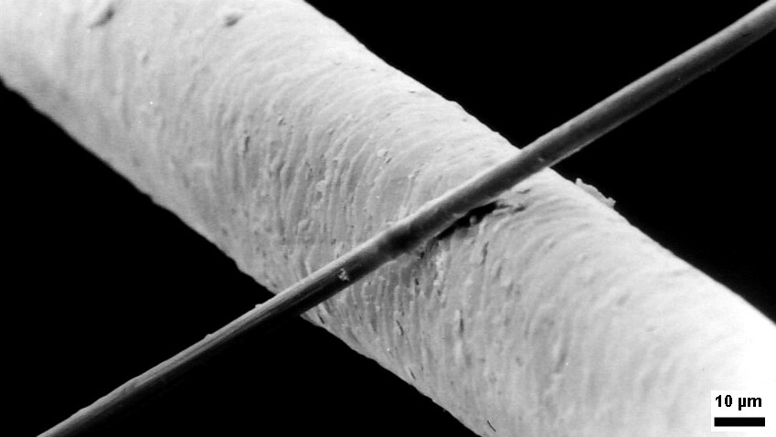
An electron microscope is a microscope that uses a beam of accelerated electrons as a source of illumination. As the wavelength of an electron can be up to 100,000 times shorter than that of visible light photons, electron microscopes have a higher resolving power than light microscopes and can reveal the structure of smaller objects. A scanning transmission electron microscope has achieved better than 50 pm resolution in annular dark-field imaging mode and magnifications of up to about 10,000,000× whereas most light microscopes are limited by diffraction to about 200 nm resolution and useful magnifications below 2000×. Electron microscopes use shaped magnetic fields to form electron optical lens systems that are analogous to the glass lenses of an optical light microscope. Electron microscopes are used to investigate the ultrastructure of a wide range of biological and inorganic specimens including microorganisms, cells, large molecules, biopsy samples, ... [...More Info...] [...Related Items...] OR: [Wikipedia] [Google] [Baidu] |
Hypha
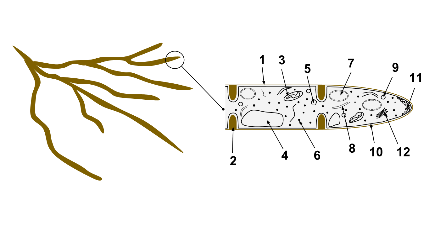
A hypha (; ) is a long, branching, filamentous structure of a fungus, oomycete, or actinobacterium. In most fungi, hyphae are the main mode of vegetative growth, and are collectively called a mycelium. Structure A hypha consists of one or more cells surrounded by a tubular cell wall. In most fungi, hyphae are divided into cells by internal cross-walls called "septa" (singular septum). Septa are usually perforated by pores large enough for ribosomes, mitochondria, and sometimes nuclei to flow between cells. The major structural polymer in fungal cell walls is typically chitin, in contrast to plants and oomycetes that have cellulosic cell walls. Some fungi have aseptate hyphae, meaning their hyphae are not partitioned by septa. Hyphae have an average diameter of 4–6 µm. Growth Hyphae grow at their tips. During tip growth, cell walls are extended by the external assembly and polymerization of cell wall components, and the internal production of new cell membrane. The S ... [...More Info...] [...Related Items...] OR: [Wikipedia] [Google] [Baidu] |
Septum
In biology, a septum (Latin for ''something that encloses''; plural septa) is a wall, dividing a cavity or structure into smaller ones. A cavity or structure divided in this way may be referred to as septate. Examples Human anatomy * Interatrial septum, the wall of tissue that is a sectional part of the left and right atria of the heart * Interventricular septum, the wall separating the left and right ventricles of the heart * Lingual septum, a vertical layer of fibrous tissue that separates the halves of the tongue. *Nasal septum: the cartilage wall separating the nostrils of the nose * Alveolar septum: the thin wall which separates the alveoli from each other in the lungs * Orbital septum, a palpebral ligament in the upper and lower eyelids * Septum pellucidum or septum lucidum, a thin structure separating two fluid pockets in the brain * Uterine septum, a malformation of the uterus * Vaginal septum, a lateral or transverse partition inside the vagina * Intermuscular sep ... [...More Info...] [...Related Items...] OR: [Wikipedia] [Google] [Baidu] |
Paraphyses
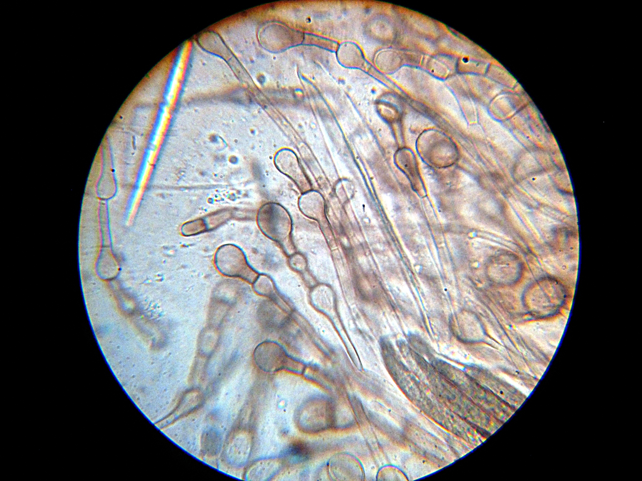
Paraphyses are erect sterile filament-like support structures occurring among the reproductive apparatuses of fungi, ferns, bryophytes and some thallophytes. The singular form of the word is paraphysis. In certain fungi, they are part of the fertile spore-bearing layer. More specifically, paraphyses are sterile filamentous hyphal end cells composing part of the hymenium of Ascomycota and Basidiomycota interspersed among either the asci or basidia respectively, and not sufficiently differentiated to be called cystidia A cystidium (plural cystidia) is a relatively large cell found on the sporocarp of a basidiomycete (for example, on the surface of a mushroom gill), often between clusters of basidia. Since cystidia have highly varied and distinct shapes that ar ..., which are specialized, swollen, often protruding cells. The tips of paraphyses may contain the pigments which colour the hymenium. In ferns and mosses, they are filament-like structures that are found on sporangia ... [...More Info...] [...Related Items...] OR: [Wikipedia] [Google] [Baidu] |
Spore Print
300px, Making a spore print of the mushroom ''Volvariella volvacea'' shown in composite: (photo lower half) mushroom cap laid on white and dark paper; (photo upper half) cap removed after 24 hours showing pinkish-tan spore print. A 3.5-centimeter glass slide placed in middle allows for examination of spore characteristics under a microscope. image:spore Print ID.gif, 300px, A printable chart to make a spore print and start identification The spore print is the powdery deposit obtained by allowing spores of a fungal sporocarp (fungi), fruit body to fall onto a surface underneath. It is an important diagnostic character in most handbooks for identifying mushrooms. It shows the colour of the mushroom spores if viewed en masse. Method A spore print is made by placing the spore-producing surface flat on a sheet of dark and white paper or on a sheet of clear, stiff plastic, which facilitates moving the spore print to a darker or lighter surface for improved contrast; for example, it ... [...More Info...] [...Related Items...] OR: [Wikipedia] [Google] [Baidu] |
Ascospore
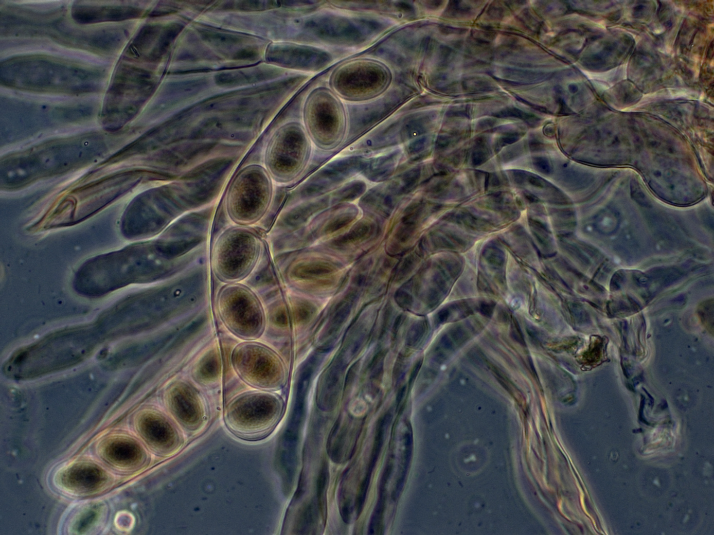
An ascus (; ) is the sexual spore-bearing cell produced in ascomycete fungi. Each ascus usually contains eight ascospores (or octad), produced by meiosis followed, in most species, by a mitotic cell division. However, asci in some genera or species can occur in numbers of one (e.g. ''Monosporascus cannonballus''), two, four, or multiples of four. In a few cases, the ascospores can bud off conidia that may fill the asci (e.g. ''Tympanis'') with hundreds of conidia, or the ascospores may fragment, e.g. some ''Cordyceps'', also filling the asci with smaller cells. Ascospores are nonmotile, usually single celled, but not infrequently may be coenocytic (lacking a septum), and in some cases coenocytic in multiple planes. Mitotic divisions within the developing spores populate each resulting cell in septate ascospores with nuclei. The term ocular chamber, or oculus, refers to the epiplasm (the portion of cytoplasm not used in ascospore formation) that is surrounded by the "bourrelet ... [...More Info...] [...Related Items...] OR: [Wikipedia] [Google] [Baidu] |
Hyaline
A hyaline substance is one with a glassy appearance. The word is derived from el, ὑάλινος, translit=hyálinos, lit=transparent, and el, ὕαλος, translit=hýalos, lit=crystal, glass, label=none. Histopathology Hyaline cartilage is named after its glassy appearance on fresh gross pathology. On light microscopy of H&E stained slides, the extracellular matrix of hyaline cartilage looks homogeneously pink, and the term "hyaline" is used to describe similarly homogeneously pink material besides the cartilage. Hyaline material is usually acellular and proteinaceous. For example, arterial hyaline is seen in aging, high blood pressure, diabetes mellitus and in association with some drugs (e.g. calcineurin inhibitors). It is bright pink with PAS staining. Ichthyology and entomology In ichthyology and entomology, ''hyaline'' denotes a colorless, transparent substance, such as unpigmented fins of fishes or clear insect wings. Resh, Vincent H. and R. T. Cardé, Eds. Encyclo ... [...More Info...] [...Related Items...] OR: [Wikipedia] [Google] [Baidu] |
Micrometre
The micrometre ( international spelling as used by the International Bureau of Weights and Measures; SI symbol: μm) or micrometer (American spelling), also commonly known as a micron, is a unit of length in the International System of Units (SI) equalling (SI standard prefix "micro-" = ); that is, one millionth of a metre (or one thousandth of a millimetre, , or about ). The nearest smaller common SI unit is the nanometre, equivalent to one thousandth of a micrometre, one millionth of a millimetre or one billionth of a metre (). The micrometre is a common unit of measurement for wavelengths of infrared radiation as well as sizes of biological cells and bacteria, and for grading wool by the diameter of the fibres. The width of a single human hair ranges from approximately 20 to . The longest human chromosome, chromosome 1, is approximately in length. Examples Between 1 μm and 10 μm: * 1–10 μm – length of a typical bacterium * 3–8 μm – width of ... [...More Info...] [...Related Items...] OR: [Wikipedia] [Google] [Baidu] |
Ascus
An ascus (; ) is the sexual spore-bearing cell produced in ascomycete fungi. Each ascus usually contains eight ascospores (or octad), produced by meiosis followed, in most species, by a mitotic cell division. However, asci in some genera or species can occur in numbers of one (e.g. ''Monosporascus cannonballus''), two, four, or multiples of four. In a few cases, the ascospores can bud off conidia that may fill the asci (e.g. ''Tympanis'') with hundreds of conidia, or the ascospores may fragment, e.g. some ''Cordyceps'', also filling the asci with smaller cells. Ascospores are nonmotile, usually single celled, but not infrequently may be coenocytic (lacking a septum), and in some cases coenocytic in multiple planes. Mitotic divisions within the developing spores populate each resulting cell in septate ascospores with nuclei. The term ocular chamber, or oculus, refers to the epiplasm (the portion of cytoplasm not used in ascospore formation) that is surrounded by the "bourrelet ... [...More Info...] [...Related Items...] OR: [Wikipedia] [Google] [Baidu] |
Asci And Ascospores Of Scutellinia Scutellata
ASCI or Asci may refer to: * Advertising Standards Council of India * Asci, the plural of ascus, in fungal anatomy * Accelerated Strategic Computing Initiative * American Society for Clinical Investigation * Argus Sour Crude Index * Association of Christian Schools International * Associazione Scouts Cattolici Italiani, co-founder of Associazione Guide e Scouts Cattolici Italiani * Administrative Staff College of India, Hyderabad * Accountable, Support, Consult, Inform (roles in a project) See also * ASCII ASCII ( ), abbreviated from American Standard Code for Information Interchange, is a character encoding standard for electronic communication. ASCII codes represent text in computers, telecommunications equipment, and other devices. Because of ... {{disambiguation pl:ASCI ... [...More Info...] [...Related Items...] OR: [Wikipedia] [Google] [Baidu] |


