|
Saccus Endolymphaticus
From the posterior wall of the saccule a canal, the endolymphatic duct, is given off; this duct is joined by the utriculosaccular duct, and then passes along the vestibular aqueduct and ends in a blind pouch, the endolymphatic sac, on the posterior surface of the petrous portion of the temporal bone, where it is in contact with the dura mater. Studies suggest that the endolymphatic duct and endolymphatic sac perform both absorptive and secretory 440px Secretion is the movement of material from one point to another, such as a secreted chemical substance from a cell or gland. In contrast, excretion is the removal of certain substances or waste products from a cell or organism. The classica ..., as well as phagocytic and immunodefensive, functions.Wackym PA, Friberg U, Linthicum FH Jr, et al. Human endolymphatic sac: morphologic evidence of immunologic function. Ann Otol Rhinol Laryngol 1987;96:276–282 Neoplasms of the endolymphatic sac are very rare tumors. References ... [...More Info...] [...Related Items...] OR: [Wikipedia] [Google] [Baidu] |
Saccule
The saccule is a bed of sensory cells in the inner ear. It translates head movements into neural impulses for the brain to interpret. The saccule detects linear accelerations and head tilts in the vertical plane. When the head moves vertically, the sensory cells of the saccule are disturbed and the neurons connected to them begin transmitting impulses to the brain. These impulses travel along the vestibular portion of the eighth cranial nerve to the vestibular nuclei in the brainstem. The vestibular system is important in maintaining balance, or equilibrium. The vestibular system includes the saccule, utricle, and the three semicircular canals. The vestibule is the name of the fluid-filled, membranous duct that contains these organs of balance. The vestibule is encased in the temporal bone of the skull. Structure The saccule, or sacculus, is the smaller of the two vestibular sacs. It is globular in form and lies in the recessus sphæricus near the opening of the vest ... [...More Info...] [...Related Items...] OR: [Wikipedia] [Google] [Baidu] |
Endolymphatic Duct
From the posterior wall of the saccule a canal, the endolymphatic duct, is given off; this duct is joined by the ductus utriculosaccularis, and then passes along the aquaeductus vestibuli and ends in a blind pouch (endolymphatic sac) on the posterior surface of the petrous portion of the temporal bone, where it is in contact with the dura mater In neuroanatomy, dura mater is a thick membrane made of dense irregular connective tissue that surrounds the brain and spinal cord. It is the outermost of the three layers of membrane called the meninges that protect the central nervous system. .... Disorders of the endolymphatic duct include Meniere's Disease and Enlarged Vestibular Aqueduct. Additional images File:Gray902.png, Transverse section through head of fetal sheep, in the region of the labyrinth. X 30. File:Gray927.png, Transverse section of a human semicircular canal and duct References External links *The Endolymphatic Duct and Sac Vestibular system ... [...More Info...] [...Related Items...] OR: [Wikipedia] [Google] [Baidu] |
Utriculosaccular Duct
The utriculosaccular duct (Latin: Ductus utriculosaccularis) is a part of the membranous labyrinth of the inner ear which connects the two parts of the vestibule, the utricle and the saccule. The utriculosaccular duct continues to the endolymphatic duct and ends in the endolymphatic sac From the posterior wall of the saccule a canal, the endolymphatic duct, is given off; this duct is joined by the utriculosaccular duct, and then passes along the vestibular aqueduct and ends in a blind pouch, the endolymphatic sac, on the poster .... Ear {{anatomy-stub ... [...More Info...] [...Related Items...] OR: [Wikipedia] [Google] [Baidu] |
Vestibular Aqueduct
At the hinder part of the medial wall of the vestibule is the orifice of the vestibular aqueduct, which extends to the posterior surface of the petrous portion of the temporal bone. It transmits a small vein, and contains a tubular prolongation of the membranous labyrinth, the ductus endolymphaticus, which ends in a cul-de-sac, the endolymphatic sac, between the layers of the dura mater within the cranial cavity. Pathology Enlargement of the vestibular aqueduct to greater than 2 mm is associated with enlarged vestibular aqueduct syndrome, a disease entity that is associated with one-sided hearing loss in children. The diagnosis can be made by high resolution CT or MRI, with comparison to the adjacent posterior semicircular canal. If the vestibular aqueduct is larger in size, and the clinical presentation is consistent, the diagnosis can be made. Treatment is with mechanical hearing implants. There is an association with Pendred syndrome Pendred syndrome is a genetic d ... [...More Info...] [...Related Items...] OR: [Wikipedia] [Google] [Baidu] |
Petrous Portion Of The Temporal Bone
The petrous part of the temporal bone is pyramid-shaped and is wedged in at the base of the skull between the sphenoid and occipital bones. Directed medially, forward, and a little upward, it presents a base, an apex, three surfaces, and three angles, and houses in its interior, the components of the inner ear. The petrous portion is among the most basal elements of the skull and forms part of the endocranium. Petrous comes from the Latin word ''petrosus'', meaning "stone-like, hard". It is one of the densest bones in the body. The petrous bone is important for studies of ancient DNA from skeletal remains, as it tends to contain extremely well-preserved DNA. Base The base is fused with the internal surfaces of the squamous and mastoid parts. Apex The apex, which is rough and uneven, is received into the angular interval between the posterior border of the great wing of the sphenoid bone and the basilar part of the occipital bone; it presents the anterior or internal openin ... [...More Info...] [...Related Items...] OR: [Wikipedia] [Google] [Baidu] |
Temporal Bone
The temporal bones are situated at the sides and base of the skull, and lateral to the temporal lobes of the cerebral cortex. The temporal bones are overlaid by the sides of the head known as the temples, and house the structures of the ears. The lower seven cranial nerves and the major vessels to and from the brain traverse the temporal bone. Structure The temporal bone consists of four parts— the squamous, mastoid, petrous and tympanic parts. The squamous part is the largest and most superiorly positioned relative to the rest of the bone. The zygomatic process is a long, arched process projecting from the lower region of the squamous part and it articulates with the zygomatic bone. Posteroinferior to the squamous is the mastoid part. Fused with the squamous and mastoid parts and between the sphenoid and occipital bones lies the petrous part, which is shaped like a pyramid. The tympanic part is relatively small and lies inferior to the squamous part, anterior to the mast ... [...More Info...] [...Related Items...] OR: [Wikipedia] [Google] [Baidu] |
Dura Mater
In neuroanatomy, dura mater is a thick membrane made of dense irregular connective tissue that surrounds the brain and spinal cord. It is the outermost of the three layers of membrane called the meninges that protect the central nervous system. The other two meningeal layers are the arachnoid mater and the pia mater. It envelops the arachnoid mater, which is responsible for keeping in the cerebrospinal fluid. It is derived primarily from the neural crest cell population, with postnatal contributions of the paraxial mesoderm. Structure The dura mater has several functions and layers. The dura mater is a membrane that envelops the arachnoid mater. It surrounds and supports the dural sinuses (also called dural venous sinuses, cerebral sinuses, or cranial sinuses) and carries blood from the brain toward the heart. Cranial dura mater has two layers called ''lamellae'', a superficial layer (also called the periosteal layer), which serves as the skull's inner periosteum, called the ... [...More Info...] [...Related Items...] OR: [Wikipedia] [Google] [Baidu] |
Secretory
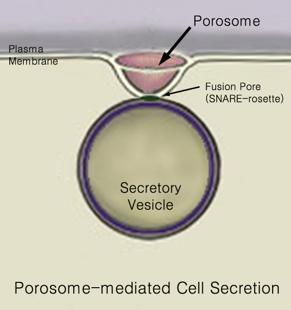
440px Secretion is the movement of material from one point to another, such as a secreted chemical substance from a cell or gland. In contrast, excretion is the removal of certain substances or waste products from a cell or organism. The classical mechanism of cell secretion is via secretory portals at the plasma membrane called porosomes. Porosomes are permanent cup-shaped lipoprotein structures embedded in the cell membrane, where secretory vesicles transiently dock and fuse to release intra-vesicular contents from the cell. Secretion in bacterial species means the transport or translocation of effector molecules for example: proteins, enzymes or toxins (such as cholera toxin in pathogenic bacteria e.g. ''Vibrio cholerae'') from across the interior (cytoplasm or cytosol) of a bacterial cell to its exterior. Secretion is a very important mechanism in bacterial functioning and operation in their natural surrounding environment for adaptation and survival. In eukaryotic cell ... [...More Info...] [...Related Items...] OR: [Wikipedia] [Google] [Baidu] |
Endolymphatic Sac Tumor
An endolymphatic sac tumor (ELST) is a very uncommon papillary epithelial neoplasm arising within the endolymphatic sac or endolymphatic duct. This tumor shows a very high association with Von Hippel–Lindau syndrome (VHL). Classification The ELST has been referred to as adenocarcinoma of endolymphatic sac, Heffner tumor, papillary adenomatous tumor, aggressive papillary adenoma, invasive papillary cystadenoma, and papillary tumor of temporal bone. However, these names are not encouraged as they do not accurately classify the current understanding of the tumor. Signs and symptoms Patients with ELST may present clinically with progressive or fluctuating, one sided sensorineural hearing loss which may mimick Ménière's disease due to the development of tumor associated endolymphatic hydrops. Patients may also experience tinnitus, vertigo, and loss of vestibular function (ataxia). Alternatively, symptom onset may be sudden, due to intralabyrinthine hemorrhage. Patients may also ... [...More Info...] [...Related Items...] OR: [Wikipedia] [Google] [Baidu] |


