|
Roundabout (gene)
The Roundabout (Robo) family of proteins are single-pass transmembrane receptors that are highly conserved across many branches of the animal kingdom, from ''C. elegans'' to humans. They were first discovered in ''Drosophila'', through a mutant screen for genes involved in axon guidance. The ''Drosophila'' roundabout mutant was named after its phenotype, which resembled the circular traffic junctions (see roundabout). The Robo receptors are most well known for their role in the development of the nervous system, where they have been shown to respond to secreted Slit ligands. One well-studied example is the requirement for Slit-Robo signaling in regulation of axonal midline crossing. Slit-Robo signaling is also critical for many neurodevelopmental processes including formation of the olfactory tract, the optic nerve, and motor axon fasciculation. In addition, Slit-Robo signaling contributes to cell migration and the development of other tissues such as the lung, kidney, liv ... [...More Info...] [...Related Items...] OR: [Wikipedia] [Google] [Baidu] |
ROBO1
Roundabout homolog 1 is a protein that in humans is encoded by the ''ROBO1'' gene. Function Bilateral symmetric nervous systems have special midline structures that establish a partition between the two mirror image halves. Some axons project toward and across the midline in response to long-range Chemotaxis, chemoattractants emanating from the midline. The protein encoded by ROBO1 is structurally similar to a Drosophila integral membrane protein which is encoded by the Drosophila roundabout gene (a member of the immunoglobulin gene superfamily) and is both an axon guidance receptor and a cell adhesion receptor, known to be involved in the decision by axons to cross the central nervous system midline. Two transcript variants encoding different Protein isoform, isoforms have been found for ROBO1. Clinical significance ''ROBO1'' was implicated in a communication disorder based on a Finland, Finnish pedigree with severe dyslexia. Analyses revealed a translocation had occurred ... [...More Info...] [...Related Items...] OR: [Wikipedia] [Google] [Baidu] |
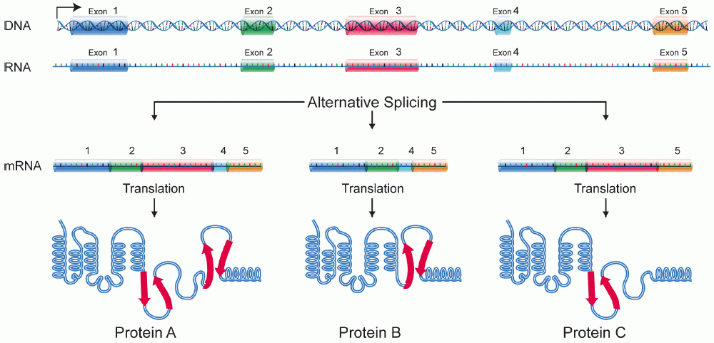
Alternative Splicing
Alternative splicing, or alternative RNA splicing, or differential splicing, is an alternative splicing process during gene expression that allows a single gene to code for multiple proteins. In this process, particular exons of a gene may be included within or excluded from the final, processed messenger RNA (mRNA) produced from that gene. This means the exons are joined in different combinations, leading to different (alternative) mRNA strands. Consequently, the proteins translated from alternatively spliced mRNAs will contain differences in their amino acid sequence and, often, in their biological functions (see Figure). Biologically relevant alternative splicing occurs as a normal phenomenon in eukaryotes, where it increases the number of proteins that can be encoded by the genome. In humans, it is widely believed that ~95% of multi-exonic genes are alternatively spliced to produce functional alternative products from the same gene but many scientists believe that most o ... [...More Info...] [...Related Items...] OR: [Wikipedia] [Google] [Baidu] |
Polymerization
In polymer chemistry, polymerization (American English), or polymerisation (British English), is a process of reacting monomer, monomer molecules together in a chemical reaction to form polymer chains or three-dimensional networks. There are many forms of polymerization and different systems exist to categorize them. In chemical compounds, polymerization can occur via a variety of reaction mechanisms that vary in complexity due to the functional groups present in the reactants and their inherent steric effects. In more straightforward polymerizations, alkenes form polymers through relatively simple free-radical reaction, radical reactions; in contrast, reactions involving substitution at a carbonyl group require more complex synthesis due to the way in which reactants polymerize. Alkanes can also be polymerized, but only with the help of strong acids. As alkenes can polymerize in somewhat straightforward radical reactions, they form useful compounds such as polyethylene and p ... [...More Info...] [...Related Items...] OR: [Wikipedia] [Google] [Baidu] |
Cytoskeleton
The cytoskeleton is a complex, dynamic network of interlinking protein filaments present in the cytoplasm of all cells, including those of bacteria and archaea. In eukaryotes, it extends from the cell nucleus to the cell membrane and is composed of similar proteins in the various organisms. It is composed of three main components, microfilaments, intermediate filaments and microtubules, and these are all capable of rapid growth or disassembly dependent on the cell's requirements. A multitude of functions can be performed by the cytoskeleton. Its primary function is to give the cell its shape and mechanical resistance to deformation, and through association with extracellular connective tissue and other cells it stabilizes entire tissues. The cytoskeleton can also contract, thereby deforming the cell and the cell's environment and allowing cells to migrate. Moreover, it is involved in many cell signaling pathways and in the uptake of extracellular material ( endocytosis), the ... [...More Info...] [...Related Items...] OR: [Wikipedia] [Google] [Baidu] |
Actin
Actin is a family of globular multi-functional proteins that form microfilaments in the cytoskeleton, and the thin filaments in muscle fibrils. It is found in essentially all eukaryotic cells, where it may be present at a concentration of over 100 μM; its mass is roughly 42 kDa, with a diameter of 4 to 7 nm. An actin protein is the monomeric subunit of two types of filaments in cells: microfilaments, one of the three major components of the cytoskeleton, and thin filaments, part of the contractile apparatus in muscle cells. It can be present as either a free monomer called G-actin (globular) or as part of a linear polymer microfilament called F-actin (filamentous), both of which are essential for such important cellular functions as the mobility and contraction of cells during cell division. Actin participates in many important cellular processes, including muscle contraction, cell motility, cell division and cytokinesis, vesicle and organelle movement, cell sign ... [...More Info...] [...Related Items...] OR: [Wikipedia] [Google] [Baidu] |
Chemotaxis
Chemotaxis (from '' chemo-'' + ''taxis'') is the movement of an organism or entity in response to a chemical stimulus. Somatic cells, bacteria, and other single-cell or multicellular organisms direct their movements according to certain chemicals in their environment. This is important for bacteria to find food (e.g., glucose) by swimming toward the highest concentration of food molecules, or to flee from poisons (e.g., phenol). In multicellular organisms, chemotaxis is critical to early development (e.g., movement of sperm towards the egg during fertilization) and development (e.g., migration of neurons or lymphocytes) as well as in normal function and health (e.g., migration of leukocytes during injury or infection). In addition, it has been recognized that mechanisms that allow chemotaxis in animals can be subverted during cancer metastasis. The aberrant chemotaxis of leukocytes and lymphocytes also contribute to inflammatory diseases such as atherosclerosis, asthma, and arthr ... [...More Info...] [...Related Items...] OR: [Wikipedia] [Google] [Baidu] |
Glioblastoma Multiforme
Glioblastoma, previously known as glioblastoma multiforme (GBM), is one of the most aggressive types of cancer that begin within the brain. Initially, signs and symptoms of glioblastoma are nonspecific. They may include headaches, personality changes, nausea, and symptoms similar to those of a stroke. Symptoms often worsen rapidly and may progress to unconsciousness. The cause of most cases of glioblastoma is not known. Uncommon risk factors include genetic disorders, such as neurofibromatosis and Li–Fraumeni syndrome, and previous radiation therapy. Glioblastomas represent 15% of all brain tumors. They can either start from normal brain cells or develop from an existing low-grade astrocytoma. The diagnosis typically is made by a combination of a CT scan, MRI scan, and tissue biopsy. There is no known method of preventing the cancer. Treatment usually involves surgery, after which chemotherapy and radiation therapy are used. The medication temozolomide is frequently used ... [...More Info...] [...Related Items...] OR: [Wikipedia] [Google] [Baidu] |
Glioma
A glioma is a type of tumor that starts in the glial cells of the brain or the spine. Gliomas comprise about 30 percent of all brain tumors and central nervous system tumours, and 80 percent of all malignant brain tumours. Signs and symptoms Symptoms of gliomas depend on which part of the central nervous system is affected. A brain glioma can cause headaches, vomiting, seizures, and cranial nerve disorders as a result of increased intracranial pressure. A glioma of the optic nerve can cause visual loss. Spinal cord gliomas can cause pain, weakness, or numbness in the extremities. Gliomas do not usually metastasize by the bloodstream, but they can spread via the cerebrospinal fluid and cause "drop metastases" to the spinal cord. Complex visual hallucinations have been described as a symptom of low-grade glioma. A child who has a subacute disorder of the central nervous system that produces cranial nerve abnormalities (especially of cranial nerve VII and the lower bulbar nerv ... [...More Info...] [...Related Items...] OR: [Wikipedia] [Google] [Baidu] |
Vascular Smooth Muscle
Vascular smooth muscle is the type of smooth muscle that makes up most of the walls of blood vessels. Structure Vascular smooth muscle refers to the particular type of smooth muscle found within, and composing the majority of the wall of blood vessels. Nerve supply Vascular smooth muscle is innervated primarily by the sympathetic nervous system through adrenergic receptors (adrenoceptors). The three types present are: alpha-1, alpha-2 and beta-2 adrenergic receptors, . The main endogenous agonist of these cell receptors is norepinephrine (NE). The adrenergic receptors exert opposite physiologic effects in the vascular smooth muscle under activation: * alpha-1 receptors. Under NE binding alpha-1 receptors cause vasoconstriction ( contraction of the vascular smooth muscle cells decreasing the diameter of the vessels). Thesea receptors are activated in response to shock or low blood pressure as a defensive reaction trying to restore the normal blood pressure. Antagonists ... [...More Info...] [...Related Items...] OR: [Wikipedia] [Google] [Baidu] |
Langerhans Cell
A Langerhans cell (LC) is a tissue-resident macrophage of the skin. These cells contain organelles called Birbeck granules. They are present in all layers of the epidermis and are most prominent in the stratum spinosum. They also occur in the papillary dermis, particularly around blood vessels, as well as in the mucosa of the mouth, foreskin, and vaginal epithelium. They can be found in other tissues, such as lymph nodes, particularly in association with the condition Langerhans cell histiocytosis (LCH). Function In skin infections, the local Langerhans cells take up and process microbial antigens to become fully functional antigen-presenting cells. Generally, tissue-resident macrophages are involved in immune homeostasis and the uptake of apoptotic bodies. However, Langerhans cells can also take on a dendritic cell-like phenotype and migrate to lymph nodes to interact with naive T-cells. Langerhans cells derive from primitive erythro-myeloid progenitors that arise in the ... [...More Info...] [...Related Items...] OR: [Wikipedia] [Google] [Baidu] |
Fibronectin Type III Domain
The Fibronectin type III domain is an evolutionarily conserved protein domain that is widely found in animal proteins. The fibronectin protein in which this domain was first identified contains 16 copies of this domain. The domain is about 100 amino acids long and possesses a beta sandwich structure. Of the three fibronectin-type domains, type III is the only one without disulfide bonding present. Fibronectin domains are found in a wide variety of extracellular proteins. They are widely distributed in animal species, but also found sporadically in yeast, plant and bacterial proteins. Human proteins containing this domain ABI3BP; ANKFN1; ASTN2; AXL; BOC; BZRAP1; C20orf75; CDON; CHL1; CMYA5; CNTFR; CNTN1; CNTN2; CNTN3; CNTN4; CNTN5; CNTN6; COL12A1; COL14A1; COL20A1; COL7A1; CRLF1; CRLF3; CSF2RB; CSF3R; DCC; DSCAM; DSCAML1; EBI3; EGFLAM; EPHA1; EPHA10; EPHA ... [...More Info...] [...Related Items...] OR: [Wikipedia] [Google] [Baidu] |


