|
Rosenthal Fiber
A Rosenthal fiber is a thick, elongated, worm-like or "corkscrew" eosinophilic (pink) bundle that is found on staining of brain tissue in the presence of long-standing gliosis, occasional tumors, and some metabolic disorders. Associated conditions Its presence is associated with either pilocytic astrocytoma (more common) or Alexander's disease (a rare leukodystrophy). They are also seen in the context of fucosidosis. Pilocytic astrocytoma is the most common primitive tumor found in pediatrics. Composition The fibers are found in astrocytic processes and are thought to be clumped intermediate filament proteins, primarily glial fibrillary acidic protein Glial fibrillary acidic protein (GFAP) is a protein that is encoded by the ''GFAP'' gene in humans. It is a type III intermediate filament (IF) protein that is expressed by numerous cell types of the central nervous system (CNS), including astroc .... Other reported constituents include alphaB crystallin, heat shock protein 27, ... [...More Info...] [...Related Items...] OR: [Wikipedia] [Google] [Baidu] |
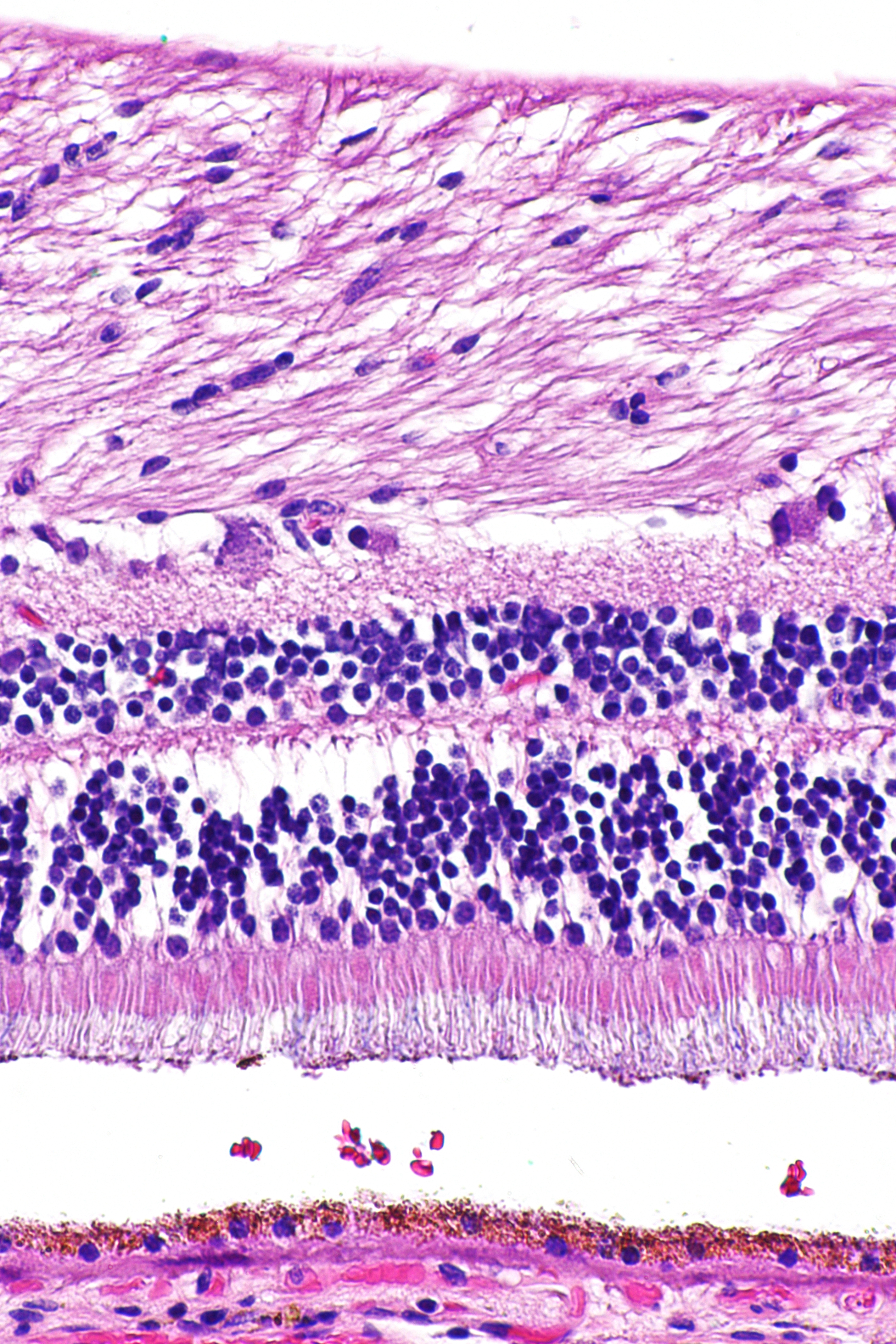
Rosenthal HE 40x
Rosenthal is a German and Jewish surname meaning "rose valley". Notable people with the name include: A * Abe M. Rosenthal (1922–2006), ''New York Times'' editor and columnist *Albert Rosenthal (1863–1939), American portrait artist *Albert J. Rosenthal (1919–2010), American legal scholar *Amy Krouse Rosenthal (1965-2017), American author *Arnold Jack Rosenthal (1923–2010), Louisiana politician; see Peter Beer *Arthur Rosenthal (1887–1959), German mathematician B *Barbara Rosenthal (born 1948), artist and writer *Ben Rosenthal (baseball) (born 1979), American baseball coach *Benjamin Stanley Rosenthal, United States Congressman (1962–1983) * Bernard "Tony" Rosenthal (1914–2009), American abstract sculptor * Bernard Rosenthal (scholar), American scholar of the Salem witchcraft trials C *Sir Charles Rosenthal (1875–1954), Australian general of World War I *Chuck Rosenthal (district attorney), Republican District Attorney of Harris County, Texas *Constantin Dan ... [...More Info...] [...Related Items...] OR: [Wikipedia] [Google] [Baidu] |
Eosinophilic
Eosinophilic (Greek suffix -phil-, meaning ''loves eosin'') is the staining of tissues, cells, or organelles after they have been washed with eosin, a dye. Eosin is an acidic dye for staining cell cytoplasm, collagen, and muscle fibers. ''Eosinophilic'' describes the appearance of cells and structures seen in histological sections that take up the staining dye eosin. Such eosinophilic structures are, in general, composed of protein. Eosin is usually combined with a stain called hematoxylin to produce a hematoxylin- and eosin-stained section (also called an H&E stain, HE or H+E section). It is the most widely used histological stain for a medical diagnosis. When a pathologist examines a biopsy of a suspected cancer, they will stain the biopsy with H&E. Some structures seen inside cells are described as being eosinophilic; for example, Lewy and Mallory bodies. [...More Info...] [...Related Items...] OR: [Wikipedia] [Google] [Baidu] |
H&E Stain
Hematoxylin and eosin stain ( or haematoxylin and eosin stain or hematoxylin-eosin stain; often abbreviated as H&E stain or HE stain) is one of the principal tissue stains used in histology. It is the most widely used stain in medical diagnosis and is often the gold standard. For example, when a pathologist looks at a biopsy of a suspected cancer, the histological section is likely to be stained with H&E. H&E is the combination of two histological stains: hematoxylin and eosin. The hematoxylin stains cell nuclei a purplish blue, and eosin stains the extracellular matrix and cytoplasm pink, with other structures taking on different shades, hues, and combinations of these colors. Hence a pathologist can easily differentiate between the nuclear and cytoplasmic parts of a cell, and additionally, the overall patterns of coloration from the stain show the general layout and distribution of cells and provides a general overview of a tissue sample's structure. Thus, pattern recogniti ... [...More Info...] [...Related Items...] OR: [Wikipedia] [Google] [Baidu] |
Gliosis
Gliosis is a nonspecific reactive change of glial cells in response to damage to the central nervous system (CNS). In most cases, gliosis involves the proliferation or hypertrophy of several different types of glial cells, including astrocytes, microglia, and oligodendrocytes. In its most extreme form, the proliferation associated with gliosis leads to the formation of a glial scar. The process of gliosis involves a series of cellular and molecular events that occur over several days. Typically, the first response to injury is the migration of macrophages and local microglia to the injury site. This process, which constitutes a form of gliosis known as microgliosis, begins within hours of the initial CNS injury. Later, after 3–5 days, oligodendrocyte precursor cells are also recruited to the site and may contribute to remyelination. The final component of gliosis is astrogliosis, the proliferation of surrounding astrocytes, which are the main constituents of the glial scar. G ... [...More Info...] [...Related Items...] OR: [Wikipedia] [Google] [Baidu] |
Pilocytic Astrocytoma
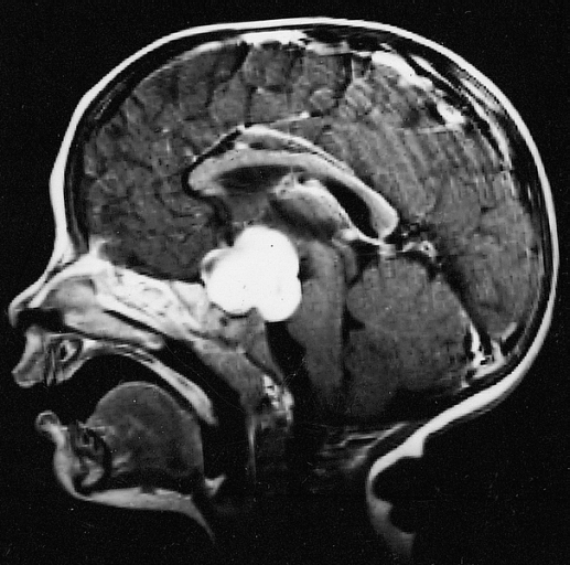
Pilocytic astrocytoma (and its variant pilomyxoid astrocytoma) is a brain tumor that occurs most commonly in children and young adults (in the first 20 years of life). They usually arise in the cerebellum, near the brainstem, in the hypothalamic region, or the optic chiasm, but they may occur in any area where astrocytes are present, including the cerebral hemispheres and the spinal cord. These tumors are usually slow growing and benign, corresponding to WHO malignancy grade 1. Signs and symptoms Children affected by pilocytic astrocytoma can present with different symptoms that might include failure to thrive (lack of appropriate weight gain/ weight loss), headache, nausea, vomiting, irritability, torticollis (tilt neck or wry neck), difficulty to coordinate movements, and visual complaints (including nystagmus). The complaints may vary depending on the location and size of the neoplasm. The most common symptoms are associated with increased intracranial pressure due to the size ... [...More Info...] [...Related Items...] OR: [Wikipedia] [Google] [Baidu] |
Alexander's Disease
Alexander disease is a very rare autosomal dominant leukodystrophy, which are neurological conditions caused by anomalies in the myelin which protects nerve fibers in the brain. The most common type is the infantile form that usually begins during the first 2 years of life. Symptoms include mental and physical developmental delays, followed by the loss of developmental milestones, an abnormal increase in head size and seizures. The juvenile form of Alexander disease has an onset between the ages of 2 and 13 years. These children may have excessive vomiting, difficulty swallowing and speaking, poor coordination, and loss of motor control. Adult-onset forms of Alexander disease are less common. The symptoms sometimes mimic those of Parkinson’s disease or multiple sclerosis, or may present primarily as a psychiatric disorder. According to the National Institute of Neurological Disorders and Stroke, the destruction of white matter is accompanied by the formation of Rosenthal fibers ... [...More Info...] [...Related Items...] OR: [Wikipedia] [Google] [Baidu] |
Leukodystrophy
Leukodystrophies are a group of usually inherited disorders characterized by degeneration of the white matter in the brain. The word ''leukodystrophy'' comes from the Greek roots ''leuko'', "white", ''dys'', "abnormal" and ''troph'', "growth". The leukodystrophies are caused by imperfect growth or development of the myelin sheath, the fatty insulating covering around nerve fibers. Leukodystrophies may be classified as hypomyelinating or demyelinating diseases, depending on whether the damage is present before birth or occurs after. Other demyelinating diseases are usually not congenital and have a toxic or autoimmune cause. When damage occurs to white matter, immune responses can lead to inflammation in the central nervous system (CNS), along with loss of myelin. The degeneration of white matter can be seen in an MRI scan and used to diagnose leukodystrophy. Leukodystrophy is characterized by specific symptoms including decreased motor function, muscle rigidity, and eventual dege ... [...More Info...] [...Related Items...] OR: [Wikipedia] [Google] [Baidu] |
Fucosidosis
Fucosidosis is a rare lysosomal storage disorder in which the FUCA1 gene experiences mutations that severely reduce or stop the activity of the alpha-L-fucosidase enzyme. The result is a buildup of complex sugars in parts of the body, which leads to death. Fucosidosis is one of nine identified glycoprotein storage diseases. The gene encoding the alpha-fucosidase, FUCA 1, was found to be located to the short arm of chromosome 1p36 - p34, by Carrit and co-workers, in 1982. Cause Fucosidosis is an autosomal recessive disorder that affects many areas of the body. Mutations in the FUCA1 gene causes fucosidosis. The FUCA1 gene provides instructions for making an enzyme called alpha-L-fucosidase. The enzyme plays a role in the breakdown of complex sugars in the body. The disorder is characterized by lysosomal accumulation of a variety of glycoproteins, glycolipids, and oligosaccharides that contain fucose moieties. The deficiency of the enzyme alpha-L-fucosidase, which is used to metab ... [...More Info...] [...Related Items...] OR: [Wikipedia] [Google] [Baidu] |
Pilocytic Astrocytoma
Pilocytic astrocytoma (and its variant pilomyxoid astrocytoma) is a brain tumor that occurs most commonly in children and young adults (in the first 20 years of life). They usually arise in the cerebellum, near the brainstem, in the hypothalamic region, or the optic chiasm, but they may occur in any area where astrocytes are present, including the cerebral hemispheres and the spinal cord. These tumors are usually slow growing and benign, corresponding to WHO malignancy grade 1. Signs and symptoms Children affected by pilocytic astrocytoma can present with different symptoms that might include failure to thrive (lack of appropriate weight gain/ weight loss), headache, nausea, vomiting, irritability, torticollis (tilt neck or wry neck), difficulty to coordinate movements, and visual complaints (including nystagmus). The complaints may vary depending on the location and size of the neoplasm. The most common symptoms are associated with increased intracranial pressure due to the size ... [...More Info...] [...Related Items...] OR: [Wikipedia] [Google] [Baidu] |
Pediatrics
Pediatrics ( also spelled ''paediatrics'' or ''pædiatrics'') is the branch of medicine that involves the medical care of infants, children, adolescents, and young adults. In the United Kingdom, paediatrics covers many of their youth until the age of 18. The American Academy of Pediatrics recommends people seek pediatric care through the age of 21, but some pediatric subspecialists continue to care for adults up to 25. Worldwide age limits of pediatrics have been trending upward year after year. A medical doctor who specializes in this area is known as a pediatrician, or paediatrician. The word ''pediatrics'' and its cognates mean "healer of children," derived from the two Greek words: (''pais'' "child") and (''iatros'' "doctor, healer"). Pediatricians work in clinics, research centers, universities, general hospitals and children's hospitals, including those who practice pediatric subspecialties (e.g. neonatology requires resources available in a NICU). History The ear ... [...More Info...] [...Related Items...] OR: [Wikipedia] [Google] [Baidu] |
Intermediate Filament
Intermediate filaments (IFs) are cytoskeletal structural components found in the cells of vertebrates, and many invertebrates. Homologues of the IF protein have been noted in an invertebrate, the cephalochordate ''Branchiostoma''. Intermediate filaments are composed of a family of related proteins sharing common structural and sequence features. Initially designated 'intermediate' because their average diameter (10 nm) is between those of narrower microfilaments (actin) and wider myosin filaments found in muscle cells, the diameter of intermediate filaments is now commonly compared to actin microfilaments (7 nm) and microtubules (25 nm). Animal intermediate filaments are subcategorized into six types based on similarities in amino acid sequence and protein structure. Most types are cytoplasmic, but one type, Type V is a nuclear lamin. Unlike microtubules, IF distribution in cells show no good correlation with the distribution of either mitochondria or endopla ... [...More Info...] [...Related Items...] OR: [Wikipedia] [Google] [Baidu] |
Glial Fibrillary Acidic Protein
Glial fibrillary acidic protein (GFAP) is a protein that is encoded by the ''GFAP'' gene in humans. It is a type III intermediate filament (IF) protein that is expressed by numerous cell types of the central nervous system (CNS), including astrocytes and ependymal cells during development. GFAP has also been found to be expressed in glomeruli and peritubular fibroblasts taken from rat kidneys, Leydig cells of the testis in both hamsters and humans, human keratinocytes, human osteocytes and chondrocytes and stellate cells of the pancreas and liver in rats. GFAP is closely related to the other three non-epithelial type III IF family members, vimentin, desmin and peripherin, which are all involved in the structure and function of the cell’s cytoskeleton. GFAP is thought to help to maintain astrocyte mechanical strength as well as the shape of cells, but its exact function remains poorly understood, despite the number of studies using it as a cell marker. The protein was named and ... [...More Info...] [...Related Items...] OR: [Wikipedia] [Google] [Baidu] |


