|
Robinow Syndrome
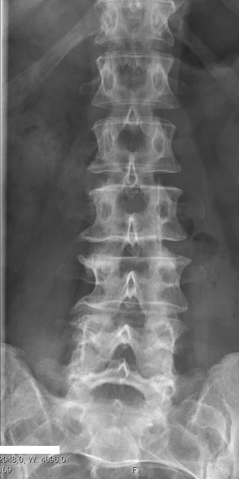
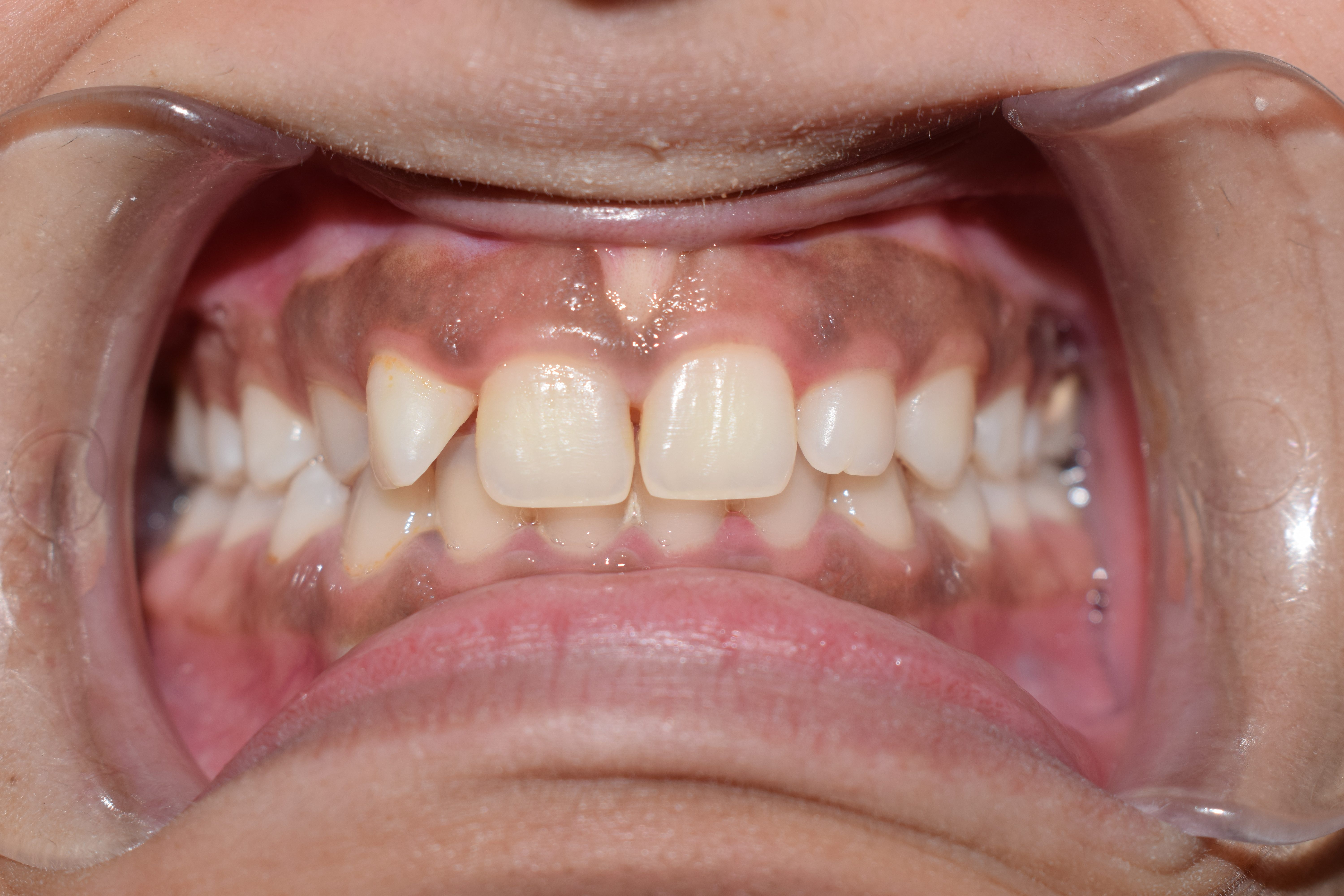
Robinow syndrome is an extremely rare genetic disorder characterized by short-limbed dwarfism, abnormalities in the head, face, and external genitalia, as well as vertebral segmentation. The disorder was first described in 1969 by human geneticist Meinhard Robinow, along with physicians Frederic N. Silverman and Hugo D. Smith, in the ''American Journal of Diseases of Children''. By 2002, over 100 cases had been documented and introduced into medical literature. Two forms of the disorder exist, dominant and recessive, of which the former is more common. Patients with the dominant version often suffer moderately from the aforementioned symptoms. Recessive cases, on the other hand, are usually more physically marked, and individuals may exhibit more skeletal abnormalities.Robinow Syndrome Foundation. General Information'. Accessed 19 May 2006. The recessive form is particularly frequent in Turkey. However, this can likely be explained by a common ancestor, as these patients' famili ... [...More Info...] [...Related Items...] OR: [Wikipedia] [Google] [Baidu] |
Genetic Disorder
A genetic disorder is a health problem caused by one or more abnormalities in the genome. It can be caused by a mutation in a single gene (monogenic) or multiple genes (polygenic) or by a chromosomal abnormality. Although polygenic disorders are the most common, the term is mostly used when discussing disorders with a single genetic cause, either in a gene or chromosome. The mutation responsible can occur spontaneously before embryonic development (a ''de novo'' mutation), or it can be Heredity, inherited from two parents who are carriers of a faulty gene (autosomal recessive inheritance) or from a parent with the disorder (autosomal dominant inheritance). When the genetic disorder is inherited from one or both parents, it is also classified as a hereditary disease. Some disorders are caused by a mutation on the X chromosome and have X-linked inheritance. Very few disorders are inherited on the Y linkage, Y chromosome or Mitochondrial disease#Causes, mitochondrial DNA (due to t ... [...More Info...] [...Related Items...] OR: [Wikipedia] [Google] [Baidu] |
Robinow Syndrome
Robinow syndrome is an extremely rare genetic disorder characterized by short-limbed dwarfism, abnormalities in the head, face, and external genitalia, as well as vertebral segmentation. The disorder was first described in 1969 by human geneticist Meinhard Robinow, along with physicians Frederic N. Silverman and Hugo D. Smith, in the ''American Journal of Diseases of Children''. By 2002, over 100 cases had been documented and introduced into medical literature. Two forms of the disorder exist, dominant and recessive, of which the former is more common. Patients with the dominant version often suffer moderately from the aforementioned symptoms. Recessive cases, on the other hand, are usually more physically marked, and individuals may exhibit more skeletal abnormalities.Robinow Syndrome Foundation. General Information'. Accessed 19 May 2006. The recessive form is particularly frequent in Turkey. However, this can likely be explained by a common ancestor, as these patients' famili ... [...More Info...] [...Related Items...] OR: [Wikipedia] [Google] [Baidu] |
Jarcho-Levin Syndrome
Spondylocostal dysostosis, also known as Jarcho-Levin syndrome (JLS), is a rare, heritable axial skeleton growth disorder. It is characterized by widespread and sometimes severe malformations of the vertebral column and ribs, shortened thorax, and moderate to severe scoliosis and kyphosis. Individuals with Jarcho-Levin typically appear to have a short trunk and neck, with arms appearing relatively long in comparison, and a slightly protuberant abdomen. Severely affected individuals may have life-threatening pulmonary complications due to deformities of the thorax. The syndrome was first described by Saul Jarcho and Paul M. Levin at Johns Hopkins University in 1938. Genetics Types include: Diagnosis Subtypes and characteristics In 1968, Dr. David Rimoin and colleagues in Baltimore first distinguished between the two major presentations of Jarcho-Levin. Both conditions were characterized as failures of proper vertebral segmentation. However, the condition within the family descr ... [...More Info...] [...Related Items...] OR: [Wikipedia] [Google] [Baidu] |
Hemivertebrae
Congenital vertebral anomalies are a collection of malformations of the spine. Most, around 85%, are not clinically significant, but they can cause compression of the spinal cord by deforming the vertebral canal or causing instability. This condition occurs in the womb. Congenital vertebral anomalies include alterations of the shape and number of vertebrae. Lumbarization and sacralization ''Lumbarization'' is an anomaly in the spine. It is defined by the nonfusion of the first and second segments of the sacrum. The lumbar spine subsequently appears to have six vertebrae or segments, not five. This sixth lumbar vertebra is known as a transitional vertebra. Conversely the sacrum appears to have only four segments instead of its designated five segments. Lumbosacral transitional vertebrae consist of the process of the last lumbar vertebra fusing with the first sacral segment. While only around 10 percent of adults have a spinal abnormality due to genetics, a sixth lumbar vertebra ... [...More Info...] [...Related Items...] OR: [Wikipedia] [Google] [Baidu] |
Butterfly Vertebrae
Congenital vertebral anomalies are a collection of malformations of the spine. Most, around 85%, are not clinically significant, but they can cause compression of the spinal cord by deforming the vertebral canal or causing instability. This condition occurs in the womb. Congenital vertebral anomalies include alterations of the shape and number of vertebrae. Lumbarization and sacralization ''Lumbarization'' is an anomaly in the spine. It is defined by the nonfusion of the first and second segments of the sacrum. The lumbar spine subsequently appears to have six vertebrae or segments, not five. This sixth lumbar vertebra is known as a transitional vertebra. Conversely the sacrum appears to have only four segments instead of its designated five segments. Lumbosacral transitional vertebrae consist of the process of the last lumbar vertebra fusing with the first sacral segment. While only around 10 percent of adults have a spinal abnormality due to genetics, a sixth lumbar verteb ... [...More Info...] [...Related Items...] OR: [Wikipedia] [Google] [Baidu] |
Ectrodactyly
Ectrodactyly, split hand, or cleft hand (derived from Greek ''ektroma'' 'abortion' and ''daktylos'' 'finger') involves the deficiency or absence of one or more central digits of the hand or foot and is also known as split hand/split foot malformation (SHFM). The hands and feet of people with ectrodactyly (ectrodactyls) are often described as "claw-like" and may include only the thumb and one finger (usually either the little finger, ring finger, or a syndactyly of the two) with similar abnormalities of the feet. It is a substantial rare form of a congenital disorder in which the development of the hand is disturbed. It is a type I failure of formation – longitudinal arrest. The central ray of the hand is affected and usually appears without proximal deficiencies of nerves, vessels, tendons, muscles and bones in contrast to the radial and ulnar deficiencies. The cleft hand appears as a V-shaped cleft situated in the centre of the hand. The digits at the borders of the cleft might ... [...More Info...] [...Related Items...] OR: [Wikipedia] [Google] [Baidu] |
Brachydactyly
Brachydactyly (Greek βραχύς = "short" plus δάκτυλος = "finger"), is a medical term which literally means "short finger". The shortness is relative to the length of other long bones and other parts of the body. Brachydactyly is an inherited, dominant trait. It most often occurs as an isolated dysmelia, but can also occur with other anomalies as part of many congenital syndromes. Brachydactyly may also be a signal that one is at risk for congenital heart disease due to the association between congenital heart disease and carpenter's syndrome and the link between carpenter's syndrome and brachydactyly Nomograms for normal values of finger length as a ratio to other body measurements have been published. In clinical genetics, the most commonly used index of digit length is the dimensionless ratio of the length of the third (middle) finger to the hand length. Both are expressed in the same units (centimeters, for example) and are measured in an open hand from the finger ... [...More Info...] [...Related Items...] OR: [Wikipedia] [Google] [Baidu] |
Mesomelic Dwarfism
Mesomelia refers to conditions in which the middle parts of limbs are disproportionately short. When applied to skeletal dysplasias, mesomelic dwarfism describes generalised shortening of the forearms and lower legs. This is in contrast to rhizomelic dwarfism in which the upper portions of limbs are short such as in achondroplasia. Forms of mesomelic dwarfism currently described include: * Langer mesomelic dysplasia * Ellis–van Creveld syndrome * Robinow syndrome * Léri–Weill dyschondrosteosis Léri–Weill dyschondrosteosis or LWD is a rare pseudoautosomal dominant genetic disorder which results in dwarfism with short forearms and legs ( mesomelic dwarfism) and a bayonet-like deformity of the forearms (Madelung's deformity). Causes It ... References Growth disorders {{congenital-malformation-stub ... [...More Info...] [...Related Items...] OR: [Wikipedia] [Google] [Baidu] |
Pinna (anatomy)
The auricle or auricula is the visible part of the ear that is outside the head. It is also called the pinna (Latin for "wing" or "fin", plural pinnae), a term that is used more in zoology. Structure The diagram shows the shape and location of most of these components: * ''antihelix'' forms a 'Y' shape where the upper parts are: ** ''Superior crus'' (to the left of the ''fossa triangularis'' in the diagram) ** ''Inferior crus'' (to the right of the ''fossa triangularis'' in the diagram) * ''Antitragus'' is below the ''tragus'' * ''Aperture'' is the entrance to the ear canal * ''Auricular sulcus'' is the depression behind the ear next to the head * ''Concha'' is the hollow next to the ear canal * Conchal angle is the angle that the back of the ''concha'' makes with the side of the head * ''Crus'' of the helix is just above the ''tragus'' * ''Cymba conchae'' is the narrowest end of the ''concha'' * External auditory meatus is the ear canal * ''Fossa triangularis'' is the depres ... [...More Info...] [...Related Items...] OR: [Wikipedia] [Google] [Baidu] |
Eyelid
An eyelid is a thin fold of skin that covers and protects an eye. The levator palpebrae superioris muscle retracts the eyelid, exposing the cornea to the outside, giving vision. This can be either voluntarily or involuntarily. The human eyelid features a row of eyelashes along the eyelid margin, which serve to heighten the protection of the eye from dust and foreign debris, as well as from perspiration. "Palpebral" (and "blepharal") means relating to the eyelids. Its key function is to regularly spread the tears and other secretions on the eye surface to keep it moist, since the cornea must be continuously moist. They keep the eyes from drying out when asleep. Moreover, the blink reflex protects the eye from foreign bodies. The appearance of the human upper eyelid often varies between different populations. The prevalence of an epicanthic fold covering the inner corner of the eye account for the majority of East Asian and Southeast Asian populations, and is also found i ... [...More Info...] [...Related Items...] OR: [Wikipedia] [Google] [Baidu] |
Organ Hypertrophy
Hypertrophy is the increase in the volume of an organ or tissue due to the enlargement of its component cells. It is distinguished from hyperplasia, in which the cells remain approximately the same size but increase in number.Updated by Linda J. Vorvick. 8/14/1Hyperplasia/ref> Although hypertrophy and hyperplasia are two distinct processes, they frequently occur together, such as in the case of the hormonally-induced proliferation and enlargement of the cells of the uterus during pregnancy. Eccentric hypertrophy is a type of hypertrophy where the walls and chamber of a hollow organ undergo growth in which the overall size and volume are enlarged. It is applied especially to the left ventricle of heart. Sarcomeres are added in series, as for example in dilated cardiomyopathy (in contrast to hypertrophic cardiomyopathy, a type of concentric hypertrophy, where sarcomeres are added in parallel). Gallery File:*+ * Photographic documentation on sexual education - Hypertrophy of brea ... [...More Info...] [...Related Items...] OR: [Wikipedia] [Google] [Baidu] |
Gingiva
The gums or gingiva (plural: ''gingivae'') consist of the mucosal tissue that lies over the mandible and maxilla inside the mouth. Gum health and disease can have an effect on general health. Structure The gums are part of the soft tissue lining of the mouth. They surround the teeth and provide a seal around them. Unlike the soft tissue linings of the lips and cheeks, most of the gums are tightly bound to the underlying bone which helps resist the friction of food passing over them. Thus when healthy, it presents an effective barrier to the barrage of periodontal insults to deeper tissue. Healthy gums are usually coral pink in light skinned people, and may be naturally darker with melanin pigmentation. Changes in color, particularly increased redness, together with swelling and an increased tendency to bleed, suggest an inflammation that is possibly due to the accumulation of bacterial plaque. Overall, the clinical appearance of the tissue reflects the underlying histology, b ... [...More Info...] [...Related Items...] OR: [Wikipedia] [Google] [Baidu] |


