|
Rigler's Triad
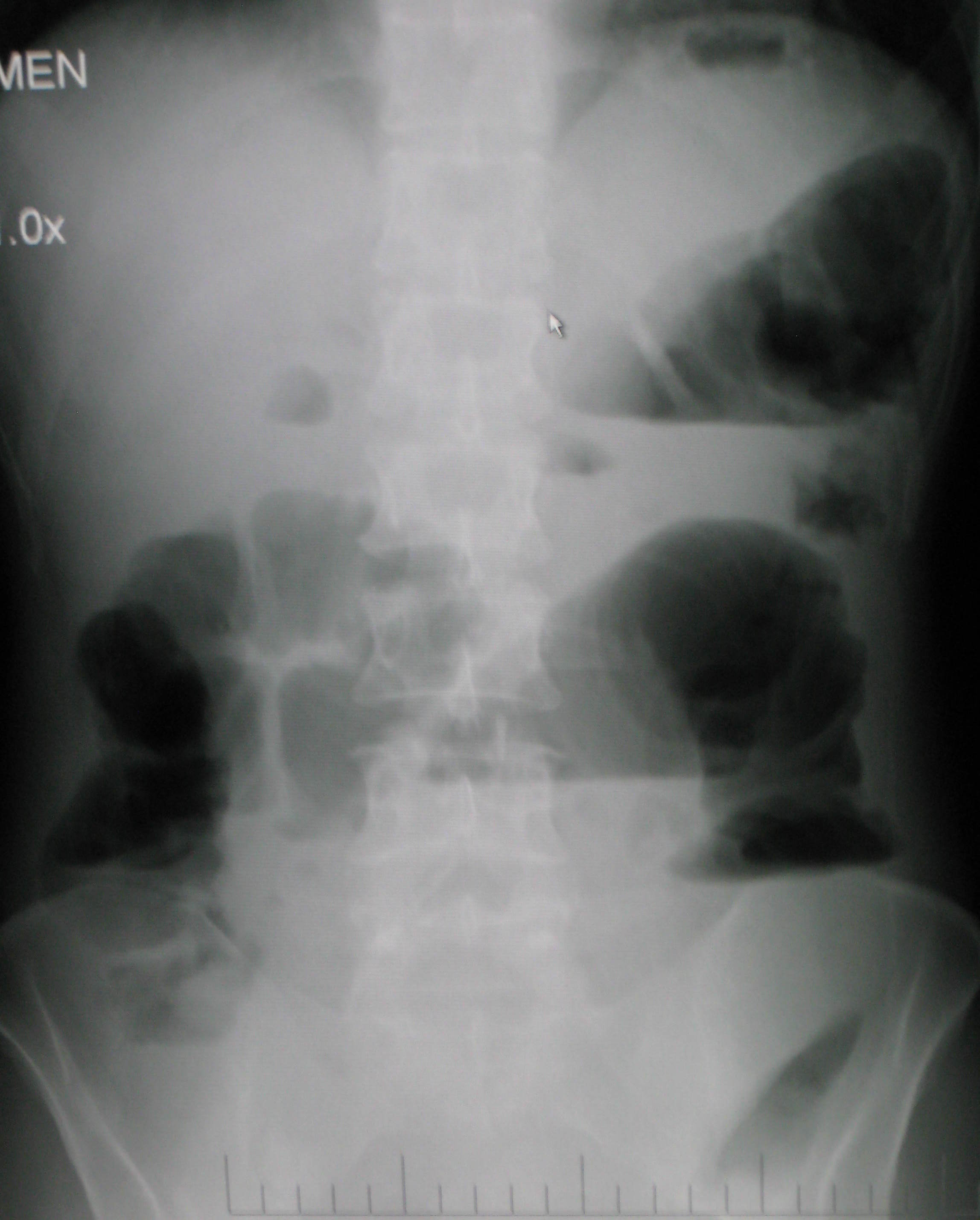
Rigler's triad is a combination of findings on an abdominal radiograph of people with gallstone ileus, a condition where a large gallstone causes bowel obstruction. Rigler's triad consists of: (1) small bowel obstruction, (2) a gallstone outside the gallbladder, and (3) air in the bile ducts. It bears the name of Leo George Rigler (1896–1979), who described it in 1941. It is not the same as Rigler's sign. It is most commonly seen in 6th to 7th decade of life and affects females more often. Most patients with gallstone ileus are asymptomatic. Due to the fistula formation between the small intestine and gallbladder, large stones can lodge in the small bowel, leading to its obstruction. Pneumobilia means air in the biliary tract. It is due to the transfer of air from bowel through the fistula A fistula (plural: fistulas or fistulae ; from Latin ''fistula'', "tube, pipe") in anatomy is an abnormal connection between two hollow spaces (technically, two epithelialized surfa ... [...More Info...] [...Related Items...] OR: [Wikipedia] [Google] [Baidu] |
Abdominal X-ray
An abdominal x-ray is an x-ray of the abdomen. It is sometimes abbreviated to AXR, or KUB (for kidneys, ureters, and urinary bladder). Indications In children, abdominal x-ray is indicated in the acute setting: *Suspected bowel obstruction or gastrointestinal perforation; Abdominal x-ray will demonstrate most cases of bowel obstruction, by showing dilated bowel loops. * Foreign body in the alimentary tract; can be identified if it is radiodense. *Suspected abdominal mass *In suspected intussusception, an abdominal x-ray does not exclude intussusception but is useful in the differential diagnosis to exclude perforation or obstruction. Yet, CT scan is the best alternative for diagnosing intra-abdominal injury. Computed tomography provides an overall better surgical strategy planning, and possibly less unnecessary laparotomies. Abdominal x-ray is therefore not recommended for adults with acute abdominal pain presenting in the emergency department. Projections The standard a ... [...More Info...] [...Related Items...] OR: [Wikipedia] [Google] [Baidu] |
Gallstone Ileus
Gallstone ileus is a rare form of small bowel obstruction caused by an impaction of a gallstone within the lumen of the small intestine. Such a gallstone enters the bowel via a cholecysto-enteric fistula. The presence of large stones, >2.5 cm in diameter, within the gallbladder are thought to predispose to fistula formation by gradual erosion through the gallbladder fundus. Once a fistula has formed, a stone may travel from the gallbladder into the bowel and become lodged almost anywhere along the gastrointestinal tract. Obstruction occurs most commonly at the near the distal ileum, within 60 cm proximally to the ileocecal valve. Rarely, gallstone ileus may recur if the underlying fistula is not treated. First described by Thomas Bartholin in 1654, the name "gallstone ileus" is a misnomer because an ileus is, by definition, a ''non-mechanical'' bowel motility failure (as opposed to a mechanical obstruction by a stone). Diagnosis Diagnosis of gallstone ileus requir ... [...More Info...] [...Related Items...] OR: [Wikipedia] [Google] [Baidu] |
Gallstone
A gallstone is a calculus (medicine), stone formed within the gallbladder from precipitated bile components. The term cholelithiasis may refer to the presence of gallstones or to any disease caused by gallstones, and choledocholithiasis refers to the presence of migrated gallstones within bile ducts. Most people with gallstones (about 80%) are asymptomatic. However, when a gallstone obstructs the bile duct and causes acute cholestasis, a reflexive smooth muscle spasm often occurs, resulting in an intense cramp-like visceral pain in the quadrant (abdomen), right upper part of the abdomen known as a biliary colic (or "gallbladder attack"). This happens in 1–4% of those with gallstones each year. Complications from gallstones may include inflammation of the gallbladder (cholecystitis), inflammation of the pancreas (pancreatitis), Jaundice#Post-hepatic, obstructive jaundice, and infection in bile ducts (ascending cholangitis, cholangitis). Symptoms of these complications may include ... [...More Info...] [...Related Items...] OR: [Wikipedia] [Google] [Baidu] |
Bowel Obstruction
Bowel obstruction, also known as intestinal obstruction, is a mechanical or Ileus, functional obstruction of the Gastrointestinal tract#Lower gastrointestinal tract, intestines which prevents the normal movement of the products of digestion. Either the Small intestine, small bowel or Large intestine, large bowel may be affected. Signs and symptoms include abdominal pain, vomiting, abdominal bloating, bloating and not passing flatulence, gas. Mechanical obstruction is the cause of about 5 to 15% of cases of acute abdomen, severe abdominal pain of sudden onset requiring admission to hospital. Causes of bowel obstruction include Adhesion (medicine), adhesions, hernias, volvulus, endometriosis, inflammatory bowel disease, appendicitis, Neoplasm, tumors, diverticulitis, ischemic colitis, ischemic bowel, tuberculosis and intussusception (medical disorder), intussusception. Small bowel obstructions are most often due to adhesions and hernias while large bowel obstructions are most often ... [...More Info...] [...Related Items...] OR: [Wikipedia] [Google] [Baidu] |
Gallbladder
In vertebrates, the gallbladder, also known as the cholecyst, is a small hollow organ where bile is stored and concentrated before it is released into the small intestine. In humans, the pear-shaped gallbladder lies beneath the liver, although the structure and position of the gallbladder can vary significantly among animal species. It receives and stores bile, produced by the liver, via the common hepatic duct, and releases it via the common bile duct into the duodenum, where the bile helps in the digestion of fats. The gallbladder can be affected by gallstones, formed by material that cannot be dissolved – usually cholesterol or bilirubin, a product of haemoglobin breakdown. These may cause significant pain, particularly in the upper-right corner of the abdomen, and are often treated with removal of the gallbladder (called a cholecystectomy). Cholecystitis, inflammation of the gallbladder, has a wide range of causes, including result from the impaction of gallstones, inf ... [...More Info...] [...Related Items...] OR: [Wikipedia] [Google] [Baidu] |
Pneumobilia
Pneumobilia is the presence of gas in the biliary system. It is typically detected by ultrasound or a radiographic imaging exam, such as CT, or MRI. It is a common finding in patients that have recently undergone biliary surgery or endoscopic biliary procedure. While the presence of air within biliary system is not harmful, this finding may alternatively suggest a pathological process, such as a biliary-enteric anastomosis, an infection of the biliary system, an incompetent sphincter of Oddi, or spontaneous biliary-enteric fistula. Causes In a healthy individual with normal anatomy, there is no air within the biliary tree. When this finding is present, it may be secondary to: * Recent surgical or endoscopic biliary procedure (e.g. ERCP, biliary enteric anastomosis) * Incompetent sphincter of Oddi (e.g. passage of large gallstone, scarring related to chronic pancreatitis) * Spontaneous biliary enteric fistula (e.g. gallstone ileus) * Infection by gas-forming organisms (e.g. emph ... [...More Info...] [...Related Items...] OR: [Wikipedia] [Google] [Baidu] |
Leo George Rigler
Leo George Rigler (16 October 1896, Minneapolis – 25 October 1979) was an American radiologist remembered for describing Rigler's sign. Biography Leo Rigler attended the University of Minnesota, receiving a B.S. in 1917, B.M. in 1919 and M.D. in 1920. He undertook his internship at the St. Louis City Hospital where he watched pioneering radiologist Dr. Leroy Sante perform fluoroscopy, and realised the potential of this new technique. He worked as a general practitioner in New England, North Dakota, but returned to the University of Minnesota after a short time. He then worked as a teaching fellow in internal medicine for a year, becoming responsible for the radiology service. He then undertook a 3-year post as radiologist at the Minneapolis General Hospital. During this time he also trained in radiology at the Battle Creek Sanatorium and the University of Michigan. In 1924 he travelled to Europe with funding from the University of Minnesota Medical School. He spent most o ... [...More Info...] [...Related Items...] OR: [Wikipedia] [Google] [Baidu] |
Rigler's Sign
Pneumoperitoneum is pneumatosis (abnormal presence of air or other gas) in the peritoneal cavity, a potential space within the abdominal cavity. The most common cause is a perforated abdominal organ, generally from a perforated peptic ulcer, although any part of the bowel may perforate from a benign ulcer, tumor or abdominal trauma. A perforated appendix seldom causes a pneumoperitoneum. Spontaneous pneumoperitoneum is a rare case that is not caused by an abdominal organ rupture. This is also called an idiopathic spontaneous pneumoperitoneum when the cause is not known. In the mid-twentieth century, an "artificial" pneumoperitoneum was sometimes intentionally administered as a treatment for a hiatal hernia. This was achieved by insufflating the abdomen with carbon dioxide. The practice is currently used by surgical teams in order to aid in performing laparoscopic surgery. Causes * Perforated duodenal ulcer – The most common cause of rupture in the abdomen. Especially of the a ... [...More Info...] [...Related Items...] OR: [Wikipedia] [Google] [Baidu] |
Asymptomatic
In medicine, any disease is classified asymptomatic if a patient tests as carrier for a disease or infection but experiences no symptoms. Whenever a medical condition fails to show noticeable symptoms after a diagnosis it might be considered asymptomatic. Infections of this kind are usually called subclinical infections. Diseases such as mental illnesses or psychosomatic conditions are considered subclinical if they present some individual symptoms but not all those normally required for a clinical diagnosis. The term clinically silent is also found. Producing only a few, mild symptoms, disease is paucisymptomatic. Symptoms appearing later, after an asymptomatic incubation period, mean a pre-symptomatic period has existed. Importance Knowing that a condition is asymptomatic is important because: * It may develop symptoms later and only then require treatment. * It may resolve itself or become benign. * It may be contagious, and the contribution of asymptomatic and pre-symptomat ... [...More Info...] [...Related Items...] OR: [Wikipedia] [Google] [Baidu] |
Small Intestine
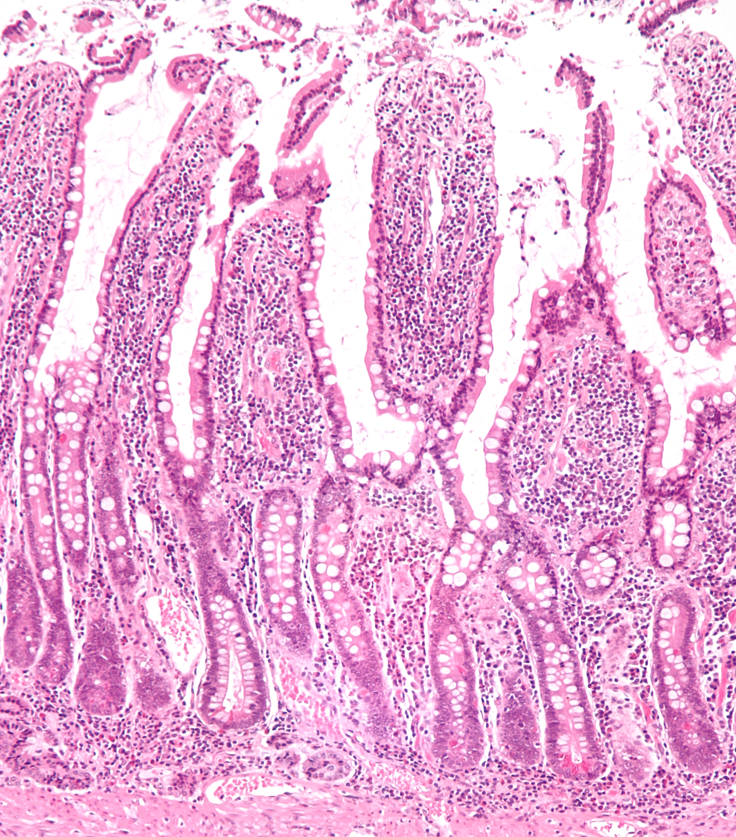
The small intestine or small bowel is an organ in the gastrointestinal tract where most of the absorption of nutrients from food takes place. It lies between the stomach and large intestine, and receives bile and pancreatic juice through the pancreatic duct to aid in digestion. The small intestine is about long and folds many times to fit in the abdomen. Although it is longer than the large intestine, it is called the small intestine because it is narrower in diameter. The small intestine has three distinct regions – the duodenum, jejunum, and ileum. The duodenum, the shortest, is where preparation for absorption through small finger-like protrusions called villi begins. The jejunum is specialized for the absorption through its lining by enterocytes: small nutrient particles which have been previously digested by enzymes in the duodenum. The main function of the ileum is to absorb vitamin B12, bile salts, and whatever products of digestion that were not absorbed by the ... [...More Info...] [...Related Items...] OR: [Wikipedia] [Google] [Baidu] |
Fistula
A fistula (plural: fistulas or fistulae ; from Latin ''fistula'', "tube, pipe") in anatomy is an abnormal connection between two hollow spaces (technically, two epithelialized surfaces), such as blood vessels, intestines, or other hollow organs. Types of fistula can be described by their location. Anal fistulas connect between the anal canal and the perianal skin. Anovaginal or rectovaginal fistulas occur when a hole develops between the anus or rectum and the vagina. Colovaginal fistulas occur between the colon and the vagina. Urinary tract fistulas are abnormal openings within the urinary tract or an abnormal connection between the urinary tract and another organ such as between the bladder and the uterus in a vesicouterine fistula, between the bladder and the vagina in a vesicovaginal fistula, and between the urethra and the vagina in urethrovaginal fistula. When occurring between two parts of the intestine, it is known as an enteroenteral fistula, between the small intest ... [...More Info...] [...Related Items...] OR: [Wikipedia] [Google] [Baidu] |
Radiologic Signs
A radiologic sign is an objective indication of some medical fact (that is, a medical sign) that is detected by a physician during radiologic examination with medical imaging (for example, via an X-ray, CT scan, MRI scan, or sonographic scan). Examples * Double decidual sac sign * Face of the giant panda sign * Football sign * Golden S sign * Hampton's hump * Hilum overlay sign * Kerley lines * Mickey Mouse sign * Omental cake * Peribronchial cuffing * Pneumatosis intestinalis * Rigler's sign * Westermark sign In chest radiography, the Westermark sign is a sign that represents a focus of oligemia (hypovolemia) (leading to collapse of vessel) seen distal to a pulmonary embolism (PE). While the chest x-ray is normal in the majority of PE cases, the Westerm ... References See also List of radiologic signs {{Radiologic signs * ... [...More Info...] [...Related Items...] OR: [Wikipedia] [Google] [Baidu] |


