|
Right Bundle Branch Block
A right bundle branch block (RBBB) is a heart block in the right bundle branch of the electrical conduction system. During a right bundle branch block, the right ventricle is not directly activated by impulses travelling through the right bundle branch. The left ventricle, however, is still normally activated by the left bundle branch. These impulses are then able to travel through the myocardium of the left ventricle to the right ventricle and depolarize the right ventricle this way. As conduction through the myocardium is slower than conduction through the Bundle of His-Purkinje fibres, the QRS complex is seen to be widened. The QRS complex often shows an extra deflection that reflects the rapid depolarisation of the left ventricle followed by the slower depolarisation of the right ventricle. Incomplete right bundle branch block Incomplete right bundle branch block (IRBBB) is an conduction abnormality in the right bundle branch block. While a complete RBBB has a QRS dura ... [...More Info...] [...Related Items...] OR: [Wikipedia] [Google] [Baidu] |
Intraventricular Septum
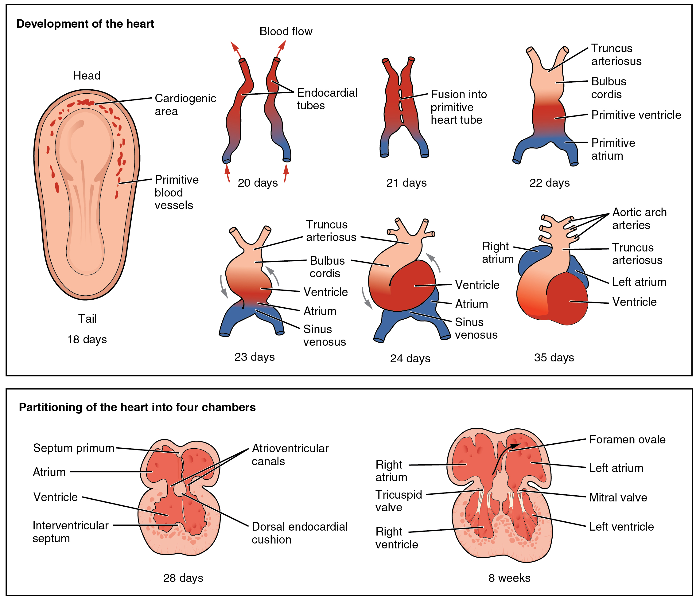
The interventricular septum (IVS, or ventricular septum, or during development septum inferius) is the stout wall separating the ventricles, the lower chambers of the heart, from one another. The ventricular septum is directed obliquely backward to the right and curved with the convexity toward the right ventricle; its margins correspond with the anterior and posterior interventricular sulci. The lower part of the septum, which is the major part, is thick and muscular, and its much smaller upper part is thin and membraneous. During each cardiac cycle the interventricular septum contracts by shortening longitudinally and becoming thicker. Structure The interventricular septum is the stout wall separating the ventricles, the lower chambers of the heart, from one another. The ventricular septum is directed obliquely backward to the right and curved with the convexity toward the right ventricle; its margins correspond with the anterior and posterior longitudinal sulci. The g ... [...More Info...] [...Related Items...] OR: [Wikipedia] [Google] [Baidu] |
Rheumatic Heart Disease
Rheumatic fever (RF) is an inflammatory disease that can involve the heart, joints, skin, and brain. The disease typically develops two to four weeks after a streptococcal throat infection. Signs and symptoms include fever, multiple painful joints, involuntary muscle movements, and occasionally a characteristic non-itchy rash known as erythema marginatum. The heart is involved in about half of the cases. Damage to the heart valves, known as rheumatic heart disease (RHD), usually occurs after repeated attacks but can sometimes occur after one. The damaged valves may result in heart failure, atrial fibrillation and infection of the valves. Rheumatic fever may occur following an infection of the throat by the bacterium ''Streptococcus pyogenes''. If the infection is left untreated, rheumatic fever occurs in up to three percent of people. The underlying mechanism is believed to involve the production of antibodies against a person's own tissues. Due to their genetics, some peop ... [...More Info...] [...Related Items...] OR: [Wikipedia] [Google] [Baidu] |
Intraventricular Block
An intraventricular block is a heart conduction disorder — heart block of the ventricles of the heart. An example is a right bundle branch block, right fascicular block, bifascicular block, trifascicular block. Types Types of intraventricular blocks are * Fascicular block **Left anterior fascicular block **Left posterior fascicular block * Trifascicular block * Bifascicular block (RBBB with fascicular block) * Right bundle branch block (RBBB) * Left bundle branch block (LBBB) Intraventricular conduction delay Intraventricular conduction delays (IVCD) are conduction disorders seen in intraventricular propagation of supraventricular impulses resulting in changes in the QRS complex duration or morphology, or both. IVCD can be caused by abnormalities in the structures of bundle of His, Purkinje fibers The Purkinje fibers (; often incorrectly ; Purkinje tissue or subendocardial branches) are located in the inner ventricular walls of the heart, just beneath the endoc ... [...More Info...] [...Related Items...] OR: [Wikipedia] [Google] [Baidu] |
Angioplasty
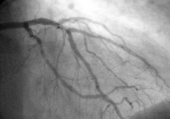
Angioplasty, is also known as balloon angioplasty and percutaneous transluminal angioplasty (PTA), is a minimally invasive endovascular procedure used to widen narrowed or obstructed arteries or veins, typically to treat arterial atherosclerosis. A deflated balloon attached to a catheter (a balloon catheter) is passed over a guide-wire into the narrowed vessel and then inflated to a fixed size. The balloon forces expansion of the blood vessel and the surrounding muscular wall, allowing an improved blood flow. A stent may be inserted at the time of ballooning to ensure the vessel remains open, and the balloon is then deflated and withdrawn. Angioplasty has come to include all manner of vascular interventions that are typically performed percutaneously. The word is composed of the combining forms of the Greek words ἀνγεῖον ' "vessel" or "cavity" (of the human body) and πλάσσω ' "form" or "mould". Uses and indications Coronary angioplasty A coronary ... [...More Info...] [...Related Items...] OR: [Wikipedia] [Google] [Baidu] |
Atrioventricular Node
The atrioventricular node or AV node electrically connects the heart's atria and ventricles to coordinate beating in the top of the heart; it is part of the electrical conduction system of the heart. The AV node lies at the lower back section of the interatrial septum near the opening of the coronary sinus, and conducts the normal electrical impulse from the atria to the ventricles. The AV node is quite compact (~1 x 3 x 5 mm).Full Size Picture triangle of-Koch.jpg Retrieved on 2008-12-22 Structure Location The AV node lies at the lower back section of the |
Atrium (heart)
The atrium ( la, ātrium, , entry hall) is one of two upper chambers in the heart that receives blood from the circulatory system. The blood in the atria is pumped into the heart ventricles through the atrioventricular valves. There are two atria in the human heart – the left atrium receives blood from the pulmonary circulation, and the right atrium receives blood from the venae cavae of the systemic circulation. During the cardiac cycle the atria receive blood while relaxed in diastole, then contract in systole to move blood to the ventricles. Each atrium is roughly cube-shaped except for an ear-shaped projection called an atrial appendage, sometimes known as an auricle. All animals with a closed circulatory system have at least one atrium. The atrium was formerly called the 'auricle'. That term is still used to describe this chamber in some other animals, such as the ''Mollusca''. They have thicker muscular walls than the atria do. Structure Humans have a four-chambered ... [...More Info...] [...Related Items...] OR: [Wikipedia] [Google] [Baidu] |
Sinoatrial Node
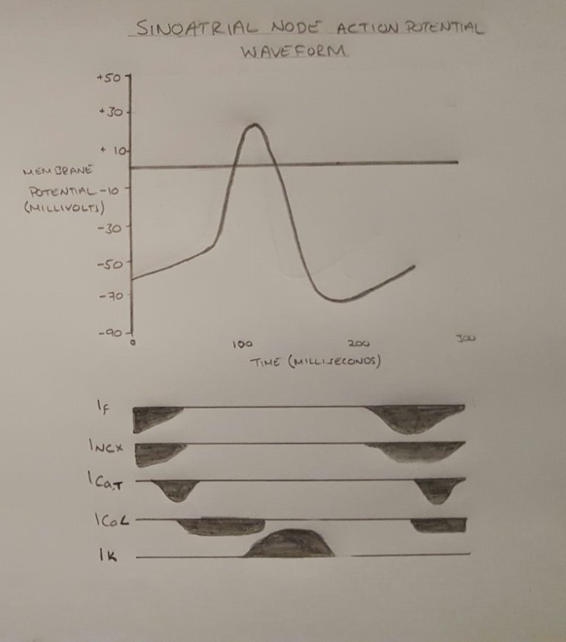
The sinoatrial node (also known as the sinuatrial node, SA node or sinus node) is an oval shaped region of special cardiac muscle in the upper back wall of the right atrium made up of cells known as pacemaker cells. The sinus node is approximately fifteen mm long, three mm wide, and one mm thick, located directly below and to the side of the superior vena cava. These cells can produce an electrical impulse an action potential known as a cardiac action potential that travels through the electrical conduction system of the heart, causing it to contract. In a healthy heart, the SA node continuously produces action potentials, setting the rhythm of the heart (sinus rhythm), and so is known as the heart's natural pacemaker. The rate of action potentials produced (and therefore the heart rate) is influenced by the nerves that supply it. Structure The sinoatrial node is a oval-shaped structure that is approximately fifteen mm long, three mm wide, and one mm thick, located directly ... [...More Info...] [...Related Items...] OR: [Wikipedia] [Google] [Baidu] |
Atrioventricular Node
The atrioventricular node or AV node electrically connects the heart's atria and ventricles to coordinate beating in the top of the heart; it is part of the electrical conduction system of the heart. The AV node lies at the lower back section of the interatrial septum near the opening of the coronary sinus, and conducts the normal electrical impulse from the atria to the ventricles. The AV node is quite compact (~1 x 3 x 5 mm).Full Size Picture triangle of-Koch.jpg Retrieved on 2008-12-22 Structure Location The AV node lies at the lower back section of the |
Sinoatrial Node
The sinoatrial node (also known as the sinuatrial node, SA node or sinus node) is an oval shaped region of special cardiac muscle in the upper back wall of the right atrium made up of cells known as pacemaker cells. The sinus node is approximately fifteen mm long, three mm wide, and one mm thick, located directly below and to the side of the superior vena cava. These cells can produce an electrical impulse an action potential known as a cardiac action potential that travels through the electrical conduction system of the heart, causing it to contract. In a healthy heart, the SA node continuously produces action potentials, setting the rhythm of the heart (sinus rhythm), and so is known as the heart's natural pacemaker. The rate of action potentials produced (and therefore the heart rate) is influenced by the nerves that supply it. Structure The sinoatrial node is a oval-shaped structure that is approximately fifteen mm long, three mm wide, and one mm thick, located directly ... [...More Info...] [...Related Items...] OR: [Wikipedia] [Google] [Baidu] |
Electrical Conduction System Of The Heart
The cardiac conduction system (CCS) (also called the electrical conduction system of the heart) transmits the signals generated by the sinoatrial node – the heart's pacemaker, to cause the heart muscle to contract, and pump blood through the body's circulatory system. The pacemaking signal travels through the right atrium to the atrioventricular node, along the bundle of His, and through the bundle branches to Purkinje fibers in the walls of the ventricles. The Purkinje fibers transmit the signals more rapidly to stimulate contraction of the ventricles. The conduction system consists of specialized heart muscle cells, situated within the myocardium. There is a skeleton of fibrous tissue that surrounds the conduction system which can be seen on an ECG. Dysfunction of the conduction system can cause irregular heart rhythms including rhythms that are too fast or too slow. Structure Electrical signals arising in the SA node (located in the right atrium) stimulate the atr ... [...More Info...] [...Related Items...] OR: [Wikipedia] [Google] [Baidu] |
Ventricular Remodeling
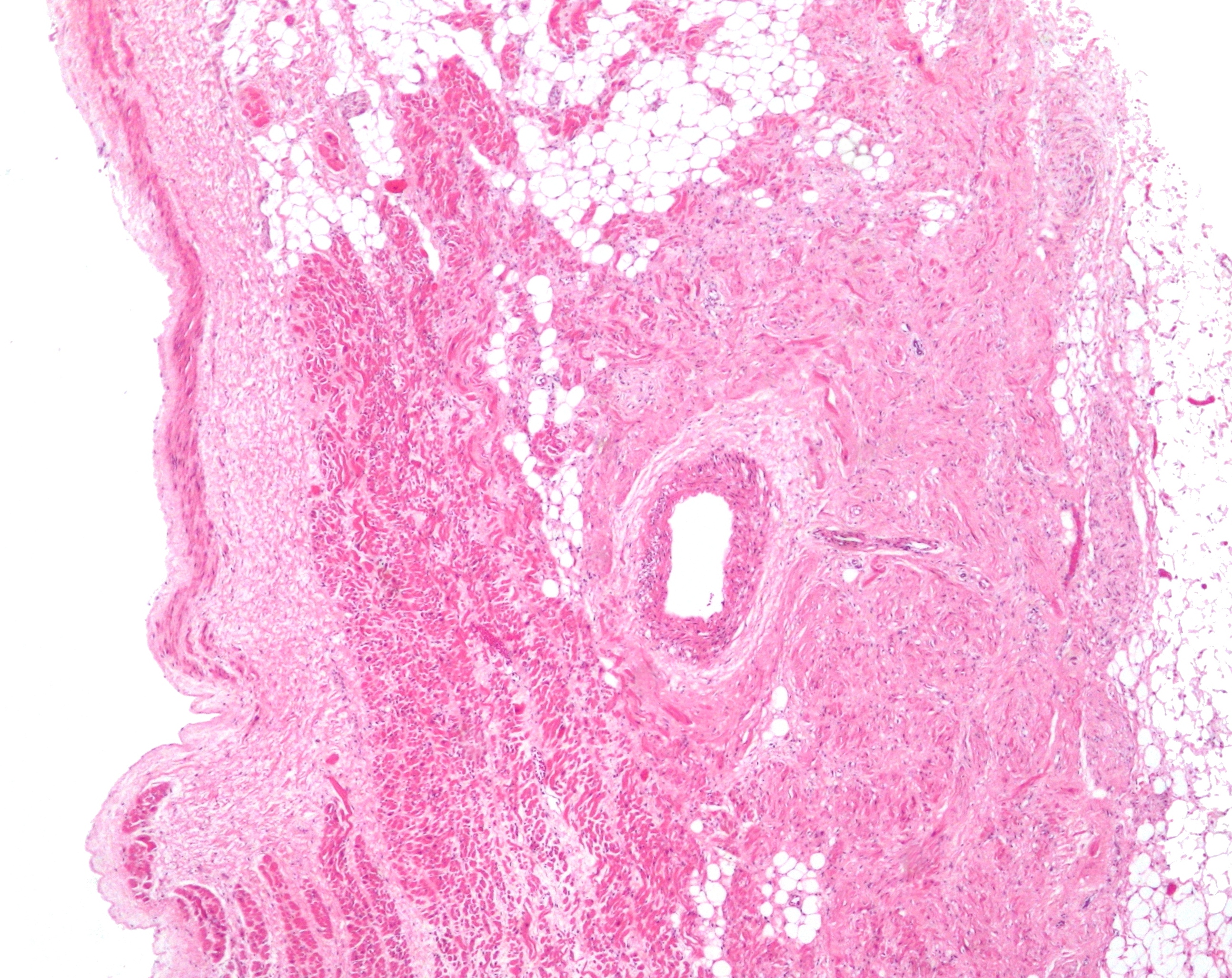
In cardiology, ventricular remodeling (or cardiac remodeling) refers to changes in the size, shape, structure, and function of the heart. This can happen as a result of exercise (physiological remodeling) or after injury to the heart muscle (pathological remodeling). The injury is typically due to acute myocardial infarction (usually transmural or ST segment elevation infarction), but may be from a number of causes that result in increased pressure or volume, causing pressure overload or volume overload (forms of strain) on the heart. Chronic hypertension, congenital heart disease with intracardiac shunting, and valvular heart disease may also lead to remodeling. After the insult occurs, a series of histopathological and structural changes occur in the left ventricular myocardium that lead to progressive decline in left ventricular performance. Ultimately, ventricular remodeling may result in diminished contractile (systolic) function and reduced stroke volume. Physiological remodeli ... [...More Info...] [...Related Items...] OR: [Wikipedia] [Google] [Baidu] |


