|
Right-sided Aortic Arch
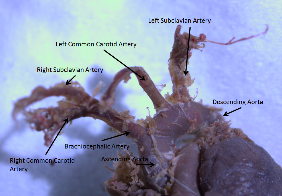
Right-sided aortic arch is a rare anatomical variant in which the aortic arch is on the right side rather than on the left. During normal embryonic development, the aortic arch is formed by the left fourth aortic arch and the left dorsal aorta. In people with a right-sided aortic arch, instead the right dorsal aorta persists and the distal left aorta disappears. Symptoms and signs A right-sided aortic arch does not cause symptoms on itself, and the overwhelming majority of people with the right-sided arch have no other symptoms. However when it is accompanied by other vascular abnormalities, it may form a vascular ring, causing symptoms due to compression of the trachea and/or esophagus. Pathophysiology The causes of right-sided aortic arch are still unknown, 22q11 deletions have been found in some people with this condition. It has also been found in association with other genetic syndromes such as Trisomy 21 (Down syndrome). Diagnosis During pregnancy, prenatal ultrasound m ... [...More Info...] [...Related Items...] OR: [Wikipedia] [Google] [Baidu] |
Chest Radiograph
A chest radiograph, called a chest X-ray (CXR), or chest film, is a projection radiograph of the chest used to diagnose conditions affecting the chest, its contents, and nearby structures. Chest radiographs are the most common film taken in medicine. Like all methods of radiography, chest radiography employs ionizing radiation in the form of X-rays to generate images of the chest. The mean radiation dose to an adult from a chest radiograph is around 0.02 mSv (2 mrem) for a front view (PA, or posteroanterior) and 0.08 mSv (8 mrem) for a side view (LL, or latero-lateral). Together, this corresponds to a background radiation equivalent time of about 10 days. Medical uses Conditions commonly identified by chest radiography * Pneumonia * Pneumothorax * Interstitial lung disease * Heart failure * Bone fracture * Hiatal hernia Chest radiographs are used to diagnose many conditions involving the chest wall, including its bones, and also structures contained within the thoracic ... [...More Info...] [...Related Items...] OR: [Wikipedia] [Google] [Baidu] |
Aortic Arch
The aortic arch, arch of the aorta, or transverse aortic arch () is the part of the aorta between the ascending and descending aorta. The arch travels backward, so that it ultimately runs to the left of the trachea. Structure The aorta begins at the level of the upper border of the second/third sternocostal articulation of the right side, behind the ventricular outflow tract and pulmonary trunk. The right atrial appendage overlaps it. The first few centimeters of the ascending aorta and pulmonary trunk lies in the same pericardial sheath. and runs at first upward, arches over the pulmonary trunk, right pulmonary artery, and right main bronchus to lie behind the right second coastal cartilage. The right lung and sternum lies anterior to the aorta at this point. The aorta then passes posteriorly and to the left, anterior to the trachea, and arches over left main bronchus and left pulmonary artery, and reaches to the left side of the T4 vertebral body. Apart from T4 vertebral body ... [...More Info...] [...Related Items...] OR: [Wikipedia] [Google] [Baidu] |
Embryonic Development
An embryo is an initial stage of development of a multicellular organism. In organisms that reproduce sexually, embryonic development is the part of the life cycle that begins just after fertilization of the female egg cell by the male sperm cell. The resulting fusion of these two cells produces a single-celled zygote that undergoes many cell divisions that produce cells known as blastomeres. The blastomeres are arranged as a solid ball that when reaching a certain size, called a morula, takes in fluid to create a cavity called a blastocoel. The structure is then termed a blastula, or a blastocyst in mammals. The mammalian blastocyst hatches before implantating into the endometrial lining of the womb. Once implanted the embryo will continue its development through the next stages of gastrulation, neurulation, and organogenesis. Gastrulation is the formation of the three germ layers that will form all of the different parts of the body. Neurulation forms the nervous syst ... [...More Info...] [...Related Items...] OR: [Wikipedia] [Google] [Baidu] |
Aortic Arches
The aortic arches or pharyngeal arch arteries (previously referred to as branchial arches in human embryos) are a series of six paired embryological vascular structures which give rise to the great arteries of the neck and head. They are ventral to the dorsal aorta and arise from the aortic sac. The aortic arches are formed sequentially within the pharyngeal arches and initially appear symmetrical on both sides of the embryo, but then undergo a significant remodelling to form the final asymmetrical structure of the great arteries. Structure Arches 1 and 2 The ''first'' and ''second arches'' disappear early. A remnant of the 1st arch forms part of the maxillary artery, a branch of the external carotid artery. The ventral end of the second develops into the ascending pharyngeal artery, and its dorsal end gives origin to the stapedial artery, a vessel which typically atrophies in humans but persists in some mammals. The stapedial artery passes through the ring of the stapes and di ... [...More Info...] [...Related Items...] OR: [Wikipedia] [Google] [Baidu] |
Vascular Ring
A vascular ring is a congenital defect in which there is an abnormal formation of the aorta and/or its surrounding blood vessels. The trachea and esophagus are completely encircled and sometimes compressed by a "ring" formed by these vessels, which can lead to breathing and digestive difficulties. Most often this is because of persistence of the double aortic arch after the second month of fetal life. Presentation The two arches surround the esophagus and trachea which, if sufficiently constrictive, may cause breathing or swallowing difficulties despite medical therapies. A less common ring is present with a right aortic arch instead of the usual left-sided aortic arch. This compresses the esophagus and trachea because of the persistence of a ductal ligament (from fetal circulation) that may connect between the aorta on the front and the left subclavian artery posteriorly going to the left arm. Symptoms 511 Diagnosis Infants with vascular rings typically present before 12 month ... [...More Info...] [...Related Items...] OR: [Wikipedia] [Google] [Baidu] |
Vertebrate Trachea
The trachea, also known as the windpipe, is a cartilaginous tube that connects the larynx to the bronchi of the lungs, allowing the passage of air, and so is present in almost all air-breathing animals with lungs. The trachea extends from the larynx and branches into the two primary bronchi. At the top of the trachea the cricoid cartilage attaches it to the larynx. The trachea is formed by a number of horseshoe-shaped rings, joined together vertically by overlying ligaments, and by the trachealis muscle at their ends. The epiglottis closes the opening to the larynx during swallowing. The trachea begins to form in the second month of embryo development, becoming longer and more fixed in its position over time. It is epithelium lined with column-shaped cells that have hair-like extensions called cilia, with scattered goblet cells that produce protective mucins. The trachea can be affected by inflammation or infection, usually as a result of a viral illness affecting other parts ... [...More Info...] [...Related Items...] OR: [Wikipedia] [Google] [Baidu] |
Esophagus
The esophagus (American English) or oesophagus (British English; both ), non-technically known also as the food pipe or gullet, is an organ in vertebrates through which food passes, aided by peristaltic contractions, from the pharynx to the stomach. The esophagus is a fibromuscular tube, about long in adults, that travels behind the trachea and heart, passes through the diaphragm, and empties into the uppermost region of the stomach. During swallowing, the epiglottis tilts backwards to prevent food from going down the larynx and lungs. The word ''oesophagus'' is from Ancient Greek οἰσοφάγος (oisophágos), from οἴσω (oísō), future form of φέρω (phérō, “I carry”) + ἔφαγον (éphagon, “I ate”). The wall of the esophagus from the lumen outwards consists of mucosa, submucosa (connective tissue), layers of muscle fibers between layers of fibrous tissue, and an outer layer of connective tissue. The mucosa is a stratified squamous epithel ... [...More Info...] [...Related Items...] OR: [Wikipedia] [Google] [Baidu] |
22q11
Q11 may refer to: * Q11 (New York City bus) * , a survey ship of the Argentine Navy * DZOE-TV, formerly Q-11 * , an armed yacht of the Royal Canadian Navy * Hud (surah) Hud ( ar, هود, ), is the 11th chapter (''Surah'') of the Quran and has 123 verses ('' ayat''). It relates in part to the prophet Hud. Regarding the timing and contextual background of the revelation (''asbāb al-nuzūl''), it is an earlier " ..., of the Quran * Motorola Q11, a Motorola smartphone {{Letter-NumberCombDisambig ... [...More Info...] [...Related Items...] OR: [Wikipedia] [Google] [Baidu] |
Sternum
The sternum or breastbone is a long flat bone located in the central part of the chest. It connects to the ribs via cartilage and forms the front of the rib cage, thus helping to protect the heart, lungs, and major blood vessels from injury. Shaped roughly like a necktie, it is one of the largest and longest flat bones of the body. Its three regions are the manubrium, the body, and the xiphoid process. The word "sternum" originates from the Ancient Greek στέρνον (stérnon), meaning "chest". Structure The sternum is a narrow, flat bone, forming the middle portion of the front of the chest. The top of the sternum supports the clavicles (collarbones) and its edges join with the costal cartilages of the first two pairs of ribs. The inner surface of the sternum is also the attachment of the sternopericardial ligaments. Its top is also connected to the sternocleidomastoid muscle. The sternum consists of three main parts, listed from the top: * Manubrium * Body (gladiolus) * ... [...More Info...] [...Related Items...] OR: [Wikipedia] [Google] [Baidu] |
X-ray Computed Tomography
An X-ray, or, much less commonly, X-radiation, is a penetrating form of high-energy electromagnetic radiation. Most X-rays have a wavelength ranging from 10 picometers to 10 nanometers, corresponding to frequencies in the range 30 petahertz to 30 exahertz ( to ) and energies in the range 145 eV to 124 keV. X-ray wavelengths are shorter than those of UV rays and typically longer than those of gamma rays. In many languages, X-radiation is referred to as Röntgen radiation, after the German scientist Wilhelm Conrad Röntgen, who discovered it on November 8, 1895. He named it ''X-radiation'' to signify an unknown type of radiation.Novelline, Robert (1997). ''Squire's Fundamentals of Radiology''. Harvard University Press. 5th edition. . Spellings of ''X-ray(s)'' in English include the variants ''x-ray(s)'', ''xray(s)'', and ''X ray(s)''. The most familiar use of X-rays is checking for fractures (broken bones), but X-rays are also used in other ways. Fo ... [...More Info...] [...Related Items...] OR: [Wikipedia] [Google] [Baidu] |
Aberrant Left Subclavian Artery
''Aberrant'' is a role-playing game created by White Wolf Game Studio in 1999, set in 2008 in a world where super-powered humans started appearing one day in 1998. It is the middle setting in the greater Trinity Universe timeline, chronologically situated about 90 years after '' Adventure!'', White Wolf's Pulp era game, and over a century before the psionic escapades of '' Trinity/Aeon''. The game deals with how the players' meta-human characters (called "novas") fit into a mundane world when they most definitely are ''not'' mundane, as well as how the mundane populace react to the sudden emergence of novas. The original ''Aberrant'' product line was discontinued in 2002, though a d20 System version was released in 2004. Onyx Path Publishing has recently acquired the rights to the Trinity Universe and released a new edition of the game, titled ''Trinity Continuum: Aberrant'', in 2021. Setting ''Aberrant'' is the middle game of the Trinity Universe, and is thus the prequel ... [...More Info...] [...Related Items...] OR: [Wikipedia] [Google] [Baidu] |


